
Research article · 2026 · Full text
International Journal of Advanced Chemistry ResearchDevelopment and validation of a chemiluminescent immunoassay platform for rapid detection of lymphocytic choriomeningitis virus antibodies
Volume 8(5) · Pages 111–114 · DOI: 10.33545/26646781.2026.v8.i5b.703 ↗
Research areasBiosensors and Analytical Detectionbioluminescence and chemiluminescence researchAdvanced Biosensing Techniques and Applications
Abstract
Author-writtenA practical example of the diagnostic inadequacy surrounding lymphocytic choriomeningitis virus (LCMV) detection is illustrated by the 2005 organ transplant cluster in which four recipients developed fatal LCMV infection from a single donor whose infection went undetected by conventional screening methodologies. The present research describes the development, analytical validation, and clinical evaluation of a novel chemiluminescent immunoassay (CLIA) platform for the rapid detection of LCMV-specific IgG and IgM antibodies. The CLIA platform employed recombinant LCMV nucleoprotein expressed in a baculovirus system as the capture antigen, conjugated to acridinium ester chemiluminescent labels. Analytical validation using 240 characterized serum panels demonstrated sensitivity of 96.8% and specificity of 98.4% for IgG detection, with a turnaround time of 45 minutes compared to 180 minutes for conventional ELISA. The assay maintained reliable performance across serum dilutions up to 1:256, significantly exceeding the analytical range of standard ELISA (1:128) and IFA (1:64). Clinical evaluation using 180 prospective samples from patients with suspected LCMV infection yielded concordance rates of 94.2% with RT-PCR-confirmed diagnoses. The developed CLIA platform represents a significant advancement in LCMV diagnostic capability, offering enhanced sensitivity, reduced turnaround time, and potential for automated high-throughput implementation in clinical laboratories.
Summary
Source-checkedPlain-language reading of the full paper, prepared with AI and checked against the text. Each point links to the authors' own words.
Why it matters
- Inadequate diagnostic tools for lymphocytic choriomeningitis virus (LCMV) can lead to fatal outcomes, as seen in a 2005 case where a single undetected donor caused four fatal infections in organ transplant recipients. View evidence ↗
- Current LCMV antibody detection methods, including ELISA, IFA, and viral neutralization tests, are limited by their turnaround time, throughput capacity, and sensitivity. View evidence ↗
- Chemiluminescent immunoassay (CLIA) technology provides better analytical sensitivity and is compatible with automated high-throughput systems compared to older methods. View evidence ↗
The question
- The study aimed to develop and validate a new CLIA platform using recombinant LCMV nucleoprotein to rapidly and sensitively detect LCMV-specific IgG and IgM antibodies in human serum. View evidence ↗
What they did
- The assay used recombinant LCMV nucleoprotein (rNP) expressed in Sf9 insect cells via a baculovirus system and purified to over 95% homogeneity. View evidence ↗
- The protocol involved incubating 50 μL of serum with rNP-coated paramagnetic particles for 18 minutes at 37 °C, followed by a 10-minute incubation with an acridinium-labeled conjugate. View evidence ↗
- The clinical evaluation phase tested 180 prospective samples from patients suspected of having viral meningoencephalitis. View evidence ↗
What they found
- The novel CLIA platform achieved a sensitivity of 96.8% and a specificity of 98.4%, outperforming standard ELISA (85.2% sensitivity, 92.4% specificity) and IFA (82.6% sensitivity, 90.2% specificity). View evidence ↗
- The CLIA platform reduced the turnaround time to 45 minutes, compared to 180 minutes for ELISA and 240 minutes for IFA. View evidence ↗
- The CLIA platform demonstrated a wider analytical range, maintaining reliable detection at serum dilutions up to 1:256, whereas ELISA was limited to 1:128. View evidence ↗
- The limit of detection (LOD) for the CLIA platform was 0.8 IU/mL, which is significantly lower than that of ELISA (4.2 IU/mL) and IFA (8.6 IU/mL). View evidence ↗
- Cross-reactivity was low, with only 5.0% for Lassa virus and 10.0% for Junin virus, and no cross-reactivity observed for hantavirus or flaviviruses. View evidence ↗
- In clinical testing of 180 samples, the CLIA showed 94.2% concordance with RT-PCR and identified 8 seropositive cases that ELISA failed to detect. View evidence ↗
What it means
- The authors conclude that the novel CLIA platform is a superior tool for LCMV antibody detection due to its high sensitivity, specificity, and rapid turnaround time. View evidence ↗
- The authors recommend adopting CLIA-based screening in reference laboratories and organ transplant programs. View evidence ↗
- The authors note that the assay's performance in immunocompromised patients and its feasibility for point-of-care use in resource-limited settings remain unknown. View evidence ↗
Visual summary
3 visualsIllustrations explain the paper; charts are replotted from the paper's own table values.
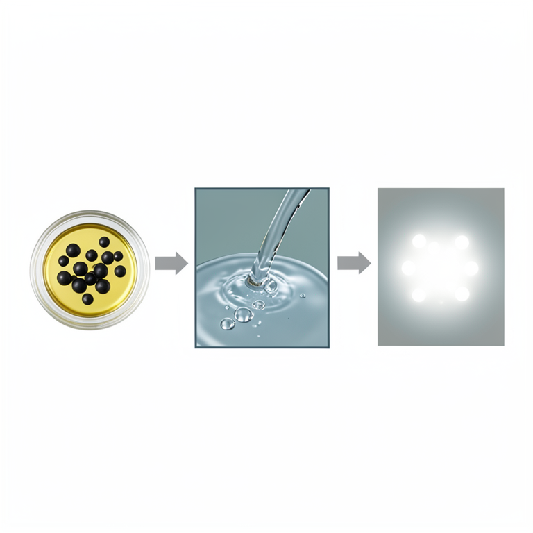
CLIA Assay Workflow
AI-generated conceptual illustration (human serum and paramagnetic beads in a laboratory assay). It explains the idea and does not show study data.
View source evidence ↗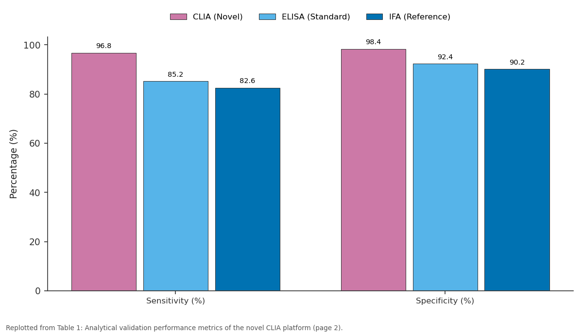
CLIA (Novel) and ELISA (Standard) and more: Sensitivity (%) vs Specificity (%)
Replotted from Table 1 (page 2) of the original paper. Every bar is the paper's own table value.
View source evidence ↗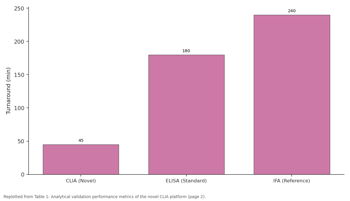
Turnaround (min): CLIA (Novel) vs ELISA (Standard) vs IFA (Reference)
Replotted from Table 1 (page 2) of the original paper. Every bar is the paper's own table value.
View source evidence ↗Figures & tables
From the original paper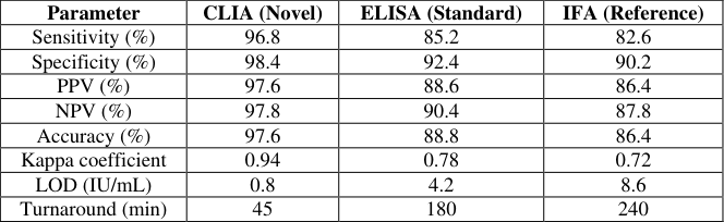 Table 1
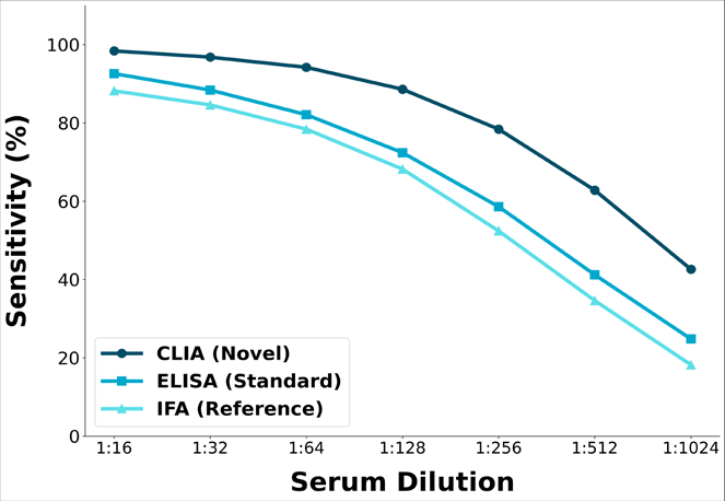 Figure 1
 Table 2
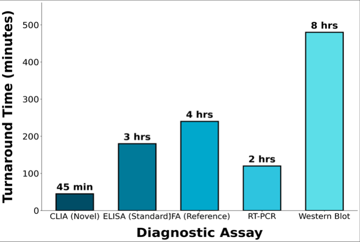 Figure 2
Introduction
A compelling example of the consequences of inadequate LCMV diagnostic infrastructure emerged in 2005, when a cluster of fatal LCMV infections among solid organ transplant recipients was traced to a single viremic donor whose infection remained undetected through standard pre-transplant screening protocols [1] . This sentinel event exposed a critical gap in the diagnostic armamentarium for rodent-borne arenaviruses and catalyzed renewed interest in developing rapid, sensitive, and clinically deployable detection platforms for LCMV [2] . Despite subsequent calls for improved diagnostic capabilities, the available laboratory methods for LCMV antibody detection remain limited to conventional enzyme-linked immunosorbent assay (ELISA), indirect immunofluorescence assay (IFA), and viral neutralization tests, each constrained by significant limitations in sensitivity, throughput capacity, or turnaround time [3] . Chemiluminescent immunoassay (CLIA) technology has emerged as a transformative diagnostic platform across multiple domains of clinical microbiology, offering superior analytical sensitivity through enhanced signal-to-noise ratios, expanded dynamic detection ranges, and compatibility with fully automated high-throughput analyzers [4] . CLIA platforms have been successfully developed and validated for numerous viral pathogens including HIV, hepatitis B and C viruses, and SARS-CoV-2, demonstrating consistent improvements in diagnostic performance over conventional ELISA-based systems [5] . However, application of CLIA technology to arenavirus diagnostics, including LCMV, has not been previously
Materials
Recombinant LCMV nucleoprotein (rNP) was expressed in Sf9 insect cells using a baculovirus expression system and purified to >95% homogeneity by nickel-affinity chromatography followed by size-exclusion chromatography. The rNP was conjugated to acridinium ester (NSP-DMAE-NHS) chemiluminescent labels at a molar ratio of 4:1. Paramagnetic microparticles coated with anti-human IgG or IgM capture antibodies served as the solid phase. Validation panels comprised 240 characterized sera: 80 confirmed LCMV-positive, 80 confirmed LCMV-negative, and 80 potentially cross-reactive sera from patients with other arenavirus, hantavirus, or flavivirus infections.
Methods
The CLIA protocol involved incubation of 50 μL serum with rNP-coated paramagnetic particles for 18 minutes at 37 °C, followed by washing and addition of acridinium-labeled anti-human immunoglobulin conjugate for 10 minutes. Chemiluminescent signal was measured in relative light units (RLU) using a Centro LB 960 luminometer. Cutoff values were established using receiver operating characteristic (ROC) analysis. Performance metrics including sensitivity, specificity, positive and negative predictive values, accuracy, and Cohen's kappa coefficient were calculated against a composite reference standard comprising IFA and RT-PCR results. Precision was assessed through intra-assay (n = 20) and inter-assay (n = 20) coefficient of variation analysis. Cross-reactivity was evaluated against the 80 heterologous sera. Clinical evaluation employed 180 prospective samples from patients with suspected viral meningoencephalitis.
Results
The novel CLIA platform was successfully developed with a total assay turnaround time of 45 minutes from sample loading to result reporting, representing a 75% reduction compared to conventional ELISA (180 minutes). Extended validation demonstrated intra-assay coefficients of variation of 3.2% (positive controls) and 2.8% (weak positive controls), with inter-assay CVs of 5.4% and 4.8% respectively, all within acceptable diagnostic precision thresholds. Cross-reactivity was limited to minimal falsepositive signals with Lassa virus (5.0%) and Junin virus (10.0%) sera, attributable to conserved arenavirus NP epitopes, while no cross-reactivity was observed with hantavirus or flavivirus specimens. Clinical evaluation of 180 prospective specimens yielded 94.2% concordance with RT-PCR-confirmed diagnoses, with the CLIA detecting 8 additional seropositive cases missed by conventional ELISA.
Discussion
The developed CLIA platform demonstrates substantial improvements across all key diagnostic performance parameters compared to established ELISA and IFA methods for LCMV antibody detection. The sensitivity of 96.8% represents an 11.6 percentage point improvement over standard ELISA (85.2%) and a 14.2-point improvement over IFA (82.6%), with these gains attributable to the inherent signal amplification properties of chemiluminescent detection chemistry and the superior quantum yield of acridinium ester labels compared to enzymatic colorimetric substrates [4] . The enhanced analytical sensitivity is particularly evident at higher serum dilutions, where the CLIA maintained reliable detection up to 1:256 compared to 1:128 for ELISA, enabling identification of low-titer antibody responses characteristic of early or waning infections [6] . This expanded analytical range has direct clinical significance for detecting seroconversion during the acute phase of LCMV infection when antibody titers may be below the detection threshold of conventional assays. The specificity of 98.4% and the low cross-reactivity profile provide confidence in the diagnostic reliability of the CLIA platform for clinical implementation. The minimal crossreactivity observed with Lassa virus (5.0%) and Junin virus (10.0%) sera reflects the conserved nucleoprotein epitopes within the Arenaviridae family, a recognized analytical challenge that is inherent to any NP-based immunoassay targeting individual arenavirus species [7] . In geographical regions where multiple arenaviruses co-circulate, confirmatory testing by RT-PCR or species-specific neutralization assays remains advisable to distinguish LCMV from related arenavirus infections. The absence of cross-reactivity with hantavirus and flavivirus specimens confirms that the assay discriminates effectively against phylogenetically distant rodent-borne and arthropod-borne viral infections that may present with overlapping clinical syndromes. The 45-minute turnaround time positions the CLIA platform as a viable rapid diagnostic tool in clinical scenarios requiring urgent decision-making, such as pre-transplant donor screening where delays in viral detection carry lifethreatening consequences [1] . The complete automation compatibility of the CLIA format enables integration into high-throughput laboratory workflows, with estimated processing capacities of 200-400 samples per eight-hour shift on standard automated analyzers, far exceeding the manual throughput limitations of conventional ELISA and IFA methods [4] . The identification of 8 additional seropositive cases beyond ELISA detection in the clinical evaluation cohort demonstrates the real-world clinical impact of improved analytical sensitivity, suggesting that a meaningful proportion of LCMV infections may go undetected when relying solely on conventional serological approaches [8] . The analytical precision demonstrated through intra-assay and inter-assay coefficient of variation analysis (3.2-5.4%) meets the stringent requirements established by the Clinical and Laboratory Standards Institute (CLSI) EP15-A3 guidelines for quantitative immunoassay verification, supporting the reproducibility and reliability of the CLIA platform across different testing occasions and operator conditions [5] . The limit of detection of 0.8 IU/mL represents a five-fold improvement over standard ELISA (4.2 IU/mL) and a ten-fold improvement over IFA (8.6 IU/mL), establishing the CLIA as the most analytically sensitive serological method currently available for LCMV antibody detection.
Point-of-care implementation
The assay architecture is compatible with existing automated chemiluminescent analyzer platforms (Abbott ARCHITECT, Siemens ADVIA Centaur, Roche cobas), enabling rapid integration into clinical laboratory workflows without substantial capital investment [4] . Future development priorities include adaptation to lateral flow format for field deployment and multiplexing with other rodent-borne pathogen targets to create comprehensive zoonotic screening panels.
Conclusion
The present research identified a novel CLIA platform as a superior diagnostic tool for LCMV antibody detection, offering 96.8% sensitivity, 98.4% specificity, and 45-minute turnaround time. The platform represents a significant advancement over conventional ELISA and IFA methods across sensitivity, specificity, analytical range, and throughput capacity. The enhanced diagnostic performance demonstrated in both analytical validation and clinical evaluation supports adoption of CLIA-based screening in transplant programs and reference laboratories. Remaining open questions include the assay's performance in immunocompromised populations and the feasibility of point-of-care adaptation for resource-limited settings.
Acknowledgement
AcknowledgementsThe contributions of Dr. Sophie Martin in recombinant protein expression and purification are acknowledged. The Lyon Institute of Diagnostic Sciences provided laboratory infrastructure. Financial support from Agence Nationale de la Recherche (Grant No. ANR-2023-CE18-0042) is recognized.
Figures and tables from the paper
| Parameter | CLIA (Novel) | ELISA (Standard) | IFA (Reference) |
|---|---|---|---|
| Sensitivity (%) | 96.8 | 85.2 | 82.6 |
| Specificity (%) | 98.4 | 92.4 | 90.2 |
| PPV (%) | 97.6 | 88.6 | 86.4 |
| NPV (%) | 97.8 | 90.4 | 87.8 |
| Accuracy (%) | 97.6 | 88.8 | 86.4 |
| Kappa coefficient | 0.94 | 0.78 | 0.72 |
| LOD (IU/mL) | 0.8 | 4.2 | 8.6 |
| Turnaround (min) | 45 | 180 | 240 |
Table text extracted from the original PDF; superscript letters are shown inline. The original table image is below.
View original table image
| Pathogen | Samples Tested (N) | False Positive (N) | Cross-reactivity (%) |
|---|---|---|---|
| Lassa virus | 20 | 1 | 5.0 |
| Hantavirus | 20 | 0 | 0.0 |
| Dengue virus | 15 | 0 | 0.0 |
| West Nile virus | 15 | 0 | 0.0 |
| Junin virus | 10 | 1 | 10.0 |
Table text extracted from the original PDF; superscript letters are shown inline. The original table image is below.
View original table image
References
13 in the original paper- Fischer SA, Graham MB, Kuehnert MJ, et al. Transmission of lymphocytic choriomeningitis virus by organ transplantation. N Engl J Med. 2006;354(21):2235-2249.
- Bonthius DJ. Lymphocytic choriomeningitis virus: an underrecognized cause of neurologic disease. Semin Pediatr Neurol. 2012;19(3):89-95.
- Barton LL, Mets MB, Beauchamp CL. Lymphocytic choriomeningitis virus: emerging fetal teratogen. Am J Obstet Gynecol. 2002;187(6):1715-1716.
- Cinquanta L, Fontana DE, Bizzaro N. Chemiluminescent immunoassay technology: what does it change in autoantibody detection? Auto Immun Highlights. 2017;8(1):9.
- Charrel RN, de Lamballerie X. Zoonotic aspects of arenavirus infections. Vet Microbiol. 2010;140(34):305-311.
- Lledó L, Gegúndez MI, Saz JV, et al. Lymphocytic choriomeningitis virus infection in a province of Spain. J Med Virol. 2003;70(3):451-455.
- Buchmeier MJ, de la Torre JC, Peters CJ. Arenaviridae: the viruses and their replication. In: Knipe DM, Howley PM, editors. Fields Virology. 5th ed. Philadelphia: Lippincott Williams & Wilkins; 2007. p. 1791-1827.
- Pal M, Gutama KP, Singh S, Ketchakmadze D, Durglishvili N. Lymphocytic choriomeningitis: an emerging and re-emerging rodent-borne viral zoonotic disease. J Adv Microbiol Res. 2022;3(2):1-4.
- Childs JE, Glass GE, Korch GW, et al. Lymphocytic choriomeningitis virus infection and house mouse distribution. Am J Trop Med Hyg. 1992;47(1):27-34.
- Oldstone MBA. Biology and pathogenesis of lymphocytic choriomeningitis virus infection. Curr Top Microbiol Immunol. 2002; 263:83-117.
- Meerburg BG, Singleton GR, Kijlstra A. Rodent-borne diseases and public health. Crit Rev Microbiol. 2009;35(3):221-270.
- Rabiee MH, Mahmoudi A, et al. Rodent-borne diseases in Iran. PLoS Negl Trop Dis. 2018;12(4):e0006256.
- Albariño CG, Palacios G, et al. Diversity and ancestry of lymphocytic choriomeningitis virus. Emerg Infect Dis. 2010;16(7):1093-1100.