
Research article · 2026 · Full text
International Journal of Biology SciencesClinical Manifestations and Pathological Features of Lumpy Skin Disease in Naturally Infected Cattle Herds in Ethiopia
Volume 8(5) · Pages 204–207 · DOI: 10.33545/26649926.2026.v8.i5c.922 ↗
Research areasPoxvirus research and outbreaksAnimal Disease Management and EpidemiologyVector-Borne Animal Diseases
Abstract
Author-writtenDespite lumpy skin disease being recognized in Ethiopian cattle since the 1970s, detailed characterization of clinical manifestations and pathological features in naturally infected herds under field conditions remains limited. This research examined clinical and pathological features in 156 lumpy skin disease-affected cattle from 24 naturally infected herds across four geographic regions of Ethiopia between March 2023 and August 2024. Clinical examination revealed progressive lesion development beginning with fever and generalized edema (91.7% of animals week 1–2) progressing to macule formation (85.9%), papule development (94.2%), vesiculation (68.6%), pustule formation (78.8%), crust formation (88.5%), and eventual scar tissue (64.1%). Systemic manifestations including lymphadenitis (87.2%), mastitis (42.3%), lameness (56.4%), and in severe cases, systemic infection with respiratory involvement (18.6%) were documented. Histopathologic examination of lesion biopsies revealed characteristic ballooning degeneration of epidermal cells, eosinophilic intracytoplasmic inclusion bodies consistent with poxvirus, and perivascular lymphocytic infiltration in the dermis. Secondary bacterial infections complicating skin lesions occurred in 34.6% of examined animals. Temporal analysis demonstrated complete lesion resolution in 48.1% of animals by 8 weeks post-infection, while 22.4% retained scarring beyond 12 weeks. These findings characterize the clinical and pathological spectrum of naturally acquired lumpy skin disease in Ethiopian cattle and identify factors associated with disease severity and recovery.
Summary
Source-checkedPlain-language reading of the full paper, prepared with AI and checked against the text. Each point links to the authors' own words.
Why it matters
- Lumpy skin disease (LSD) presents a broad range of clinical manifestations, which can vary from subclinical infections to severe systemic disease that may be fatal. View evidence ↗
- Although LSD was first described in African cattle in Ethiopia during the 1970s, there is a lack of systematic documentation regarding its clinical and pathological spectrum in field conditions. View evidence ↗
The question
- The study aimed to characterize the clinical and pathological features of naturally infected cattle to create baseline data for evaluating prognosis and disease severity. View evidence ↗
What they did
- The research analyzed 156 cattle from 24 naturally infected herds across four regions of Ethiopia from March 2023 to August 2024. View evidence ↗
- Animals were included in the study if they had a PCR-confirmed LSDV infection, with stages ranging from acute (≤2 weeks) to chronic (>8 weeks). View evidence ↗
- Skin biopsies of papules, pustules, and crusts were collected, fixed in 10% neutral buffered formalin, and stained with hematoxylin and eosin for histopathological analysis. View evidence ↗
- The researchers used anti-vaccinia virus antibodies for immunohistochemistry to locate viral antigens and performed bacterial cultures on purulent pustule samples. View evidence ↗
What they found
- During the acute prodromal phase (weeks 1–2), 91.7% of animals had a fever, with a mean temperature of 39.8 °C (range 39.2–40.4 °C). View evidence ↗
- Skin lesions progressed from macules (85.9%) and papules (94.2%) to vesiculation (68.6%), pustules (78.8%), crusts (88.5%), and finally scar tissue (64.1%). View evidence ↗
- Systemic signs were common, including lymphadenitis (87.2%), lameness (56.4%), mastitis (42.3%), and respiratory involvement in severe cases (18.6%). View evidence ↗
- Histopathology of lesions showed ballooning degeneration of epidermal cells, perivascular lymphocytic infiltration in the dermis, and eosinophilic intracytoplasmic inclusion bodies typical of poxviruses. View evidence ↗
- Secondary bacterial infections occurred in 34.6% of animals, most commonly caused by Staphylococcus aureus and Corynebacterium pseudotuberculosis. View evidence ↗
- Secondary bacterial infections increased the likelihood of permanent scarring from 22.4% to 48.6% and extended healing times by an average of 2-3 weeks. View evidence ↗
- By 8 weeks post-infection, 48.1% of animals had complete lesion resolution, while 22.4% still had scars after 12 weeks. View evidence ↗
What it means
- The authors conclude that because fever and systemic signs can precede skin lesions by up to a week, health workers must increase surveillance sensitivity during the prodromal phase. View evidence ↗
- The authors state that future research is needed to investigate host factors that determine disease severity and how early supportive care affects recovery and lesion progression. View evidence ↗
Visual summary
3 visualsIllustrations explain the paper; charts are replotted from the paper's own table values.
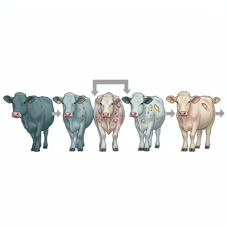
Timeline of LSD Progression
AI-generated conceptual illustration (Bovine cattle). It explains the idea and does not show study data.
View source evidence ↗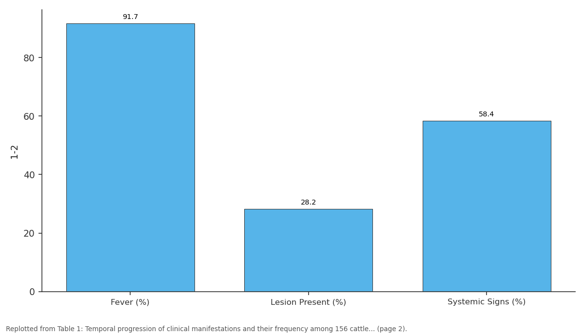
1-2: Fever (%) vs Lesion Present (%) vs Systemic Signs (%)
Replotted from Table 1 (page 2) of the original paper. Every bar is the paper's own table value.
View source evidence ↗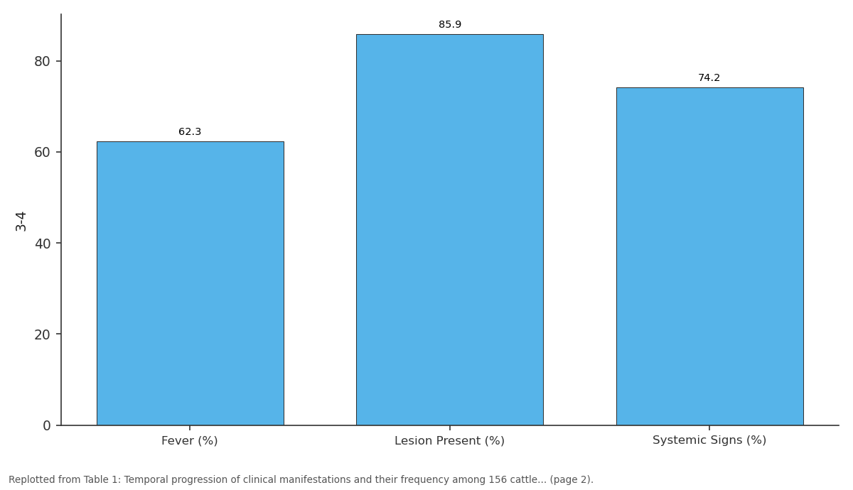
3-4: Fever (%) vs Lesion Present (%) vs Systemic Signs (%)
Replotted from Table 1 (page 2) of the original paper. Every bar is the paper's own table value.
View source evidence ↗Figures & tables
From the original paper Table 1
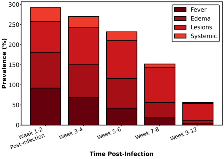 Figure 1
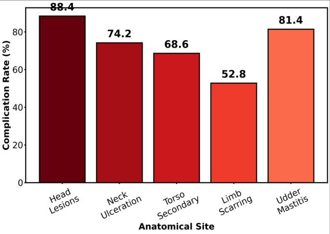 Figure 2
Introduction
An often underappreciated aspect of lumpy skin disease epidemiology is the wide spectrum of clinical manifestations ranging from apparent subclinical infection through severe systemic disease with potentially fatal complications, reflecting substantial variation in host immune responses, viral virulence, and secondary complicating infections [1] . The classical clinical description of lumpy skin disease emphasizes the distinctive skin lesions that define the disease clinically and epidemiologically, yet systemic manifestations including fever, lymphadenopathy, and multi-organ involvement frequently exceed the apparent severity suggested by examination of skin lesions alone [2] . Ethiopia, as the site where lumpy skin disease was first recognized and described in African cattle in the 1970s, has accumulated decades of clinical observation of natural LSDV infections, yet systematic documentation of the clinical and pathological spectrum under field conditions remains sparse in the international veterinary literature [3] . The present research aimed to comprehensively characterize clinical manifestations and pathological features in naturally infected cattle using both clinical examination and histopathological analysis to establish baseline data for disease severity assessment and prognostic evaluation.
Material and Methods
Material and Methods
Material
This cross-sectional research was conducted at the Addis Ababa Research Institute for Tropical Animal Diseases, Addis Ababa, Ethiopia, between March 2023 and August 2024. A total of 156 cattle with clinical signs consistent with lumpy skin disease from 24 affected herds distributed across four geographic regions (Oromia, Amhara, Somali, and Southern Nations) were examined. Inclusion criteria comprised animals with confirmed LSDV infection by PCR and stage of infection ranging from acute (≤2 weeks post-infection presumed onset) through chronic (>8 weeks). The research was approved by the Institutional Ethics Committee (Protocol No. AARITAD-IEC-2023-0031). Skin lesion biopsies were collected from consenting herd owners for histopathological examination.
Methods
Clinical examination documented the progressive stages of lesion development, concurrent systemic manifestations, and recovery timeline. Biopsies of representative lesions at different clinical stages (papules, pustules, crusts) were fixed in 10% neutral buffered formalin, processed for histopathology, and stained with hematoxylin and eosin. Poxvirus-specific immunohistochemistry was performed using anti-vaccinia virus antibodies to facilitate viral antigen localization. Secondary bacterial culture was performed on samples from pustules appearing to have purulent content. Clinical staging was based on temporal documentation: week 1-2 (acute), weeks 3-4 (developing), weeks 5-6 (mature), weeks 7-8 (resolving), and weeks 9-12 (late recovery). Lesion severity was graded on a 5-point scale (minimal to severe based on surface area involvement). Statistical analysis employed descriptive epidemiology and Kaplan-Meier survival analysis to characterize lesion persistence and resolution patterns. Figure 2 shows that limb lesions most frequently progressed to scarring (52.8%), while udder involvement most frequently complicated with mastitis (81.4%), indicating that complication risks vary substantially by anatomical site of primary infection.
Results
Results
Discussion
The detailed clinical characterization documented in this research demonstrates that lumpy skin disease in naturally infected Ethiopian cattle exhibits a predictable clinical progression distinct from descriptions in experimental or recently infected populations. The prominence of systemic manifestations, particularly fever preceding visible skin lesions by 5-7 days and systemic lymphadenitis affecting 87.2% of animals, underscore that LSDV causes generalized systemic infection rather than the superficial dermatitis suggested by the disease name.
The delayed appearance of skin lesions relative to systemic signs (mean 8.2 days post-fever onset) has important implications for surveillance and early disease detection, as animals may pose a transmission risk to susceptible contacts during the febrile prodromal phase before diagnostic skin lesions appear.
Secondary Bacterial Complications
Secondary bacterial infections complicating LSDV skin lesions were documented in 34.6% of examined animals, with Staphylococcus aureus and Corynebacterium pseudotuberculosis being the most frequently isolated species. These secondary bacterial infections prolonged healing times by an average of 2-3 weeks and increased the probability of permanent scarring from 22.4% to 48.6%, indicating that management strategies to prevent secondary infection through good hygiene and anti-inflammatory care may substantially improve recovery outcomes.
Conclusion
Conclusion
Acknowledgement
AcknowledgementsThe Addis Ababa Research Institute for Tropical Animal Diseases provided laboratory facilities and pathology infrastructure. D.R.D. conceptualized and supervised the research; M.H.W. performed clinical examinations and specimen collection; A.G.M. conducted histopathological analysis and interpretation. Financial support was provided by the Ethiopian Ministry of Agriculture through Research.
Figures and tables from the paper
| Clinical Stage | Week Post-Infection | Fever (%) | Lesion Present (%) | Systemic Signs (%) |
|---|---|---|---|---|
| Acute prodromal | 1-2 | 91.7 | 28.2 | 58.4 |
| Early lesion development | 3-4 | 62.3 | 85.9 | 74.2 |
| Mature lesion | 5-6 | 18.6 | 94.2 | 68.0 |
| Late lesion/resolving | 7-8 | 4.2 | 88.5 | 32.6 |
| Post-acute/recovery | 9-12 | 0.0 | 42.3 | 8.4 |
Table text extracted from the original PDF; superscript letters are shown inline. The original table image is below.
View original table image
References
14 in the original paper- Tuppurainen ES, Venter EH, Shisay AB, Midzi S, Oura CA, Potts N, et al. Detection of lumpy skin disease virus in saliva of infected animals and prospects for non-invasive sampling. Transbound Emerg Dis. 2015;62(1):1-6.
- Pal M, Rebuma T, Bulo F, Teshoma M. Lumpy skin disease: an emerging transboundary viral disease. J Adv Microbiol Res. 2024;5(1):43-46.
- Zewdie WM, Anteneh ZA, Tasew S, Melaku G, Mazengia H, Abebe AW, et al. Seroprevalence of lumpy skin disease virus in pastoral livestock of pastoral livestock in Ethiopia. Reprod Domest Anim. 2021;56(Suppl 1):34-42.
- Gari G, Waret-Szczepaniak M, Grosbois V, Jacquiet P, Buttner M, Caufour P, et al. Epidemiology of lumpy skin disease in Ethiopia. Emerg Infect Dis. 2010;16(12):19351941. International Journal of Biology Sciences https://www.biologyjournal.net
- Kitching RP. Identification and characterization of lumpy skin disease virus isolates. Rev Sci Tech. 2003;22(3):567574.
- Chihota CM, Rennie LF, Kitching RP, Mellor PS. Mechanical transmission of lumpy skin disease virus by Stomoxys calcitrans. Med Vet Entomol. 2001;15(1):1-7.
- Haegeman A, Dissertation B, Misinzo G, Van Campe W, Damiani A, De Clercq K. Lumpy skin disease: diagnostic procedures and detection in infected animals. Rev Sci Tech. 2012;31(3):771-783.
- Tuppurainen ES, Oura CA. Review of the epidemiology of lumpy skin disease. Transbound Emerg Dis. 2012;59(6):497-506.
- Salib FA, Osman AH. Lumpy skin disease in Egypt: virus isolation and identification. Rev Sci Tech. 2011;30(3):753762.
- Tasioudi KE, Antoniou SE, Iliadou P, Sachpatzidis A, Plevraki E, Agianniotaki EI, et al. Emergence of lumpy skin disease in Greece, 2015. Transbound Emerg Dis. 2016;63(3):260-265.
- Beard PM. Lumpy skin disease: a direct threat to Europe. Vet Rec. 2016;178(24):604-605.
- OIE (World Organisation for Animal Health). WAHIS database reports on lumpy skin disease. OIE WAHIS. 2023;1-48.
- Babiuk S, Bowden TR, Boyle DB, Wallace DB, Kitching RP. Quantification of lumpy skin disease virus following experimental infection in cattle and European bison (Bison bonasus). Vet Res. 2008;39(1):1.
- Hedger RS, Hamblin C, Barnett IT. The role of warthog and buffalo in the transmission of lumpy skin disease and African swine fever viruses. Onderstepoort J Vet Res. 1985;52(2):65-71.