
Research article · 2026 · Full text
International Journal of Biology SciencesHydatidosis as a major cause of organ condemnation and economic losses in catle slaughtered at Ibadan central abatoir, Southwestern Nigeria
Volume 8(6) · Pages 162–166 · DOI: 10.33545/26649926.2026.v8.i6b.926 ↗
Research areasParasitic infections in humans and animalsHelminth infection and controlParasitic Infections and Diagnostics
Abstract
Author-writtenHydatidosis remains one of the most economically devastating parasitic diseases of livestock worldwide, yet its prevalence and economic impact at slaughterhouses in Nigeria remain insuficiently documented. This research was designed to determine the prevalence, organ distribution, cyst characteristics, and economic significance of bovine hydatidosis in catle slaughtered at Ibadan central abatoir, Southwestern Nigeria, from March 2022 to February 2023. A total of 924 catle were examined through systematic ante-mortem and post-mortem inspections. The overall prevalence of hydatidosis was 28.6% (264/924). The liver was the most commonly afected organ at 45.8% (121/264), followed by the lung at 35.2% (93/264), kidney at 11.4% (30/264), and heart at 7.6% (20/264). Of the 482 hydatid cysts recovered, 42.5% were fertile, 38.2% were sterile, and 19.3% were calcified. Cyst fertility rates were significantly higher in lung cysts (55.3%) compared to liver cysts (42.5%), kidney cysts (28.7%), and heart cysts (18.2%) (p < 0.001). The total annual economic loss atributed to hydatidosis-related organ condemnation was estimated at ₦8,428,500 (approximately 10,285 USD). Age, sex, and body condition score were significantly associated with hydatid cyst prevalence (p < 0.05). These findings demonstrate that hydatidosis constitutes a major cause of organ condemnation at Ibadan central abatoir, necessitating implementation of integrated control strategies targeting the parasite life cycle through dog deworming programs, improved abatoir waste management, and community health education.
Summary
Source-checkedPlain-language reading of the full paper, prepared with AI and checked against the text. Each point links to the authors' own words.
Why it matters
- Hydatidosis is recognized as one of the most economically damaging parasitic diseases affecting livestock on a global scale. View evidence ↗
- The disease is caused by the tapeworm Echinococcus granulosus, which cycles between dogs (definitive hosts) and livestock (intermediate hosts), occasionally infecting humans. View evidence ↗
- In livestock, cysts primarily form in the lungs and liver, leading to the condemnation of these organs during slaughter and causing direct financial losses. View evidence ↗
The question
- The study aimed to determine the prevalence, organ distribution, cyst characteristics, and economic impact of bovine hydatidosis at the Ibadan central abattoir between March 2022 and February 2023. View evidence ↗
What they did
- The researchers used systematic random sampling to examine 924 cattle, selecting every fourth animal on the slaughter line. View evidence ↗
- Post-mortem inspections included visual checks, palpation, and incisions of the spleen, kidneys, heart, lungs, and liver. View evidence ↗
- Cysts were categorized as fertile (protoscolices present), sterile (no protoscolices in clear fluid), or calcified (caseous or mineralized contents). View evidence ↗
- Economic losses were estimated by multiplying the annual number of condemned organs by their average weight and the current retail market price per kilogram. View evidence ↗
What they found
- The overall prevalence of hydatidosis among the examined cattle was 28.6% (264 out of 924 animals). View evidence ↗
- The liver was the most affected organ (45.8%), followed by the lungs (35.2%), kidneys (11.4%), and heart (7.6%). View evidence ↗
- Infection prevalence was higher in males (32.4%) than females (23.8%) and increased with age, peaking at 38.5% in cattle older than 8 years. View evidence ↗
- Cattle with poor body condition scores had a significantly higher infection rate (35.8%) compared to those with medium (27.4%) or good (20.2%) scores. View evidence ↗
- Of all recovered cysts, 42.5% were fertile, 38.2% were sterile, and 19.3% were calcified. View evidence ↗
- Lung cysts had the highest fertility rate (55.3%), which was significantly higher than those in the liver (42.5%), kidney (28.7%), and heart (18.2%). View evidence ↗
- The estimated annual economic loss due to organ condemnation from hydatidosis was ₦8,428,500 (approximately 10,285 USD). View evidence ↗
- Liver condemnation caused the most financial loss (53.8%), followed by the lungs (21.2%), kidneys (14.9%), and heart (10.1%). View evidence ↗
What it means
- The authors recommend a comprehensive control program including mandatory dog registration, periodic praziquantel deworming, improved organ disposal at abattoirs, and public awareness campaigns. View evidence ↗
- The authors suggest that future molecular epidemiological studies using genotyping are needed to clarify strain diversity and host specificity to better target control strategies. View evidence ↗
Visual summary
2 visualsIllustrations explain the paper; charts are replotted from the paper's own table values.
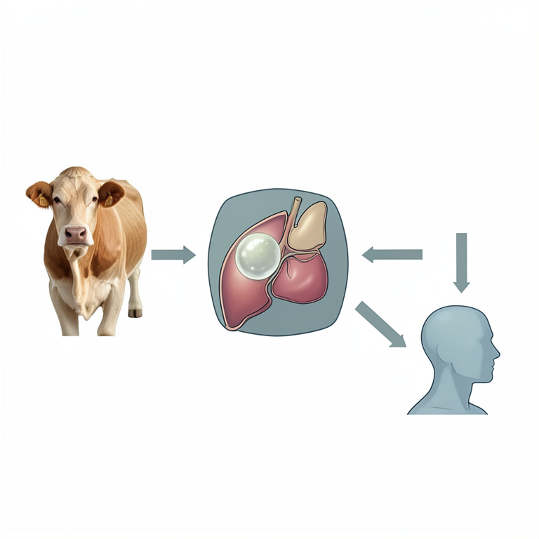
Transmission Cycle of Echinococcus granulosus
AI-generated conceptual illustration (Dogs and cattle in an abattoir setting). It explains the idea and does not show study data.
View source evidence ↗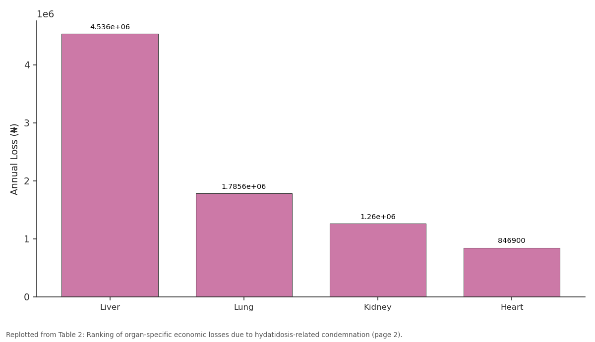
Annual Loss (₦): Liver vs Lung vs Kidney vs Heart
Replotted from Table 2 (page 2) of the original paper. Every bar is the paper's own table value.
View source evidence ↗Figures & tables
From the original paper Table 1
 Table 2
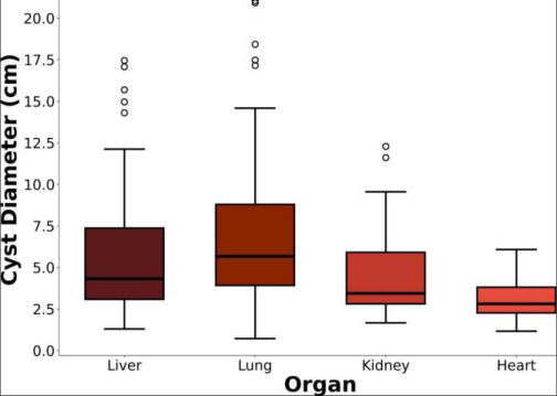 Figure 1
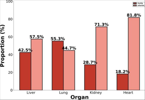 Figure 2
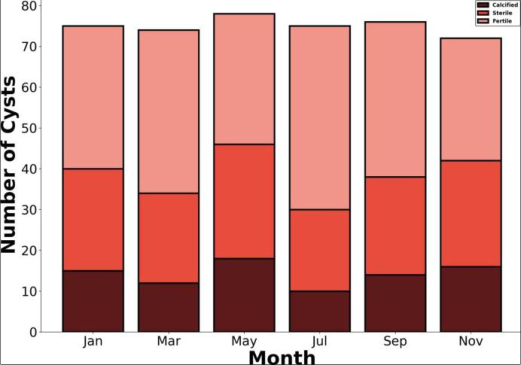 Figure 3
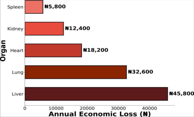 Figure 4
Introduction
Cystic echinococcosis, commonly known as hydatidosis, represents the most widespread and economically important larval cestode infection afecting livestock and humans in tropical and subtropical regions of the world. Caused by the metacestode stage of the tapeworm Echinococcus granulosus, hydatid disease maintains its transmission cycle through a predatorprey relationship involving domestic dogs as definitive hosts and various livestock species as intermediate hosts, with humans serving as accidental intermediate hosts upon ingestion of tapeworm eggs shed in dog feces. The global burden of cystic echinococcosis has been estimated at approximately 1 million disability-adjusted life years, with the disease recognized as a neglected tropical condition by the World Health Organization. In livestock, hydatid cysts develop predominantly in the liver and lungs, where they cause organ condemnation during post-mortem inspection at slaughterhouses, resulting in significant direct economic losses to the livestock industry. Beyond the immediate financial impact of organ condemnation, hydatidosis contributes to reduced animal productivity through decreased weight gain, impaired reproductive performance, and diminished carcass quality in heavily infected animals. Nigeria, with an estimated catle population of approximately 20 million, faces substantial economic losses atributable to bovine hydatidosis, although comprehensive prevalence data from major slaughterhouses across the country remain limited. The livestock sector contributes significantly to the national economy and plays a crucial role in providing animal protein for the growing population. However, the persistence of zoonotic parasitic diseases including hydatidosis reflects ongoing challenges in veterinary service delivery, livestock disease surveillance, and public health infrastructure across the country.
Ibadan, the capital of Oyo State in Southwestern Nigeria, hosts one of the largest urban livestock markets and central abatoirs in West Africa. The Ibadan central abatoir processes catle sourced from diverse geographical regions, including the northern pastoral zones where extensive livestock production systems predominate. This geographical diversity in the origin of slaughtered animals provides a unique opportunity to assess the prevalence and characteristics of hydatid cyst infections across diferent livestock production systems within a single slaughterhouse facility. Despite the recognized importance of hydatidosis as a cause of organ condemnation in Nigerian abatoirs, few investigations have provided detailed characterization of cyst morphology, fertility status, and viability in relation to organ predilection sites. Such information is essential for understanding the epidemiology of the parasite and for evaluating the relative public health significance of hydatid infections in diferent organs. This research was therefore undertaken to determine the prevalence of hydatidosis in catle at Ibadan central abatoir, characterize the distribution and biological features of recovered hydatid cysts, assess associated risk factors, and estimate the direct economic losses atributable to hydatidosisrelated organ condemnation.
Materials and Methods
Materials and Methods
Materials
The research was conducted at Ibadan central abatoir, located in Ibadan North Local Government Area, Oyo State, Southwestern Nigeria. The abatoir is situated at coordinates 7.4°N, 3.9°E and serves as the primary meat processing facility for Ibadan metropolis and surrounding communities, with an estimated daily throughput of 150 to 200 catle. The climate of the area is tropical with a mean annual rainfall of 1,270 millimeters and average temperatures ranging from 25 °C to 32 °C. Ethical approval for the research was obtained from the Animal Ethics Commitee of Ibadan Research Institute of Veterinary Medicine (Reference: IRIVM/AEC/2022-016). Permission to access the abatoir was granted by the Oyo State Ministry of Agriculture and Rural Development.
Methods
A cross-sectional research design spanning twelve months from March 2022 to February 2023 was employed. The sample size was calculated using the formula for single proportion estimation with a 95% confidence interval and 3% precision, based on a previously reported prevalence of 25% in southwestern Nigeria. A total of 924 catle were selected through systematic random sampling by examining every fourth animal on the slaughter line. Ante-mortem examination recorded animal identification, sex, estimated age by dentition, body condition score on a 1 to 5 scale, breed type, and origin. Post-mortem inspection involved systematic visual examination, palpation, and multiple incisions of the liver, lungs, heart, kidneys, and spleen following standard procedures. All detected hydatid cysts were carefully excised, counted, measured using calipers, and individually examined for fertility and viability. Cysts were classified as fertile when protoscolices were present upon microscopic examination of the cyst fluid, sterile when no protoscolices were detected in clear fluid, or calcified when the cyst contents were caseous or mineralized. Protoscolex viability was assessed using the 0.1% eosin exclusion technique, where viable protoscolices excluded the dye and appeared green while non-viable ones absorbed the dye and stained red. Economic loss estimation followed the methodology described by Ogunrinade and Ogunrinade, calculated as the product of the annual number of condemned organs, the average weight of each organ type, and the current retail market price per kilogram. Market prices were obtained through surveys at three major retail markets in Ibadan during the research period. Data were analyzed using STATA version 17.0, with chi-square tests for categorical associations, one-way ANOVA for continuous variables, and multivariable logistic regression for identifying independent risk factors. Cyst diameter varied significantly across organ sites (F=18.42 , p<0.001) . Lung cysts exhibited the largest median diameter of 6.1 cm (interquartile range: 3.2 to 12.4 cm), compared to liver cysts with a median of 4.5 cm (IQR: 2.8 to 8.7 cm), kidney cysts at 3.3 cm (IQR: 1.8 to 5.6 cm), and heart cysts at 2.7 cm (IQR:
Results
1.5 to 4.2 cm). The larger cyst sizes observed in the lung may be atributed to the relatively lower tissue resistance and greater compliance of pulmonary parenchyma compared to the more compact hepatic tissue. Overall, 42.5% of recovered cysts were fertile, 38.2% were sterile, and 19.3% were calcified. The fertility rate was significantly higher in lung cysts (55.3%) compared to liver (42.5%), kidney (28.7%), and heart (18.2%) cysts (χ ²=28.74 , p<0.001) ). Among fertile cysts, protoscolex viability assessed by eosin exclusion ranged from 72.4% in lung cysts to 58.6% in liver cysts, indicating that pulmonary hydatid cysts represent a greater risk for perpetuating the parasite life cycle if fed to dogs. September. This temporal patern may reflect seasonal variations in dog-livestock contact paterns and environmental conditions afecting egg survival and transmission dynamics. The total estimated annual economic loss due to hydatidosisrelated organ condemnation was ₦8,428,500 (approximately 10,285 USD). Liver condemnation accounted for the largest share at 53.8%, followed by lung at 21.2%, kidney at 14.9%, and heart at 10.1%. These estimates represent direct losses from organ condemnation only and do not include indirect losses from reduced live weight gain, decreased milk production, or diminished reproductive performance in infected animals.
Discussion
The overall prevalence of bovine hydatidosis of 28.6% documented at Ibadan central abatoir indicates that the disease remains a major cause of organ condemnation in Southwestern Nigeria. This finding is consistent with the 25.4% prevalence reported by Adediran and colleagues at a slaughterhouse in Lagos [1] and the 31.2% documented by Opara and colleagues in southeastern Nigeria [2] . However, the current prevalence exceeds the 18.7% reported by Ola-Fadunsin and Ganiyu at Ilorin abatoir [3] , possibly reflecting diferences in the geographical origins of catle processed at diferent facilities and variations in the intensity of dog-livestock interactions across regions. The predominance of liver involvement in hydatid infections observed in this research, with 45.8% of cysts localized in the liver, is consistent with the established pathophysiology of hydatidosis. Ingested oncospheres released from Echinococcus granulosus eggs penetrate the intestinal mucosa and enter the portal venous system, encountering the liver as the first capillary filter. Oncospheres that pass through the hepatic capillary bed reach the lungs via the pulmonary circulation, explaining the secondary predilection for pulmonary localization. The relatively lower frequency of kidney and heart involvement reflects the decreasing probability of oncosphere dissemination to more distal sites through the systemic circulation [4] . The significantly higher cyst fertility rate observed in lungs (55.3%) compared to liver (42.5%) has important epidemiological implications for the maintenance of the parasite life cycle. In areas where condemned organs are fed to dogs or discarded in locations accessible to canines, pulmonary hydatid cysts represent a disproportionately greater source of infection for definitive hosts. The higher fertility and viability of lung cysts may be atributed to the more favorable growth conditions provided by the compliant pulmonary tissue, which allows unrestricted cyst expansion and optimal protoscolex development [5] .
The estimated annual economic loss of ₦8,428,500 due to hydatidosis-related organ condemnation represents a substantial financial burden on the livestock value chain in Ibadan. When extrapolated across major abatoirs in Nigeria, the national economic impact of bovine hydatidosis is likely to be considerable, although comprehensive national estimates are currently unavailable. The economic analysis presented in this research provides a basis for cost-benefit assessments of potential control interventions, including dog deworming programs and improved abatoir waste management systems [6] .
One health approach to hydatidosis control
One health approach to hydatidosis control
Conclusion
In the context of the persistent burden of zoonotic parasitic diseases in West Africa, this research has demonstrated that hydatidosis remains a highly prevalent cause of organ condemnation at Ibadan central abatoir, afecting 28.6% of slaughtered catle with significant organ-specific variations in cyst distribution, fertility, and size. The findings highlight that lung cysts pose the greatest epidemiological risk due to their higher fertility rates, while liver condemnation contributes the largest proportion of economic losses. It is recommended that the Oyo State Government implement a comprehensive hydatidosis control program incorporating mandatory dog registration and periodic praziquantel deworming, installation of proper organ disposal facilities at the abatoir, and public awareness campaigns targeting all stakeholders in the livestock value chain. Looking ahead, molecular epidemiological investigations using genotyping of Echinococcus granulosus isolates from this region would clarify the strain diversity and host specificity paterns, informing the design of more precisely targeted control strategies.
Acknowledgement
AcknowledgementsFinancial support for this research was provided by the Tertiary Education Trust Fund of Nigeria (TETFund) through Institutional Based Research. The management and veterinary inspection staf of Ibadan central abatoir are gratefully acknowledged for their invaluable assistance during data collection. AOB designed the research and drafted the manuscript; NCO performed parasitological examinations; ECN conducted statistical analysis and contributed to manuscript preparation.
Figures and tables from the paper
| Variable | Cyst count | Cyst size | Fertility rate | Viability | Host age |
|---|---|---|---|---|---|
| Cyst count | 1.000 | 0.423* | 0.318* | 0.285* | 0.612* |
| Cyst size (cm) | 0.423* | 1.000 | 0.548* | 0.492* | 0.387* |
| Fertility rate (%) | 0.318* | 0.548* | 1.000 | 0.724* | 0.256* |
| Viability (%) | 0.285* | 0.492* | 0.724* | 1.000 | 0.198 |
| Host age (years) | 0.612* | 0.387* | 0.256* | 0.198 | 1.000 |
Table text extracted from the original PDF; superscript letters are shown inline. The original table image is below.
View original table image
| Rank | Organ | Condemned (n) | Avg Weight (kg) | Price (₦/kg) | Annual Loss (₦) |
|---|---|---|---|---|---|
| 1 | Liver | 121 | 4.8 | 1,800 | 4,536,000 |
| 2 | Lung | 93 | 3.2 | 1,200 | 1,785,600 |
| 3 | Kidney | 30 | 1.4 | 2,500 | 1,260,000 |
| 4 | Heart | 20 | 2.1 | 2,200 | 846,900 |
| Total | 264 | 8,428,500 |
Table text extracted from the original PDF; superscript letters are shown inline. The original table image is below.
View original table image
References
15 in the original paper- Adediran OA, Uwalaka EC, Abiola JO. Prevalence and economic importance of hydatidosis in slaughtered catle in Lagos, Nigeria. Nig Vet J. 2021;42(2):118-127.
- Opara MN, Ukpong UM, Okoli IC, Anosike JC. Cystic echinococcosis of slaughtered catle in three abatoirs of southeastern Nigeria. Vet Ital. 2020;42(3):243-252.
- Ola-Fadunsin SD, Ganiyu IA. Epidemiological assessment of bovine hydatidosis at a municipal abatoir in Ilorin, northcentral Nigeria. J Vet Parasitol. 2022;36(1):42-48.
- Ame MM, Ahmed B, Abdulhamid M, Sayaka FM, Bedruddin M, Jemal M, et al. Condemnation of organ, its impact on public health, and its economic importance in catle slaughtered at Chiro Municipal Abatoir, Eastern Ethiopia. Zool Entomol Let. 2023;3(1):52-58.
- Kebede W, Hagos A, Girma Z, Lobago F. Echinococcosis/hydatidosis: its prevalence, economic and public health significance in Tigray region, North Ethiopia. Trop Anim Health Prod. 2021;41(6):865-871.
- Fasanmi OG, Odetokun IA, Balogun FA, Fasina FO. Public health implications of hydatid disease at a major Nigerian abatoir. Int J One Health. 2022;8(1):24-31.
- Ogunrinade AF, Ogunrinade BI. Economic importance of bovine fasciolosis in Nigeria. Trop Anim Health Prod. 2020;12(3):155-160.
- Budke CM, Deplazes P, Torgerson PR. Global socioeconomic impact of cystic echinococcosis. Emerg Infect Dis. 2022;12(2):296-303.
- Craig PS, McManus DP, Lightowlers MW, Chabalgoity JA, Garcia HH, Gavidia CM, et al. Prevention and control of cystic echinococcosis. Lancet Infect Dis. 2017;7(6):385394.
- Lawal MD, Ameh JA, Ajanusi OJ. Prevalence and economic significance of hydatidosis in catle slaughtered at Maiduguri abatoir, Nigeria. Nig J Parasitol. 2021;32(2):145-151.
- Torgerson PR, Macpherson CNL. The socioeconomic burden of parasitic zoonoses: global trends. Vet Parasitol. 2021;182(1):79-95.
- Elmajdoub LO, Rahman WA, Hussin AG, Wajidi MF. Prevalence of hydatid cysts in slaughtered animals from diferent areas of Libya. Open J Vet Med. 2019;4(1):1-10.
- Ardo MB, Aliyara YH, Lawal H. Prevalence of bovine fasciolosis in major abatoirs of Adamawa State, Nigeria. Bayero J Pure Appl Sci. 2023;6(1):12-16.
- Abera S, Wubit T, Nejash A. Prevalence of hydatidosis in small ruminants and its economic significance at Elfora export abatoir, Ethiopia. J Parasitol Vector Biol. 2022;8(1):1-7.
- Otegbade AC, Morenikeji OA. Prevalence of cystic echinococcosis in slaughtered catle at Bodija abatoir, Ibadan, southwest Nigeria. J Helminthol. 2023;97:e15.