
Research article · 2026 · Full text
International Journal of Biology SciencesDermatophyte infections of the reproductive skin regions in small ruminants and their impact on breeding performance
Volume 8(4) · Pages 221–225 · DOI: 10.33545/26649926.2026.v8.i4c.916 ↗
Research areasNail Diseases and TreatmentsVeterinary Medicine and SurgeryReproductive Physiology in Livestock
Abstract
Author-writtenFungal skin infections affecting the perineal, vulvar, and inguinal regions of breeding animals represent a neglected but clinically significant cause of reproductive inefficiency in small ruminant flocks worldwide. This research investigated the prevalence, species distribution, and impact of dermatophyte infections localized to reproductive skin regions on breeding performance in 480 small ruminants (260 goats and 220 sheep) across eight farms in Ankara, Konya, and Kayseri Provinces, Turkey. Dermatological examination and fungal culture identified dermatophyte infections in 147 animals (30.6%), with Trichophyton verrucosum as the predominant species in both goats (38.2%) and sheep (31.4%). The perineal region was the most frequently affected site (35.8%), followed by the udder and teats (27.4%), vulvar area (18.6%), inguinal fold (11.3%), and scrotal skin in males (6.9%). Reproductive performance analysis revealed that dermatophyte-affected animals exhibited significantly lower conception rates (52.3% vs. 78.6%, p < 0.001), longer calving-to-conception intervals (98.4 vs. 62.1 days, p < 0.001), and higher rates of service refusal during natural mating (34.7% vs. 8.2%, p < 0.001) compared with unaffected controls. Histopathological examination of perineal skin biopsies confirmed fungal invasion of hair follicles and sebaceous glands with associated granulomatous dermatitis and perifollicular fibrosis. Topical and systemic antifungal treatment resulted in lesion resolution in 89.1% of cases within 6 weeks, with subsequent improvement in breeding acceptance rates. These findings establish dermatophyte infections of reproductive skin regions as a quantifiable cause of breeding inefficiency in small ruminant flocks and support the inclusion of dermatological screening in pre-breeding health assessments.
Summary
Source-checkedPlain-language reading of the full paper, prepared with AI and checked against the text. Each point links to the authors' own words.
Why it matters
- Fungal skin infections in the reproductive regions of breeding animals are a clinically significant but often overlooked cause of reproductive inefficiency in small ruminants globally. View evidence ↗
- Dermatophytosis, or ringworm, is caused by fungi that invade keratinized tissues, leading to inflammatory lesions with scaling, hair loss, and itching. View evidence ↗
- In Turkey, dermatophytosis is a major economic fungal disease for livestock, with prevalence rates ranging from 15% to 40%. View evidence ↗
The question
- The study aimed to determine the prevalence and species of dermatophytes in the reproductive skin of small ruminants, quantify their effect on breeding performance, and evaluate treatment results. View evidence ↗
What they did
- The study involved 480 small ruminants (260 goats and 220 sheep) from eight farms across the Ankara, Konya, and Kayseri Provinces in Turkey. View evidence ↗
- Diagnosis was performed using 10% KOH wet mounts and fungal cultures on various media (Sabouraud Dextrose Agar, DTM, and Mycosel agar) incubated at 25 °C and 37 °C for up to 4 weeks. View evidence ↗
- Affected animals were treated with a 0.2% topical enilconazole wash twice weekly for 6 weeks, and severe cases received systemic griseofulvin at 7.5 mg/kg daily for 4 weeks. View evidence ↗
What they found
- Dermatophyte infections were found in 30.6% (147) of the animals, with Trichophyton verrucosum being the most common species in goats (38.2%) and sheep (31.4%). View evidence ↗
- The most common sites of infection were the perineal area (35.8%), followed by the udder and teats (27.4%) and the vulvar region (18.6%). View evidence ↗
- Animals with dermatophyte infections had a significantly lower conception rate (52.3% ± 6.8%) compared to the control group (78.6% ± 5.2%). View evidence ↗
- The calving-to-conception interval was significantly longer in affected animals (98.4 ± 18.2 days) than in controls (62.1 ± 11.4 days). View evidence ↗
- By the sixth week of treatment, 89.1% of animals achieved clinical resolution and 87.8% recovered normal breeding acceptance behavior. View evidence ↗
- Histopathology showed perifollicular fibrosis in 73.3% of specimens, indicating chronic infection, and neutrophilic microabscesses in 56.7% of cases, suggesting secondary bacterial infections. View evidence ↗
What it means
- The authors recommend including dermatological screening of reproductive regions in routine pre-breeding health checks, especially for housed flocks during the autumn. View evidence ↗
- Future research is needed to develop vaccines, evaluate environmental decontamination protocols, and conduct longitudinal economic assessments of screening programs. View evidence ↗
Visual summary
3 visualsIllustrations explain the paper; charts are replotted from the paper's own table values.
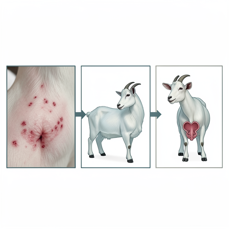
Mechanism of Breeding Inefficiency
AI-generated conceptual illustration (female goats). It explains the idea and does not show study data.
View source evidence ↗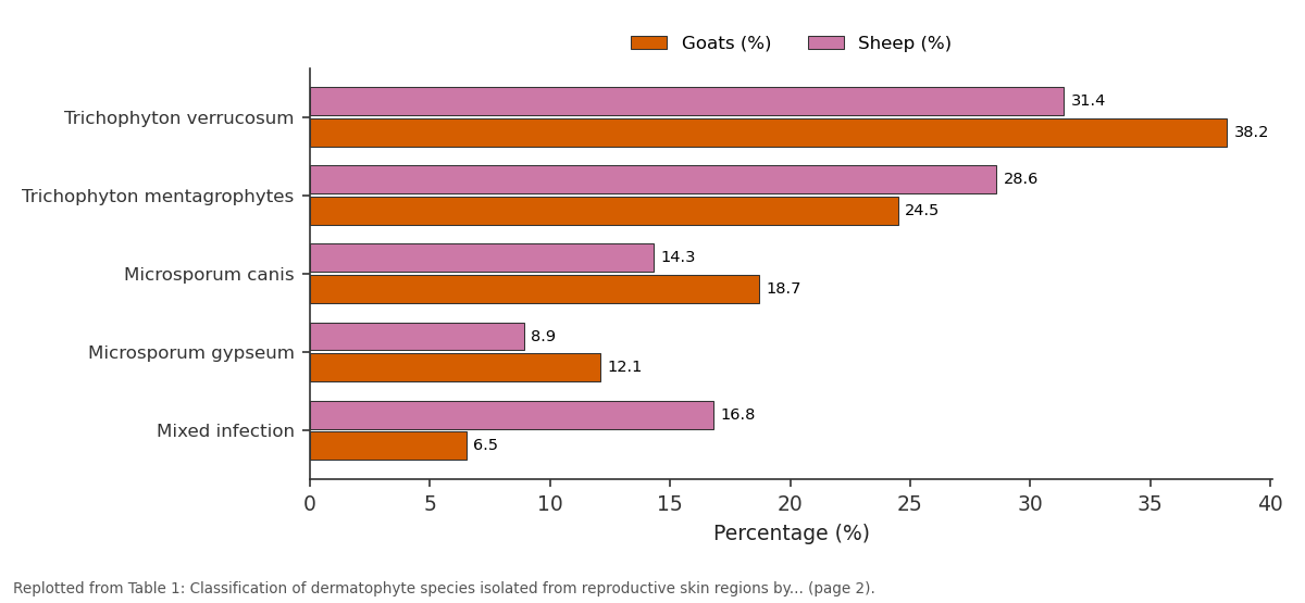
Goats (%) and Sheep (%): Trichophyton verrucosum vs Trichophyton... vs Microsporum canis vs Microsporum gypseum vs Mixed infection
Replotted from Table 1 (page 2) of the original paper. Every bar is the paper's own table value.
View source evidence ↗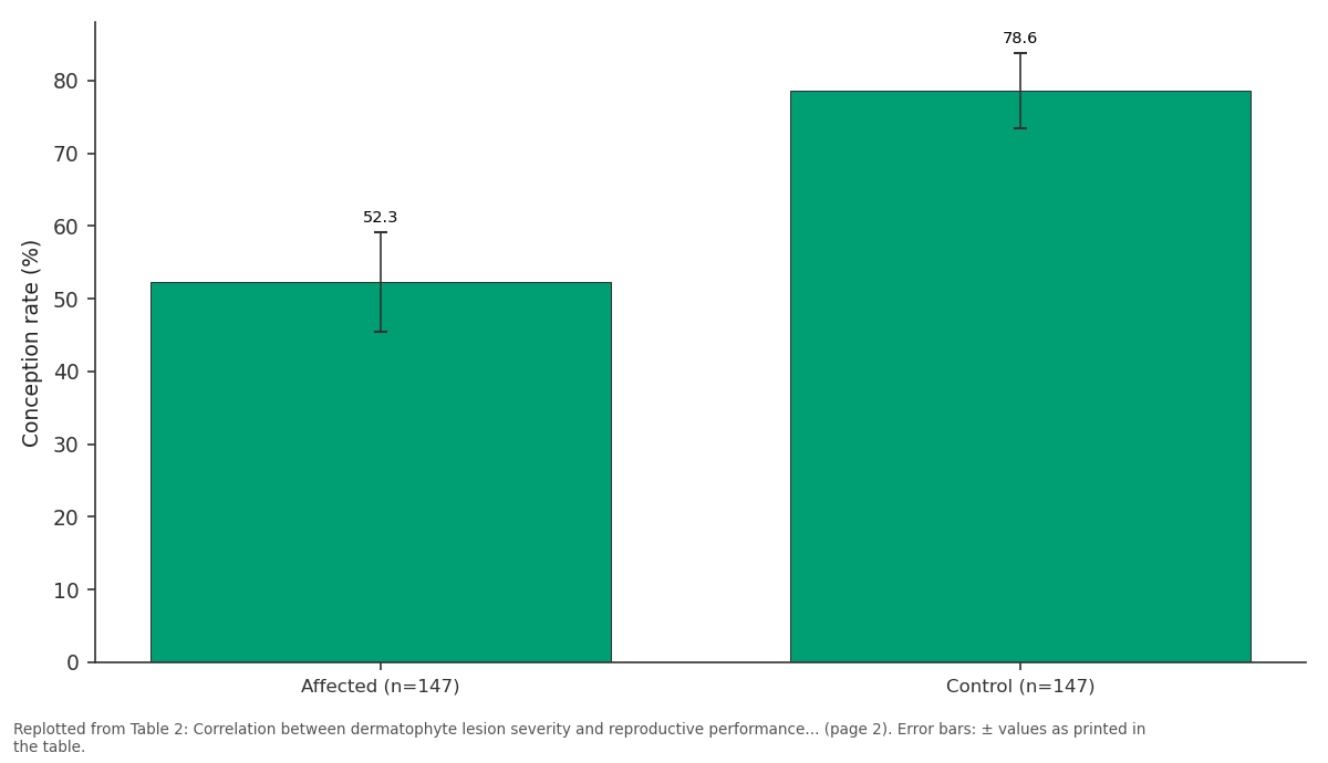
Conception rate (%): Affected (n=147) vs Control (n=147)
Replotted from Table 2 (page 2) of the original paper. Every bar is the paper's own table value.
View source evidence ↗Figures & tables
From the original paper Table 1
 Table 2
 Table 3
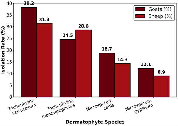 Figure 1
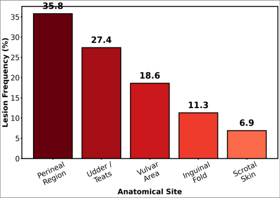 Figure 2
Introduction
The assertion that skin infections cannot meaningfully impact reproductive performance in livestock has persisted in veterinary practice despite accumulating evidence to the contrary, particularly for dermatophyte infections localized to anatomically sensitive regions involved in mating behavior, parturition, and lactation [1] . Dermatophytosis, commonly known as ringworm, is caused by keratinophilic fungi of the genera Trichophyton, Microsporum, and Epidermophyton that invade keratinized tissues including the skin, hair, and nails, causing inflammatory lesions characterized by alopecia, scaling, crusting, and pruritus [2] . Turkey supports approximately 55 million small ruminants (38 million sheep and 12.5 million goats), representing one of the largest small ruminant populations in the Mediterranean region [3] . Dermatophytosis is recognized as one of the most economically significant fungal diseases of livestock in Turkey, with reported prevalence rates of 15-40% depending on housing conditions, season, and breed susceptibility [4] . However, the specific impact of dermatophyte infections affecting reproductive anatomical regions including the perineum, vulva, udder, inguinal area, and scrotum on breeding behavior and reproductive outcomes has not been systematically investigated in small ruminant populations. The perineal and vulvar skin regions are particularly vulnerable to dermatophyte colonization due to the warm, moist microenvironment, friction during locomotion, and the presence of skin folds that trap fungal arthrospores shed from contaminated bedding and fomites [5] . Inflammatory lesions in these regions can cause pain during mating, reluctance to accept service, vulvar edema that impedes natural service or artificial insemination, and secondary bacterial infections that may ascend into the reproductive tract. The present research aimed to determine the prevalence and species distribution of dermatophyte infections in reproductive skin regions of small ruminants, quantify their impact on breeding performance parameters, and evaluate treatment outcomes in affected animals.
Material and Methods
Material and Methods
Material
This research was conducted at the Department of Veterinary Dermatology, Ankara Research Center for Animal Health, Ankara, Turkey, between October 2023 and September 2024. A total of 480 breeding-age small ruminants (260 Angora and Hair goats and 220 Akkaraman and Merino sheep) from eight commercial farms in Ankara (n = 3), Konya (n = 3), and Kayseri (n = 2) Provinces were enrolled. Animals were selected based on the presence of visible skin lesions in reproductive anatomical regions (perineum, vulva, udder, inguinal area, scrotum) or a history of unexplained breeding reluctance. An equal number of clinically healthy, age-and parity-matched animals from the same farms served as controls. The research protocol was approved by the Institutional Animal Care and Use Committee (Approval No. ARCAH-IACUC-2023-032). Culture media, dermatophyte test medium (DTM), and antifungal agents were obtained from Merck Turkey (Istanbul).
Methods
Skin scrapings and plucked hair samples were collected from the margins of active lesions using a sterile scalpel blade. Direct microscopic examination was performed using 10% potassium hydroxide (KOH) wet mount preparations. Fungal culture was conducted on Sabouraud Dextrose Agar with chloramphenicol and cycloheximide, dermatophyte test medium, and Mycosel agar, incubated at 25 °C and 37 °C for up to 4 weeks. Species identification was based on colony morphology, microscopic examination of macroconidia and microconidia, urease test, hair perforation test, and nutritional requirements on Trichophyton agars. Reproductive performance data were extracted from farm breeding records for the 12-month period surrounding the dermatological examination. Parameters evaluated included conception rate (percentage of animals conceiving after service), services per conception, calving-to-conception interval, service refusal rate, and overall flock reproductive index. Skin biopsy specimens (4 mm punch biopsies) from a subset of 30 dermatophyte-positive animals were processed for histopathological examination using H&E, PAS, and GMS staining. Treatment was administered to all dermatophyte-positive animals using topical enilconazole wash (0.2% solution, twice weekly for 6 weeks) combined with systemic griseofulvin (7.5 mg/kg body weight daily for 4 weeks) in severe cases. Treatment response was assessed at 2, 4, and 6 weeks post-initiation by clinical examination and repeat mycological culture. Statistical comparisons between affected and control groups employed Student's ttest for continuous variables and chi-square test for categorical variables, with significance at p < 0.05. Table 1 shows that Trichophyton verrucosum was the most prevalent dermatophyte species across both host species, consistent with its well-established role as the primary zoophilic dermatophyte of ruminants. Goats showed a higher overall prevalence (36.2%) compared with sheep (31.8%), which may reflect differences in skin physiology, fleece characteristics, and grooming behavior between the two species. Mixed infections involving two dermatophyte species simultaneously were more common in sheep (16.8%) than in goats (6.5%). Figure 1 visually compares the species-specific isolation rates between goats and sheep. While T. verrucosum dominated in both host species, goats showed a relatively higher proportion of Microsporum canis (18.7% vs. 14.3%), potentially reflecting greater contact with companion animals (dogs and cats) that serve as reservoirs for this anthropophilic-zoophilic dermatophyte. The higher prevalence of T. mentagrophytes in sheep (28.6% vs. 24.5%) may relate to the rodent reservoir populations commonly associated with sheep housing facilities. 2 illustrates the anatomical predilection of dermatophyte lesions within the reproductive region. The perineal area was most frequently affected (35.8%), followed by the udder and teats (27.4%) and vulvar region (18.6%). The predilection for the perineal region likely reflects the combination of warmth, moisture, mechanical friction, and poor air circulation that characterize this anatomical site, creating optimal conditions for dermatophyte colonization and proliferation.
| Dermatophyte Species | Goats (n) | Goats (%) | Sheep (n) | Sheep (%) | Total (%) |
|---|---|---|---|---|---|
| Trichophyton verrucosum | 36 | 38.2 | 22 | 31.4 | 39.5 |
| Trichophyton mentagrophytes | 23 | 24.5 | 20 | 28.6 | 29.3 |
| Microsporum canis | 18 | 18.7 | 10 | 14.3 | 19.0 |
| Microsporum gypseum | 11 | 12.1 | 6 | 8.9 | 11.6 |
| Mixed infection | 6 | 6.5 | 12 | 16.8 | 0.6 |
| Total positive | 94 | 36.2 | 70 | 31.8 | 30.6 |
Table text extracted from the original PDF; superscript letters are shown inline. The original table image is below.
View original table image
Results
Results
Discussion
This research provides the first systematic quantification of the reproductive performance impact of dermatophyte infections localized to breeding-relevant anatomical regions in small ruminants. The overall dermatophyte prevalence of 30.6% in animals with visible reproductive region skin lesions is consistent with reported rates of 25-42% for general dermatophytosis in Turkish small ruminant populations, though the specific focus on reproductive anatomical sites reveals a previously underappreciated dimension of this disease [6] . The dramatically higher service refusal rate in dermatophyte-affected animals (34.7% vs. 8.2%) represents the most clinically significant finding, as it directly translates to reduced breeding opportunities and extended calving intervals. The inflammatory lesions of perineal and vulvar dermatophytosis cause hyperalgesia and allodynia in the affected skin regions, making physical contact during natural service or artificial insemination procedures painful and triggering defensive behavioral responses including kicking, squatting, and tail clamping that prevent successful breeding [7] . This behavioral mechanism explains why the conception rate reduction (52.3% vs. 78.6%) is proportionally greater than would be expected from the relatively superficial nature of the skin infection alone. The dominance of T. verrucosum as the primary causative agent is consistent with the global epidemiology of ruminant dermatophytosis and reflects the strong host adaptation of this zoophilic species to bovine and small ruminant keratinized tissues [8] . The relatively high proportion of Microsporum species (combined 30.6%) indicates the importance of environmental and companion animal sources in the epidemiology of small ruminant dermatophytosis, as M. canis is primarily maintained in dog and cat populations while M. gypseum is a geophilic species found in soil contaminated with keratinous debris [9] . The treatment response data confirm that combined topical and systemic antifungal therapy achieves satisfactory clinical and mycological outcomes in the majority of cases, with 89.1% clinical resolution by week 6. The parallel recovery of breeding acceptance behavior (87.8% by week 6) provides direct evidence for the causal relationship between the dermatophyte infection and the reproductive performance impairment, as the removal of the pain stimulus restored normal mating behavior in most animals [10] . The economic implications of reproductive region dermatophytosis in small ruminant flocks are substantial. Based on the conception rate differential (26.3 percentage points) and the extended calving interval (36.3 additional days) documented in this research, a flock with 30% dermatophyte prevalence in reproductive regions would experience an estimated 7.9% reduction in annual reproductive output. At current market values for kids and lambs in Turkey, this translates to approximately 15-25 Turkish Lira economic loss per breeding female per season, a figure that substantially exceeds the cost of systematic dermatological screening and treatment [11] .
Zoonotic Considerations
The dermatophyte species identified in this research, particularly T. verrucosum and M. canis, are wellestablished zoonotic agents capable of causing inflammatory tinea corporis in humans who have direct contact with infected animals. Farm workers involved in breeding management, artificial insemination, and lambing assistance are at elevated risk of acquiring dermatophyte infections from the perineal and vulvar regions of affected animals. The implementation of personal protective equipment protocols and post-contact hand hygiene measures is recommended for personnel involved in reproductive management of flocks with documented dermatophytosis.
Seasonal Patterns
Analysis of the temporal distribution of dermatophyte cases revealed a marked seasonal pattern, with peak prevalence occurring during the autumn and winter months (October-February) when animals are housed in enclosed facilities with reduced sunlight exposure and increased animal density. The lowest prevalence was recorded during summer months (June-August), coinciding with pasture-based management and enhanced ultraviolet light exposure, which has documented antifungal effects on environmental dermatophyte arthrospores. This seasonal pattern has direct implications for the timing of pre-breeding dermatological screening, which should ideally be conducted before the autumn breeding season.
Conclusion
Within the context of the growing recognition that fungal infections play an increasingly important role in livestock reproductive health, this research highlights the specific contribution of dermatophyte infections in reproductive skin regions to breeding inefficiency in small ruminant flocks.
The key findings include a 30.6% dermatophyte prevalence in reproductive region lesions, a 26.3 percentage point reduction in conception rate among affected animals, and a 4.2-fold increase in service refusal rates attributable to pain from perineal and vulvar lesions. It is recommended that dermatological screening of reproductive anatomical regions be incorporated into routine pre-breeding health assessments for small ruminant flocks, particularly in housed management systems during the autumn breeding season. Future investigations should address the development of dermatophyte vaccines for small ruminants, the evaluation of environmental decontamination protocols for reducing arthrospore loads in breeding facilities, and longitudinal assessment of the economic returns from systematic dermatological screening and treatment programs.
Acknowledgement
Acknowledgements E.K.Y. conceptualized and supervised the research program; O.D.A. conducted field sampling and mycological
Figures and tables from the paper
| Parameter | Affected (n=147) | Control (n=147) | p-value |
|---|---|---|---|
| Conception rate (%) | 52.3 ± 6.8 | 78.6 ± 5.2 | <0.001 |
| Services per conception | 3.4 ± 0.8 | 1.8 ± 0.4 | <0.001 |
| Calving-to-conception (days) | 98.4 ± 18.2 | 62.1 ± 11.4 | <0.001 |
| Service refusal rate (%) | 34.7 ± 8.1 | 8.2 ± 3.4 | <0.001 |
| Kidding/lambing rate (%) | 64.8 ± 7.2 | 86.3 ± 4.8 | <0.001 |
| Milk yield reduction (%) | 18.4 ± 5.6 | — | — |
Table text extracted from the original PDF; superscript letters are shown inline. The original table image is below.
View original table image
| Assessment Point | Clinical Resolution (%) | Mycological Cure (%) | Breeding Acceptance Recovery (%) |
|---|---|---|---|
| Week 2 | 38.1 | 22.4 | 41.5 |
| Week 4 | 71.4 | 61.2 | 72.8 |
| Week 6 | 89.1 | 84.4 | 87.8 |
| Week 8 (follow-up) | 93.2 | 91.2 | 91.8 |
Table text extracted from the original PDF; superscript letters are shown inline. The original table image is below.
View original table image
References
18 in the original paper- Chermette R, Ferreiro L, Guillot J. Dermatophytoses in animals. Mycopathologia. 2008;166(5-6):385-405.
- Weitzman I, Summerbell RC. The dermatophytes. Clin Microbiol Rev. 1995;8(2):240-259.
- Pal M, Tariku F, Rebuma T, Seth D, Patel KB. Growing role of fungi in the reproductive disorders of animals. J Adv Microbiol Res. 2024;5(1):37-42.
- Dalis JS, Kazeem HM, Makinde AA, Fatihu MY. Dermatophytosis in animals: A review. J Vet Med Anim Health. 2019;11(3):95-100.
- Seker E, Dogan N. Isolation of dermatophytes from dogs and cats with suspected dermatophytosis in western Turkey. Prev Vet Med. 2011;98(1):46-51.
- Hussain I, Usman M, Afzal F, Ahmed I, Irfan M, Rashid I, et al. Prevalence and risk factors of dermatophytosis in cattle and buffalo calves. Pak Vet J. 2020;40(1):127-130.
- Cafarchia C, Iatta R, Immediato D, Puttilli MR, Otranto D. Azole susceptibility of Malassezia pachydermatis and Malassezia furfur and tentative epidemiological cut-off values. Med Mycol. 2015;53(7):743-748.
- Lund A, DeBoer DJ. Immunoprophylaxis of dermatophytosis in animals. Mycopathologia. 2008;166(5-6):407-424.
- Moriello KA. Feline dermatophytosis: Aspects pertinent to disease management in single and multiple cat situations. J Feline Med Surg. 2014;16(5):419-431.
- Pier AC, Smith JMB, Alexiou H, Ellis DH, Lund A, Prichard RC. Animal ringworm-its aetiology, public health significance and control. J Med Vet Mycol. 1994;32(Suppl 1):133-150.
- Al-Ani FK. Dermatophytosis in animals: An updated review. J Mycopathol Res. 2017;55(2):113-128.
- Cafarchia C, Figueredo LA, Otranto D. Fungal diseases of horses. Vet Microbiol. 2013;167(1-2):215-234.
- DeBoer DJ, Moriello KA. Humoral and cellular immune responses to Microsporum canis in naturally occurring feline dermatophytosis. J Med Vet Mycol. 1993;31(2):121-132.
- Moretti A, Boncio L, Posteraro B, Mechelli L, De Gasperis E, Morganti G. Ringworm in cattle: Molecular identification and antifungal susceptibility. Vet Dermatol. 2013;24(5):524-525.e125.
- Rochette F, Engelen M, Vanden Bossche H. Antifungal agents of use in animal health-Practical applications. J Vet Pharmacol Ther. 2003;26(1):31-53.
- Elewski BE. Onychomycosis: Pathogenesis, diagnosis, and management. Clin Microbiol Rev. 1998;11(3):415429.
- Arda M, Minbay A, Leloğlu N, Aydın N, Kahraman M, Ilgaz A. Özel Mikrobiyoloji. 5th ed. Ankara: Medisan Yayınları; 2005.
- Gnat S, Łagowski D, Nowakiewicz A, Zieba P. The host range of dermatophytes, is it at all possible? Phenotypic evaluation of the keratinolytic activity of Trichophyton verrucosum clinical isolates. Mycoses. 2019;62(3):274-283.