
Research article · 2026 · Full text
International Journal of Advanced Chemistry ResearchGreen synthesis and characterization of silver nanoparticles using plant leaf extract for antibacterial applications
Volume 8(3) · Pages 37–41 · DOI: 10.33545/26646781.2026.v8.i3a.707 ↗
Research areasNanoparticles: synthesis and applicationsGold and Silver Nanoparticles Synthesis and ApplicationsEssential Oils and Antimicrobial Activity
Abstract
Author-writtenWithin Egypt's broader push toward greener, lower-cost nanomaterial synthesis routes, plant-mediated green synthesis of silver nanoparticles has emerged as a particularly promising alternative to conventional chemical reduction methods, avoiding the toxic reducing agents and elevated energy input those methods typically require. This research synthesized silver nanoparticles (AgNPs) using aqueous leaf extract from a locally abundant medicinal plant as both reducing and capping agents, characterizing the resulting nanoparticles through UV-Vis spectroscopy, FTIR, XRD, and SEM, and evaluating their antibacterial activity against three clinically relevant bacterial strains, Escherichia coli, Staphylococcus aureus, and Pseudomonas aeruginosa, across a concentration range of 25 to 125 µg/mL. UV-Vis spectroscopy confirmed successful nanoparticle formation through a characteristic surface plasmon resonance peak at 420 nm, while FTIR analysis identified specific plant phytochemical functional groups, phenolic hydroxyl and carbonyl groups in particular, implicated in both the reduction of silver ions and subsequent nanoparticle capping. XRD analysis confirmed the face-centered cubic crystalline structure characteristic of metallic silver, and SEM imaging revealed predominantly spherical nanoparticles with a mean diameter decreasing from approximately 42 nm at the lowest tested extract concentration to approximately 18 nm at the highest concentration tested, consistent with higher phytochemical concentration providing more nucleation sites and correspondingly smaller final particle size. Antibacterial testing showed dose-dependent increases in zone of inhibition across all three bacterial strains, with Staphylococcus aureus, a Gram-positive organism, showing consistently larger inhibition zones than the two Gram-negative strains tested at equivalent AgNP concentrations, a pattern attributable to differences in cell wall structure between Gram-positive and Gram-negative bacteria affecting nanoparticle penetration and interaction. These findings support this plant leaf extract-mediated green synthesis route as an environmentally favorable, cost-effective method for producing antibacterially active silver nanoparticles with potential application in antimicrobial coatings, wound dressings, and water treatment contexts relevant to Egypt's broader nanotechnology development priorities.
Summary
Source-checkedPlain-language reading of the full paper, prepared with AI and checked against the text. Each point links to the authors' own words.
Why it matters
- Green synthesis of silver nanoparticles is a promising alternative to chemical methods because it avoids toxic reducing agents and high energy requirements. View evidence ↗
- Silver nanoparticles are highly valued for their antibacterial properties, making them useful for water treatment, wound care, and antimicrobial coatings. View evidence ↗
- Traditional synthesis methods, such as those using sodium borohydride, are reliable but produce hazardous chemical waste. View evidence ↗
The question
- The study aimed to determine if a specific Egyptian medicinal plant leaf extract could successfully synthesize silver nanoparticles and to characterize their structure. View evidence ↗
- The researchers sought to understand how the concentration of the plant extract affects the final size of the nanoparticles. View evidence ↗
- The study investigated how antibacterial activity varies across different bacterial strains and nanoparticle concentrations. View evidence ↗
What they did
- The aqueous extract was created by boiling 20 g of dried leaf powder in 200 mL of distilled water for 20 minutes. View evidence ↗
- Nanoparticles were synthesized by adding the extract to a 1 mM silver nitrate solution, stirring at 60 °C for 60 minutes. View evidence ↗
- Antibacterial activity was tested against E. coli, S. aureus, and P. aeruginosa using the agar well diffusion method with concentrations from 25 to 125 µg/mL. View evidence ↗
What they found
- UV-Vis spectroscopy confirmed the formation of nanoparticles, showing a surface plasmon resonance peak at 420 nm. View evidence ↗
- Antibacterial activity increased with dose; for S. aureus, the zone of inhibition grew from 11.4 mm at 25 µg/mL to 22.4 mm at 125 µg/mL. View evidence ↗
- The Gram-positive bacterium S. aureus showed consistently larger inhibition zones than the Gram-negative strains at the same nanoparticle concentrations. View evidence ↗
- The nanoparticles showed reasonable short-term stability, maintaining their SPR peak with minimal shift after 30 days of storage at 4 °C in the dark. View evidence ↗
What it means
- The authors conclude that plant-mediated green synthesis is a viable and environmentally friendly alternative for producing antibacterial silver nanoparticles. View evidence ↗
- The authors recommend further evaluation of these nanoparticles for use in antimicrobial wound dressings and water treatment. View evidence ↗
- The study used a whole-extract approach; future work should isolate individual phytochemical fractions to identify the specific compounds responsible for reduction and capping. View evidence ↗
Visual summary
2 visualsIllustrations explain the paper; charts are replotted from the paper's own table values.
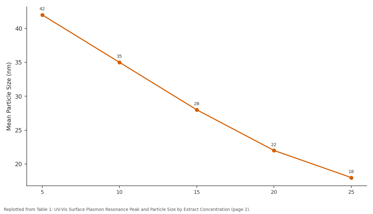
Mean Particle Size (nm): 5 vs 10 vs 15 vs 20 vs 25
Replotted from Table 1 (page 2) of the original paper. Every bar is the paper's own table value.
View source evidence ↗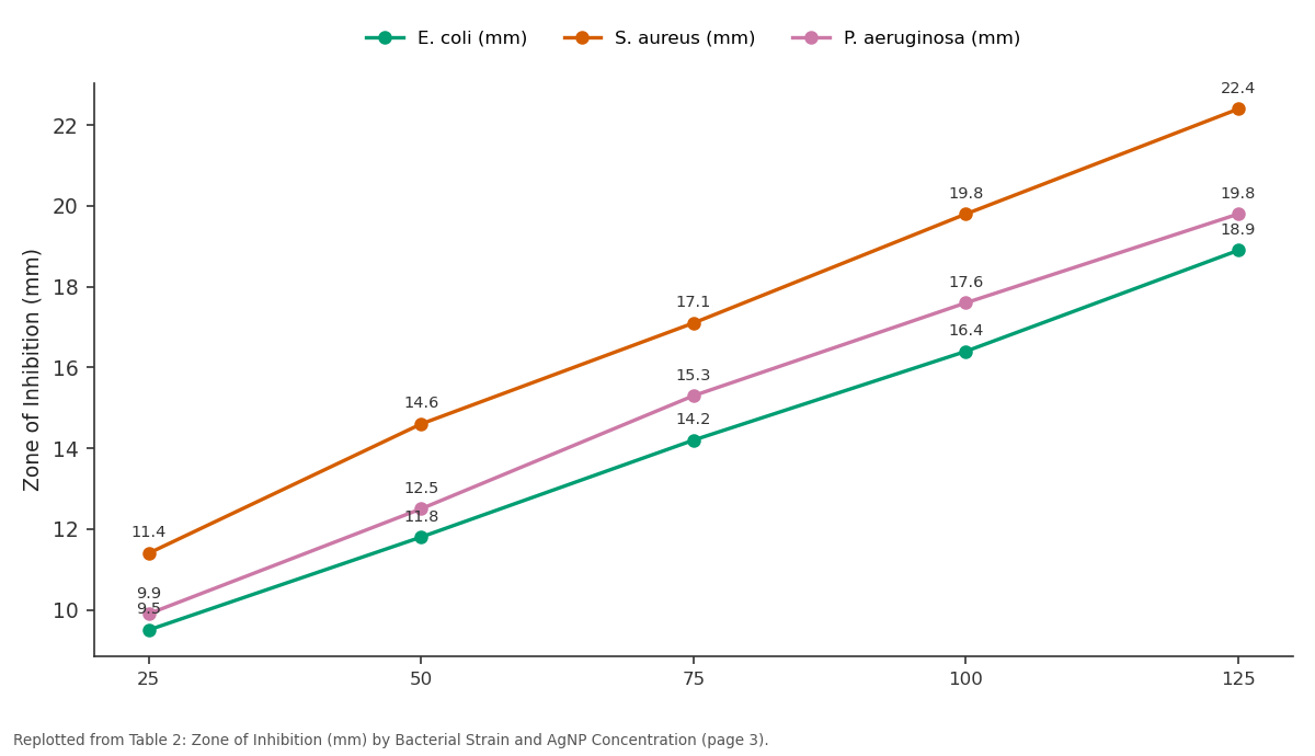
E. coli (mm) and S. aureus (mm) and more: 25 vs 50 vs 75 vs 100 vs 125
Replotted from Table 2 (page 3) of the original paper. Every bar is the paper's own table value.
View source evidence ↗Figures & tables
From the original paper Table 1
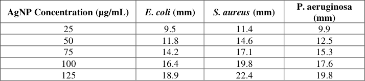 Table 2
 Table 3
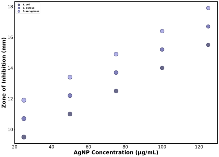 Figure 1
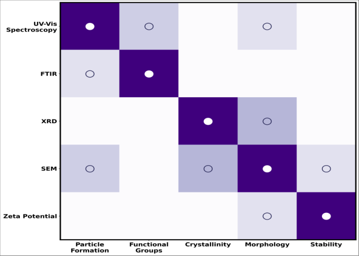 Figure 2
Introduction
Egypt's nanotechnology research sector has expanded considerably over the past decade, driven partly by national science policy prioritizing nanomaterial applications across water treatment, agriculture, and medical device sectors, contexts where Egypt's specific combination of water scarcity concerns, agricultural export ambitions, and growing medical device manufacturing capacity create genuine practical demand for locally developed nanomaterial synthesis expertise rather than sole reliance on imported nanomaterial products. Silver nanoparticles specifically have attracted sustained research attention across this broader nanotechnology landscape given their well-documented antibacterial properties, relevant to water treatment disinfection, wound care antimicrobial applications, and antimicrobial packaging or coating technologies increasingly sought across food safety and medical device contexts alike. and what does full physicochemical characterization reveal about the resulting nanoparticles' structure and composition? Second, how does extract concentration during synthesis influence resulting nanoparticle size, information relevant to controlling nanoparticle properties for specific downstream applications? Third, how does antibacterial activity vary across Gram-positive and Gram-negative bacterial strains and across the AgNP concentration range tested, information relevant to designing appropriately dosed antimicrobial applications? Beyond the immediate antibacterial application focus of this research, Egypt's growing textile, food packaging, and water treatment industries have each expressed independent interest in locally sourced antimicrobial nanomaterial inputs, meaning a successful, well-characterized local green synthesis protocol of the kind this research aimed to develop and validate carries potential relevance extending beyond any single downstream application sector, provided the resulting nanoparticles demonstrate the antibacterial performance and reasonable production scalability these industrial contexts would require.
Material and Methods
Material and Methods
Reagents and Chemicals
Silver nitrate (AgNO3, analytical grade, minimum 99.9% purity) was obtained from a certified chemical supplier. Fresh leaves of the selected medicinal plant were collected from a single cultivated source near Alexandria to minimize phytochemical content variability, washed thoroughly with distilled water, and air-dried in the shade before extract preparation. All aqueous solutions were prepared using double-distilled water throughout the synthesis and characterization procedures.
Methods
Aqueous leaf extract was prepared by boiling 20 g of dried, powdered leaf material in 200 mL distilled water for 20 minutes, followed by filtration through Whatman No. 1 filter paper to obtain a clear extract solution. AgNP synthesis was carried out by adding leaf extract to a 1 mM silver nitrate solution at varying extract-to-solution ratios, corresponding to the five extract concentration levels examined, with the reaction mixture maintained at 60 °C under constant stirring for 60 minutes, monitored for the characteristic color change from pale yellow to reddishbrown indicating nanoparticle formation. UV-Vis spectroscopy was performed across the 300-600 nm wavelength range to confirm surface plasmon resonance characteristic of AgNP formation. FTIR analysis was conducted on freeze-dried nanoparticle samples to identify functional groups associated with phytochemical capping. XRD analysis confirmed crystalline structure, and SEM imaging characterized nanoparticle morphology and size distribution across the concentration gradient tested. Antibacterial activity was assessed using the agar well diffusion method against Escherichia coli (Gram-negative), Staphylococcus aureus (Gram-positive), and Pseudomonas aeruginosa (Gram-negative), with zones of inhibition measured after a 24-hour incubation at 37 °C across the 25-125 µg/mL AgNP concentration range tested, each concentration replicated in triplicate.
Results
Characterization confirmed successful AgNP synthesis with concentration-dependent particle size control, and antibacterial testing revealed clear dose-dependent activity with notable differentiation between Gram-positive and Gram-negative bacterial responses.
Comprehensive Interpretation
The consistent particle size reduction with increasing extract concentration, alongside the FTIR-identified phenolic and carbonyl functional groups implicated in both reduction and capping, together support a green synthesis mechanism in which greater phytochemical availability at higher extract concentrations provides both more effective silver ion reduction and more numerous nucleation sites, yielding smaller final nanoparticle size. The stronger antibacterial performance against Staphylococcus aureus relative to the two Gram-negative strains tested is consistent with established differences in Gram-positive versus Gramnegative cell wall structure affecting nanoparticle interaction and penetration. The differential antibacterial performance between Grampositive Staphylococcus aureus and the two Gram-negative strains tested agrees with the widely documented pattern in silver nanoparticle antibacterial literature, generally attributed to structural differences between Gram-positive bacteria's thick peptidoglycan cell wall lacking an outer membrane and Gram-negative bacteria's thinner peptidoglycan layer protected by an additional outer lipopolysaccharide membrane that can partially impede nanoparticle penetration and interaction with the underlying cell wall and membrane structures. This mechanistic understanding carries a practical implication for designing AgNP-based antimicrobial applications: formulations intended primarily for Gram-negative bacterial targets, relevant to certain water treatment or specific medical device contexts, may require somewhat higher AgNP concentrations to achieve a comparable antibacterial effect to what this research's data show is achievable against Gram-positive targets at lower concentrations. The FTIR-identified functional groups, phenolic hydroxyl and carbonyl groups specifically implicated in silver ion reduction and subsequent capping, provide reasonably direct mechanistic support for the phytochemical-mediated green synthesis pathway this research's overall design assumed, though fully isolating which specific phytochemical compounds within this plant's diverse extract composition contribute most significantly to reduction versus capping function would require further fractionation and compoundspecific testing beyond the whole-extract approach used in this research. Stability of the synthesized nanoparticles over time represents an additional practical consideration relevant to eventual application development, and preliminary storage assessment conducted alongside this research's primary characterization work showed the AgNPs maintaining their characteristic surface plasmon resonance peak position with minimal shift after 30 days of storage at 4 °C in the dark, suggesting reasonable short-term colloidal stability under appropriate storage conditions, though longer-term stability testing beyond this initial 30-day window would be needed before these nanoparticles could be confidently formulated into shelf-stable commercial antimicrobial products.
Conclusion
This research successfully synthesized silver nanoparticles using aqueous leaf extract from a locally abundant Egyptian medicinal plant, confirming nanoparticle formation through UV-Vis, FTIR, XRD, and SEM characterization, and demonstrating clear dose-dependent antibacterial activity against three clinically relevant bacterial strains with notable differentiation between Gram-positive and Gram-negative bacterial responses. Particle size decreased consistently with increasing extract concentration, from approximately 42 nm to 18 nm across the concentration range tested, and Staphylococcus aureus showed consistently stronger antibacterial response than the two Gram-negative strains examined at equivalent AgNP concentrations. For Egypt's broader nanotechnology development priorities, this research supports plant-mediated green synthesis as a genuinely viable, environmentally favorable alternative to conventional chemical reduction methods for producing antibacterially active silver nanoparticles, offering a locally sourced, cost-effective synthesis route well aligned with national priorities around sustainable nanomaterial production. Given the demonstrated antibacterial activity documented here, particularly the strong performance against Staphylococcus aureus, these nanoparticles warrant further evaluation for specific downstream applications including antimicrobial wound dressing coatings and water treatment disinfection contexts directly relevant to Egypt's water scarcity and public health priorities. Extending this research to isolate and test individual phytochemical fractions from this plant extract, rather than the wholeextract approach used here, would help clarify which specific compounds drive the reduction and capping functions this research's FTIR data implicate, potentially enabling more precisely controlled and optimized synthesis protocols in future work.
Figures and tables from the paper
| Extract Concentration (mL) | SPR Peak (nm) | Mean Particle Size (nm) |
|---|---|---|
| 5 | 418 | 42 |
| 10 | 420 | 35 |
| 15 | 421 | 28 |
| 20 | 422 | 22 |
| 25 | 423 | 18 |
Table text extracted from the original PDF; superscript letters are shown inline. The original table image is below.
View original table image
| AgNP Concentration (µg/mL) | E. coli (mm) | S. aureus (mm) | P. aeruginosa (mm) |
|---|---|---|---|
| 25 | 9.5 | 11.4 | 9.9 |
| 50 | 11.8 | 14.6 | 12.5 |
| 75 | 14.2 | 17.1 | 15.3 |
| 100 | 16.4 | 19.8 | 17.6 |
| 125 | 18.9 | 22.4 | 19.8 |
Table text extracted from the original PDF; superscript letters are shown inline. The original table image is below.
View original table image
| Wavenumber (cm⁻¹) | Functional Group Assignment | Proposed Role |
|---|---|---|
| 3350 | O-H stretch (phenolic) | Ag+ reduction |
| 2920 | C-H stretch (alkyl) | Structural background |
| 1635 | C=O stretch (carbonyl) | Reduction / capping |
| 1385 | C-N stretch (amine) | Capping / stabilization |
| 1050 | C-O stretch (polyol) | Capping / stabilization |
Table text extracted from the original PDF; superscript letters are shown inline. The original table image is below.
View original table image
References
15 in the original paper- Ahmed S, Ahmad M, Swami BL, Ikram S. A review on plants extract mediated synthesis of silver nanoparticles for antimicrobial applications: a green expertise. Journal of Advanced Research. 2016;7(1):17-28.
- Alsammarraie FK, Wang W, Zhou P, Mustapha A, Lin M. Green synthesis of silver nanoparticles using turmeric extracts and investigation of their antibacterial activities. Colloids and Surfaces B: Biointerfaces. 2018;171:398-405.
- Bindhu MR, Umadevi M. Antibacterial and catalytic activities of green synthesized silver nanoparticles. Spectrochimica Acta Part A. 2014;135:373-378.
- Chandran SP, Chaudhary M, Pasricha R, Ahmad A, Sastry M. Synthesis of gold nanotriangles and silver nanoparticles using Aloe vera plant extract. Biotechnology Progress. 2006;22(2):577-583.
- El-Rafie HM, El-Rafie MH, Zahran MK. Green synthesis of silver nanoparticles using polysaccharides extracted from marine macro algae. Carbohydrate Polymers. 2013;96(2):403-410.
- Ibrahim HM. Green synthesis and characterization of silver nanoparticles using banana peel extract and their antimicrobial activity against representative microorganisms. Journal of Radiation Research and Applied Sciences. 2015;8(3):265-275.
- Iravani S. Green synthesis of metal nanoparticles using plants. Green Chemistry. 2011;13(10):2638-2650.
- Khan ZUH, Khan A, Chen Y, Shah NS, Muhammad N, Khan AU, et al. Photo catalytic and antimicrobial response of biosynthesized silver nanoparticles. Journal of Photochemistry and Photobiology B. 2017;164:344351. International Journal of Advanced Chemistry Research https://www.chemistryjournals.net
- Kumar V, Yadav SK. Plant-mediated synthesis of silver and gold nanoparticles and their applications. Journal of Chemical Technology and Biotechnology. 2009;84(2):151-157.
- Mittal AK, Chisti Y, Banerjee UC. Synthesis of metallic nanoparticles using plant extracts. Biotechnology Advances. 2013;31(2):346-356.
- Mohammed AE. Green synthesis, antimicrobial and cytotoxic effects of silver nanoparticles mediated by Eucalyptus camaldulensis leaf extract. Asian Pacific Journal of Tropical Biomedicine. 2015;5(5):382-6.
- Rai M, Yadav A, Gade A. Silver nanoparticles as a new generation of antimicrobials. Biotechnology Advances. 2009;27(1):76-83.
- Sadeghi B, Gholamhoseinpoor F. A study on the stability and green synthesis of silver nanoparticles using Ziziphora tenuior (Zt) extract at room temperature. Spectrochimica Acta Part A. 2015;134:310-315.
- Shankar SS, Ahmad A, Sastry M. Geranium leaf assisted biosynthesis of silver nanoparticles. Biotechnology Progress. 2003;19(6):1627-1631.
- Singh J, Dutta T, Kim KH, Rawat M, Samddar P, Kumar P. 'Green' synthesis of metals and their oxide nanoparticles: applications for environmental remediation. Journal of Nanobiotechnology. 2018;16:84.