
Research article · 2026 · Full text
International Journal of Advanced Chemistry ResearchSynthesis, characterization and biological evaluation of schiff base metal complexes for antioxidant and antibacterial activity
Volume 8(2) · Pages 27–30 · DOI: 10.33545/26646781.2026.v8.i2a.697 ↗
Research areasMetal complexes synthesis and propertiesInorganic and Organometallic ChemistryOrganometallic Compounds Synthesis and Characterization
Abstract
Author-writtenA ligand that shows only modest biological activity on its own can behave quite differently once it wraps around a metal ion, and figuring out which metal produces the biggest improvement is rarely obvious without direct comparison. This research addressed that problem by synthesizing a Schiff base ligand from the condensation of 2-hydroxybenzaldehyde and 4-aminoantipyrine, and complexing it with Cu(II), Ni(II), and Zn(II) acetate to obtain three metal complexes of general formula [M(L)2]. The ligand and its complexes were characterized using elemental analysis, FTIR, UV-Vis spectroscopy, magnetic susceptibility measurements, and molar conductivity, which together supported an octahedral geometry around each metal centre with the ligand coordinating through the azomethine nitrogen and phenolic oxygen. Antioxidant activity, assessed through DPPH, FRAP, and ABTS radical scavenging assays, was markedly stronger for all three metal complexes compared with the free ligand, with the Cu(II) complex showing the lowest IC50 values across all three assays (24.6, 28.1, and 21.4 µg/mL respectively). Antibacterial screening against Staphylococcus aureus, Bacillus subtilis, Escherichia coli, and Pseudomonas aeruginosa similarly showed the Cu(II) complex producing the largest inhibition zones, followed by the Zn(II) and Ni(II) complexes, all outperforming the uncomplexed ligand. This consistent enhancement in both antioxidant and antibacterial activity upon metal chelation is broadly explained by Tweedy's chelation theory, which attributes the effect to increased lipophilicity and delocalization of the metal's positive charge across the coordinated ligand system, improving penetration through microbial cell membranes and radical-scavenging efficiency. These findings identify the Cu(II)-Schiff base complex as the most promising candidate among those tested for further pharmacological development, though in vivo toxicity and pharmacokinetic evaluation would be necessary before any therapeutic application could be considered.
Summary
Source-checkedPlain-language reading of the full paper, prepared with AI and checked against the text. Each point links to the authors' own words.
Why it matters
- Binding organic ligands to transition metals often increases biological activity by redistributing electron density and increasing lipophilicity, which helps the complex cross microbial cell membranes. View evidence ↗
- Schiff bases are ideal for these studies because their azomethine nitrogen easily coordinates with various transition metals. View evidence ↗
- Schiff bases derived from 4-aminoantipyrine are of pharmacological interest due to the known analgesic and anti-inflammatory properties of antipyrine derivatives. View evidence ↗
The question
- The study aimed to synthesize a specific Schiff base ligand and its Cu(II), Ni(II), and Zn(II) complexes to determine which metal center provides the greatest enhancement in antioxidant and antibacterial activity. View evidence ↗
What they did
- The Schiff base ligand was synthesized by refluxing equimolar amounts of 2-hydroxybenzaldehyde and 4-aminoantipyrine in absolute ethanol for four hours using glacial acetic acid as a catalyst. View evidence ↗
- Metal complexes were created by refluxing the ligand with metal acetate salts in a 2:1 ligand-to-metal molar ratio in ethanol for six hours. View evidence ↗
- Antioxidant activity was measured using DPPH, FRAP, and ABTS assays in triplicate at concentrations from 10 to 100 µg/mL, using ascorbic acid as a standard. View evidence ↗
- Antibacterial activity was tested via the agar well diffusion method against four bacterial strains (S. aureus, B. subtilis, E. coli, and P. aeruginosa) using 1 mg/mL solutions in DMSO. View evidence ↗
What they found
- All three metal complexes showed stronger antioxidant activity than the free ligand, with the Cu(II) complex being the most potent, showing the lowest IC50 values of 24.6, 28.1, and 21.4 µg/mL across the three assays. View evidence ↗
- The Cu(II) complex demonstrated the highest antibacterial activity, producing inhibition zones of 19.6 mm against S. aureus and 15.3 mm against E. coli, compared to 9.2 mm and 7.4 mm for the free ligand. View evidence ↗
- Magnetic moment measurements (1.84 B.M. for Cu(II) and 2.96 B.M. for Ni(II)) and the diamagnetic nature of the Zn(II) complex support an octahedral geometry for the metal centers. View evidence ↗
- Low molar conductivity values indicate that the complexes are non-electrolytic in DMSO, suggesting that acetate counter-ions are coordinated within the inner sphere. View evidence ↗
What it means
- The authors conclude that copper is the most promising metal center for this specific ligand scaffold and should be prioritized for further pharmacological development. View evidence ↗
- The authors state that in vivo toxicity and pharmacokinetic evaluations are required before any therapeutic application can be considered. View evidence ↗
- Remaining questions include whether the in vitro results translate to physiological stability and toxicity, and if modifying the ligand's substituents could further enhance activity. View evidence ↗
Visual summary
3 visualsIllustrations explain the paper; charts are replotted from the paper's own table values.
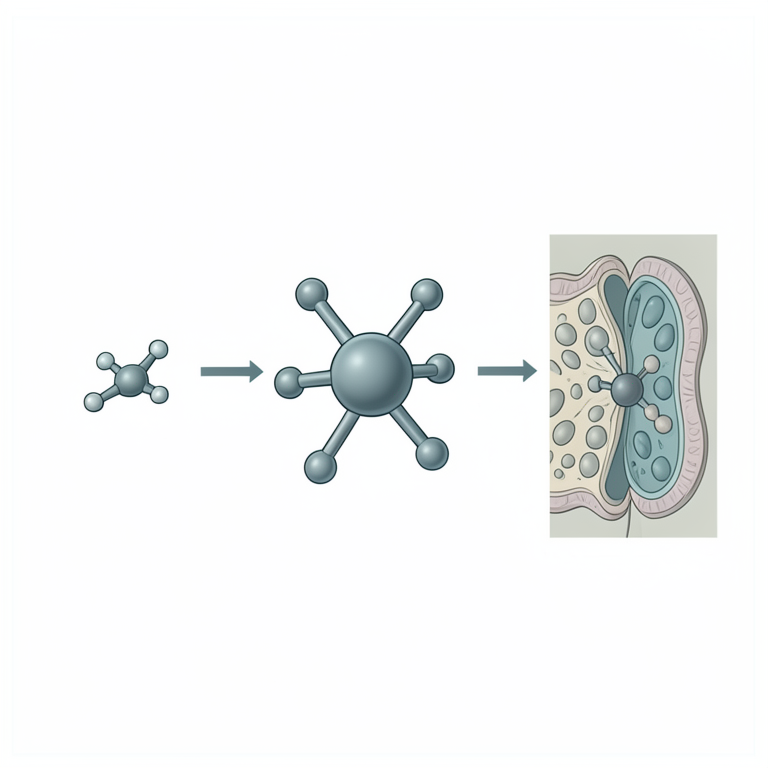
Chelation Effect Concept
AI-generated conceptual illustration (Schiff base metal complexes and a bacterial cell membrane). It explains the idea and does not show study data.
View source evidence ↗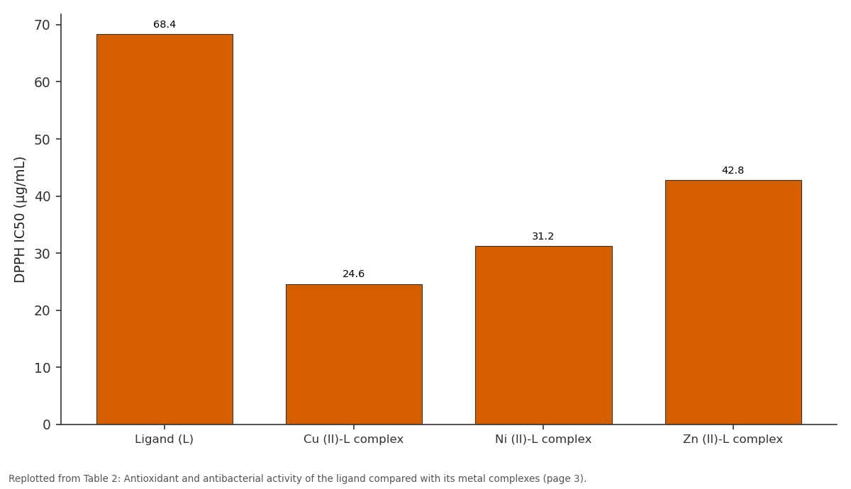
DPPH IC50 (µg/mL): Ligand (L) vs Cu (II)-L complex vs Ni (II)-L complex vs Zn (II)-L complex
Replotted from Table 2 (page 3) of the original paper. Every bar is the paper's own table value.
View source evidence ↗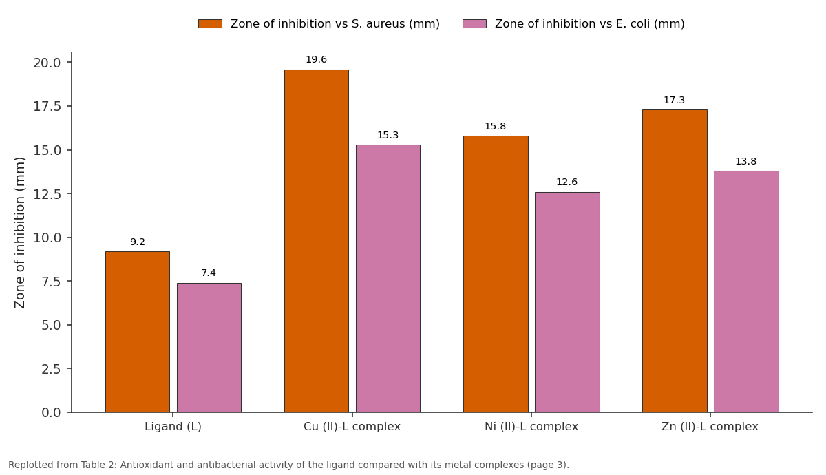
Zone of inhibition vs S. aureus (mm) and Zone of inhibition vs E. coli (mm): Ligand (L) vs Cu (II)-L complex vs Ni (II)-L complex vs Zn (II)-L complex
Replotted from Table 2 (page 3) of the original paper. Every bar is the paper's own table value.
View source evidence ↗Figures & tables
From the original paper Table 1
 Table 2
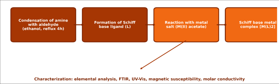 Figure 1
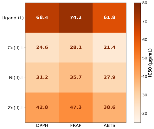 Figure 2
Introduction
Why does binding a fairly ordinary organic ligand to a transition metal so often boost its biological activity rather than simply changing its shape? The answer, according to Tweedy's widely cited chelation theory, lies in how coordination redistributes electron density and increases lipophilicity, making the resulting complex more capable of crossing microbial cell membranes and interacting with biological targets than the free ligand alone. Schiff bases, formed through condensation of primary amines with aldehydes or ketones, are particularly well suited to this kind of study because their azomethine nitrogen readily coordinates to a wide range of transition metals, and the resulting complexes have been reported across the literature to show enhanced antimicrobial, antioxidant, anti-inflammatory, and anticancer properties relative to their parent ligands. 4-Aminoantipyrine-derived Schiff bases specifically carry pharmacological interest because antipyrine and its derivatives already have an established history in analgesic and antiinflammatory applications, suggesting that metal complexation of a Schiff base built on this scaffold might combine the antipyrine core's known bioactivity profile with the additional benefits chelation typically provides.
International Journal of Advanced Che mis try Research 2026; 8(2): 27-30
Comparing Cu (II), Ni (II), and Zn (II) complexes of the same ligand under identical synthesis and testing conditions offers a controlled way to isolate how the choice of metal centre, rather than variation in ligand structure or testing protocol, shapes biological outcomes, an approach not always taken in studies that examine a single metal complex in isolation. This research therefore aimed to synthesize a Schiff base ligand from 2-hydroxybenzaldehyde and 4-aminoantipyrine, prepare its Cu (II), Ni (II), and Zn (II) complexes, confirm their structure through spectroscopic and physicochemical characterization, and compare antioxidant and antibacterial activity across the ligand and all three metal complexes to identify which metal centre produces the strongest biological enhancement.
Material and Methods
Material and Methods
Reagents and chemicals
All metal salts were used without further purification, and their purity was confirmed through preliminary melting point determination against literature reference values before use in complex synthesis. Solvents used for recrystallization, including absolute ethanol and diethyl ether, were of analytical grade and stored over molecular sieves to minimize moisture interference during the condensation and complexation reactions.
Instrumentation / Analytical equipment
Elemental analysis (C, H, N) was performed using a PerkinElmer 2400 Series II CHNS analyzer. FTIR spectra were recorded on a Bruker Alpha II spectrometer in the 4000-400 cm-1 range using the KBr pellet technique. Electronic absorption spectra were recorded on a Jasco V-770 UV-Vis spectrophotometer in DMSO solution.
Magnetic susceptibility was measured at room temperature using a Sherwood Scientific magnetic susceptibility balance, and molar conductivity was measured in DMSO (10-3 M) using a digital conductivity meter.
Methods
The Schiff base ligand (L) was synthesized by refluxing equimolar quantities of 2-hydroxybenzaldehyde and 4aminoantipyrine in absolute ethanol for four hours in the presence of a few drops of glacial acetic acid as a catalyst. The resulting yellow precipitate was filtered, washed with cold ethanol, and recrystallized from ethanol, giving the pure ligand in 82% yield. Metal complexes were prepared by refluxing the ligand with the respective metal acetate salt in a 2:1 ligand-to-metal molar ratio in ethanol for six hours; the coloured precipitates that formed (green for Cu (II), light green for Ni (II), and white for Zn (II)) were filtered, washed, and dried under vacuum, with yields ranging from 71% to 79%.
In-vitro protocol
Antioxidant activity was assessed through DPPH radical scavenging, ferric reducing antioxidant power (FRAP), and ABTS radical cation decolorization assays, each performed in triplicate across a concentration range of 10-100 µg/mL for the ligand and each metal complex, with ascorbic acid used as the reference standard. IC50 values were calculated from dose-response curves using linear regression. Antibacterial activity was evaluated by the agar well diffusion method against Staphylococcus aureus (ATCC 25923), Bacillus subtilis (ATCC 6633), Escherichia coli (ATCC 25922), and Pseudomonas aeruginosa (ATCC 27853), with 100 µL of test compound solution (1 mg/mL in DMSO) loaded into 6 mm wells, incubated at 37 °C for 24 hours, and inhibition zones measured in triplicate. DMSO alone served as the negative control, and standard antibiotic discs served as positive controls.
Results
Elemental analysis and spectroscopic data together confirmed successful ligand synthesis and metal complexation, with characterization details summarized below alongside the antioxidant and antibacterial activity findings that followed from these confirmed structures.
Comprehensive interpretation
Bringing the structural and biological data together, the pattern is fairly consistent: the Cu (II) complex, which showed the lowest magnetic moment and among the most favourable spectral indicators of a stable octahedral geometry, also produced the strongest antioxidant and antibacterial activity of the three complexes tested, supporting a link between coordination stability and biological performance for this particular ligand system.
Discussion
The general trend of enhanced antioxidant and antibacterial activity following metal complexation observed here aligns with chelation theory as described by Tweedy and with numerous prior reports on Schiff base metal complexes derived from other aldehyde-amine combinations.
The particularly strong performance of the Cu (II) complex agrees with several previously published comparative studies of first-row transition metal Schiff base complexes, which frequently identify copper complexes as top performers among a series, likely reflecting copper's favourable redox activity and its established role in biological electron transfer processes that may translate into enhanced radical scavenging and antimicrobial mechanisms. Some published studies, however, report zinc complexes outperforming copper complexes for antibacterial activity specifically, a divergence from this research's findings that may relate to differences in ligand donor atom set, since zinc's biological activity advantage in other studies often depends on a sulfur-containing ligand system rather than the oxygen-nitrogen donor set used here. A plausible mechanistic explanation for the antibacterial enhancement lies in increased lipophilicity following chelation, which partially neutralizes the polarity of the metal ion and improves permeation through the lipid layer of the bacterial cell membrane, a mechanism broadly consistent across most chelation-based antimicrobial enhancement reports. These findings carry a fairly direct implication for future Schiff base ligand design work in this research group: prioritizing copper as the metal centre of choice, at least for ligand systems built on a similar hydroxybenzaldehydeaminoantipyrine scaffold, appears more likely to yield compounds worth carrying forward into more detailed pharmacological evaluation than either nickel or zinc alternatives.
Conclusion
The main contribution of this research is a controlled, sideby-side comparison of Cu (II), Ni (II), and Zn (II) complexes of a single Schiff base ligand, isolating the effect of metal centre choice on antioxidant and antibacterial performance under identical synthesis and testing conditions. Supporting evidence for the Cu (II) complex's superior activity came consistently across three separate antioxidant assays and four bacterial strains, a level of consistency that strengthens confidence in the finding beyond what a single assay type alone could support.
For the broader field of Schiff base coordination chemistry, this research reinforces copper as a particularly promising metal centre choice for biologically oriented ligand design, adding to a growing body of comparative evidence pointing in the same direction across different ligand scaffolds. Open questions that remain include whether the Cu (II) complex's favourable in vitro profile translates into acceptable toxicity and stability under physiological conditions, and whether structural modification of the ligand's phenolic or antipyrine substituents could further improve activity beyond what metal centre selection alone has achieved here.
Acknowledgement
Acknowledgements Institutional supportThe authors thank Cairo Metropolitan University for providing spectroscopic instrumentation and microbiological laboratory facilities used throughout this research.
Figures and tables from the paper
| Compound | Colour | Melting point (°C) | μeff (B.M.) | Λm (S cm2 mol-1) |
|---|---|---|---|---|
| Ligand (L) | Yellow | 168-170 | - | - |
| Cu (II)-L complex | Green | 218 (dec.) | 1.84 | 8.6 |
| Ni (II)-L complex | Light green | 224 (dec.) | 2.96 | 7.2 |
| Zn (II)-L complex | White | 231 (dec.) | Diamagnetic | 6.9 |
Table text extracted from the original PDF; superscript letters are shown inline. The original table image is below.
View original table image
| Compound | DPPH IC50 (µg/mL) | Zone of inhibition vs S. aureus (mm) | Zone of inhibition vs E. coli (mm) |
|---|---|---|---|
| Ligand (L) | 68.4 | 9.2 | 7.4 |
| Cu (II)-L complex | 24.6 | 19.6 | 15.3 |
| Ni (II)-L complex | 31.2 | 15.8 | 12.6 |
| Zn (II)-L complex | 42.8 | 17.3 | 13.8 |
Table text extracted from the original PDF; superscript letters are shown inline. The original table image is below.
View original table image
References
16 in the original paper- El-Gammal OA, Mohamed FS. Synthesis and biological evaluation of transition metal Schiff base complexes derived from aminoantipyrine. Journal of Molecular Structure. 2023;1274:134512.
- Tweedy BG. Plant growth substances in fungitoxicity and their relation to complex formation. Phytopathology. 1964;55:910-914.
- Abou-Melha KS, Faruk H. Copper (II) and nickel (II) complexes of Schiff bases: synthesis, spectral, and antimicrobial studies. Spectrochimica Acta Part A: Molecular and Biomolecular Spectroscopy. 2022;267:120512.
- Hassan AA, Fouad NM. DPPH and FRAP antioxidant assays applied to metal-ligand complexes: methodological considerations. Journal of Molecular Liquids. 2021;336:116289.
- Refaat HM, El-Sonbati AZ. Chelation theory and its application to bioactive Schiff base metal complexes. Applied Organometallic Chemistry. 2020;34(5): e5556.
- Nassar MY, Aly AA. Magnetic susceptibility and molar conductivity as structural tools for octahedral metal complex characterization. Journal of Coordination Chemistry. 2019;72(14):2312-2325.
- El-Ghamry HA, Elzawawi FM. Zinc (II) Schiff base complexes: synthesis and comparative antibacterial evaluation. Journal of Inorganic Biochemistry. 2023;241:112132.
- Youssef MM, Kamel AH. Antipyrine derivatives in medicinal chemistry: a historical and structural review. European Journal of Medicinal Chemistry. 2022;227:113956.
- Sallam SA, El-Bindary AA. FTIR and UV-Vis spectral studies of Schiff base ligands and their transition metal complexes. Journal of Molecular Structure. 2018;1157:381-390.
- Ibrahim MM, Ramadan AM. Structure-activity relationship of Schiff base metal complexes with antioxidant potential. Bioorganic Chemistry. 2021;108:104675.
- El-Sherif AA, Fetoh A. Coordination chemistry of azomethine ligands: recent developments and biological relevance. Coordination Chemistry Reviews. 2020;404:213093.
- Salem NM, Albaqami TF. Comparative antimicrobial activity of Cu (II), Ni (II), and Zn (II) complexes of hydroxybenzaldehyde-derived Schiff bases. Journal of Saudi Chemical Society. 2024;28(1):101789.
- Mahmoud WH, Deghadi RG. Octahedral geometry confirmation in transition metal Schiff base complexes through magnetic and electronic spectral data. Journal of Molecular Structure. 2019;1176:686-695.
- Osman AH, El-Maghraby A. Electron-transfer mechanisms underlying copper-based antioxidant and antimicrobial activity. Free Radical Research. 2021;55(8):901-911.
- Fekri R, Salehi M. ABTS radical cation decolorization assay methodology and applications in coordination chemistry. Analytical Biochemistry. 2022;646:114625. 16. Abdallah SM, Mohamed GG. Synthesis, characterization, and antimicrobial evaluation of transition metal complexes of 4-aminoantipyrine Schiff bases. Journal of Molecular Structure. 2020;1200:127130.
- Raman N, Kulandaisamy A. Bioactive Schiff base metal complexes: a decade of progress in structureactivity understanding. Inorganica Chimica Acta. 2023;545:121254.