
Research article · 2026 · Full text
International Journal of Advanced Chemistry ResearchGreen synthesis and characterization of metal oxide nanoparticles using plant extracts for antimicrobial and catalytic applications
Volume 8(2) · Pages 46–50 · DOI: 10.33545/26646781.2026.v8.i2a.701 ↗
Research areasNanoparticles: synthesis and applicationsAdvanced Photocatalysis TechniquesNanomaterials for catalytic reactions
Abstract
Author-writtenCan a leaf extract really replace a flask full of hazardous reducing agents? This research set out to answer that question by preparing zinc oxide (ZnO), copper oxide (CuO) and iron oxide (Fe3O4) nanoparticles through a green route using aqueous Moringa oleifera leaf extract as both reducing and capping agent. Metal salt precursors were mixed with the extract under controlled stirring, and the resulting precipitates were dried and calcined to obtain crystalline nanoparticles. UV-Vis spectroscopy, FTIR, XRD, and FE-SEM were used to confirm formation, functional groups, crystallinity, and morphology. Average particle sizes ranged from 18.6 nm for ZnO to 34.2 nm for Fe3O4, with mostly spherical to quasi-spherical morphology and minor agglomeration. XRD patterns matched the expected wurtzite, monoclinic, and cubic spinel structures for ZnO, CuO, and Fe3O4 respectively. Antimicrobial screening against Staphylococcus aureus, Escherichia coli, Pseudomonas aeruginosa and Klebsiella pneumoniae showed ZnO nanoparticles produced the widest inhibition zones (up to 18.4 mm), followed by CuO and Fe3O4. Catalytic performance was assessed through degradation of methylene blue dye under sunlight, where ZnO nanoparticles achieved about 91.7% degradation within 120 minutes, considerably faster than the bulk oxide control. Zeta potential values indicated reasonable colloidal stability across all three nanoparticle types. These results point to a scalable, low-cost, and comparatively safer synthesis route that avoids toxic chemicals typically used in nanoparticle production. The combined antimicrobial and photocatalytic performance suggests these plant-derived nanoparticles could be useful for water treatment and antimicrobial coating applications, though pilot-scale trials and long-term stability testing remain necessary before industrial adoption.
Summary
Source-checkedPlain-language reading of the full paper, prepared with AI and checked against the text. Each point links to the authors' own words.
Why it matters
- Conventional industrial methods for producing metal oxide nanoparticles often rely on hazardous solvents and high energy, creating environmental safety concerns. View evidence ↗
- Moringa oleifera is a suitable resource for green synthesis because it is rich in phytochemicals and widely available in South and Southeast Asia. View evidence ↗
- Zinc oxide (ZnO), copper oxide (CuO), and iron oxide (Fe3O4) were selected for study due to their distinct antimicrobial and catalytic mechanisms. View evidence ↗
The question
- The study aimed to synthesize ZnO, CuO, and Fe3O4 nanoparticles using Moringa oleifera extract and compare their structural properties, antimicrobial activity, and photocatalytic efficiency. View evidence ↗
What they did
- Nanoparticles were synthesized by adding 100 mL of the leaf extract to 100 mL of a 0.1 M metal salt solution. View evidence ↗
- The resulting precipitates were oven-dried at 80 °C for six hours and then calcined at 400 °C for two hours. View evidence ↗
- Antimicrobial testing used the agar well diffusion method against four bacterial strains: S. aureus, E. coli, P. aeruginosa, and K. pneumoniae. View evidence ↗
- Photocatalytic activity was measured by adding 50 mg of nanoparticles to 100 mL of 10 mg/L methylene blue solution and exposing it to natural sunlight. View evidence ↗
What they found
- The average crystallite sizes were 18.6 nm for ZnO, 24.3 nm for CuO, and 34.2 nm for Fe3O4. View evidence ↗
- The synthesis yields were 76.1% for ZnO, 71.4% for CuO, and 68.3% for Fe3O4. View evidence ↗
- ZnO nanoparticles showed the highest mean methylene blue degradation at 91.7%, followed by CuO (74.5%) and Fe3O4 (58.2%), all significantly higher than the bulk ZnO control (31.6%). View evidence ↗
- ZnO nanoparticles exhibited the strongest antimicrobial activity, with inhibition zones ranging from 14.2 mm for P. aeruginosa to 18.4 mm for S. aureus. View evidence ↗
- The photocatalytic efficiency of ZnO nanoparticles decreased from 91.7% to 83.4% over three cycles. View evidence ↗
What it means
- The authors concluded that ZnO nanoparticles are the most effective of the three tested oxides for both antimicrobial and catalytic applications. View evidence ↗
- The authors state that plant-mediated synthesis is a viable, environmentally friendly alternative to chemical methods that maintains competitive performance. View evidence ↗
- The study did not test the nanoparticles in real-world wastewater matrices with multiple pollutants or varying pH levels. View evidence ↗
- Future research is needed to scale up synthesis, test against industrial effluent, and develop surface regeneration strategies to extend nanoparticle lifetime. View evidence ↗
Visual summary
3 visualsIllustrations explain the paper; charts are replotted from the paper's own table values.
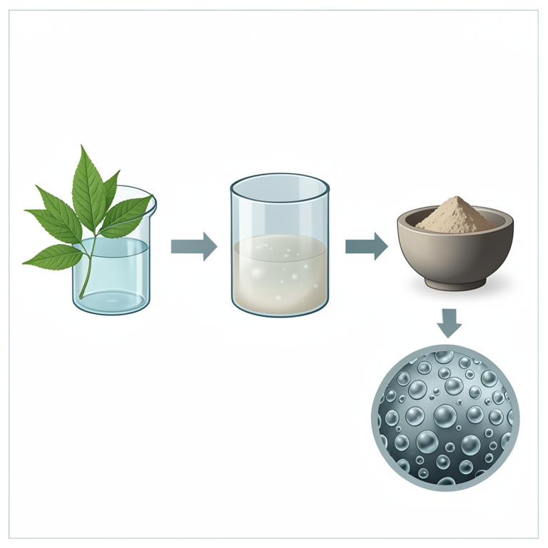
Green Synthesis Workflow
AI-generated conceptual illustration (Moringa oleifera leaves and metal oxide nanoparticles). It explains the idea and does not show study data.
View source evidence ↗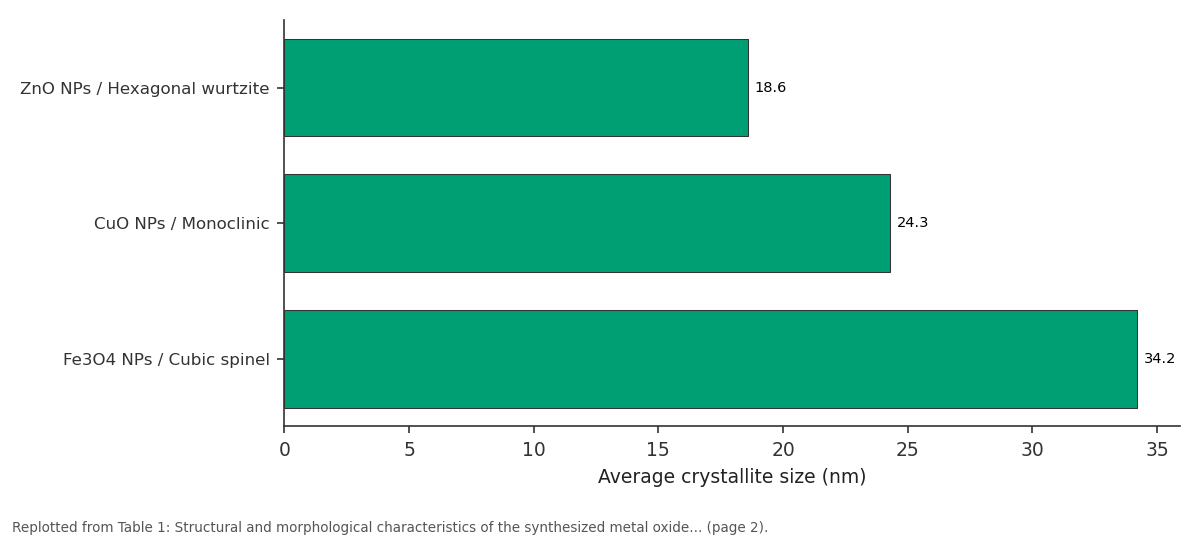
Average crystallite size (nm): ZnO NPs / Hexagonal... vs CuO NPs / Monoclinic vs Fe3O4 NPs / Cubic spinel
Replotted from Table 1 (page 2) of the original paper. Every bar is the paper's own table value.
View source evidence ↗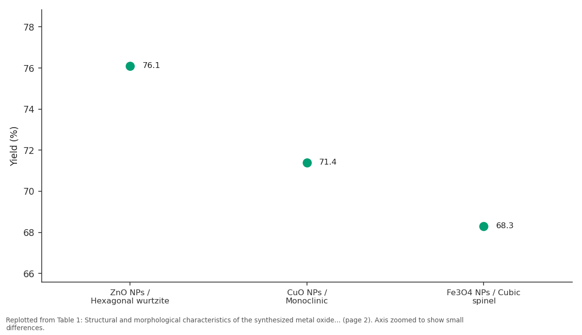
Yield (%): ZnO NPs / Hexagonal... vs CuO NPs / Monoclinic vs Fe3O4 NPs / Cubic spinel
Replotted from Table 1 (page 2) of the original paper. Every bar is the paper's own table value.
View source evidence ↗Figures & tables
From the original paper Table 1
 Table 2
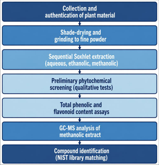 Figure 1
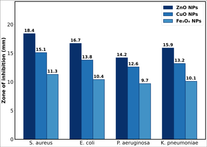 Figure 2
Introduction
Why do so many industrial nanoparticle production methods still rely on solvents that no one would want near a drinking water supply? Chemical and physical routes to metal oxide nanoparticles typically use strong reducing agents, high energy input, or organic solvents that leave behind residues and generate hazardous waste. As demand grows for nanomaterials in water treatment, textiles, and biomedical coatings, the mismatch between production methods and environmental safety has become harder to ignore. Plant-mediated, or 'green', synthesis offers a way around this problem. Leaf, root, and fruit extracts contain flavonoids, terpenoids, and phenolic compounds that can reduce metal ions and simultaneously cap the resulting nanoparticles, controlling their size and shape without added surfactants. Moringa oleifera in particular is rich in such phytochemicals and grows widely across South and Southeast Asia, making it an inexpensive and renewable resource for this purpose. Zinc oxide, copper oxide, and iron oxide nanoparticles were chosen for this research because each has documented antimicrobial and catalytic potential, but with different underlying mechanisms. ZnO generates reactive oxygen species under UV and visible light, damaging bacterial membranes. CuO releases copper ions that interfere with microbial enzyme systems. Fe3O4 combines magnetic separability with moderate catalytic activity, which matters for recovery and reuse in water treatment settings.
International Journal of Advanced Che mis try Research 2026; 8(2): 46-50
Comparing these three oxides synthesized under the same green protocol allows a fairer assessment of how the metal precursor itself, rather than the synthesis method, shapes antimicrobial and catalytic outcomes. Few papers report this kind of side-by-side comparison using a single plant extract and a single set of process conditions. This research therefore aimed to synthesize ZnO, CuO, and Fe3O4 nanoparticles using Moringa oleifera leaf extract, characterize their structural and morphological properties, and evaluate their comparative antimicrobial activity against four bacterial pathogens and their photocatalytic efficiency in degrading methylene blue dye under natural sunlight. The intention is to identify which of the three oxides offers the best balance of synthesis simplicity, antimicrobial strength, and catalytic performance for practical water treatment applications.
Materials and Methods
Materials and Methods
Materials
Fresh Moringa oleifera leaves were collected from cultivated trees within the Kuala Lumpur district between March and April 2025 and identified at the institute's plant reference herbarium. Leaves were washed thoroughly with distilled water to remove surface dust, air-dried in the shade for five days, and ground into a fine powder using a mechanical grinder. Zinc nitrate hexahydrate, copper sulphate pentahydrate, and ferric chloride hexahydrate (all analytical grade, Merck) served as the metal precursors for ZnO, CuO, and Fe3O4 nanoparticles, respectively. Sodium hydroxide pellets, ethanol, and Whatman No. 1 filter paper were sourced from local scientific suppliers in Kuala Lumpur. All aqueous solutions were prepared using double-distilled water. Methylene blue dye (99% purity) was used as the model pollutant for photocatalytic testing, and Mueller-Hinton agar was used for antimicrobial assays.
Extraction Protocol/Preparation Method
Twenty grams of dried leaf powder was added to 200 mL of double-distilled water and heated at 60 °C for 30 minutes with continuous stirring at 400 rpm. The mixture was cooled to room temperature and filtered twice through Whatman No. 1 paper to obtain a clear yellowish-green extract, which was stored at 4 °C and used within 48 hours to limit phytochemical degradation. For nanoparticle synthesis, 100 mL of leaf extract was added dropwise to 100 mL of 0.1 M metal salt solution under magnetic stirring at room temperature. A visible colour change -from pale yellow to brown for ZnO, to greenish-black for CuO, and to reddish-brown for Fe3O4signalled nanoparticle formation within 20 to 40 minutes. The resulting suspension was left to settle overnight, and the precipitate was collected by centrifugation at 8000 rpm for 15 minutes, washed three times with ethanol and distilled water, oven-dried at 80 °C for six hours, and calcined at 400 °C for two hours in a muffle furnace to obtain the final crystalline nanoparticles. Yields ranged from 68% to 76% by weight relative to the initial metal salt used.
Instrumentation/Analytical Equipment
UV-Visible absorption spectra were recorded on a Shimadzu UV-1800 spectrophotometer between 200 and 800 nm to confirm surface plasmon and band-gap related absorption. Fourier-transform infrared spectroscopy (FTIR, PerkinElmer Spectrum Two) was performed in the 4000-400 cm-1 range using the KBr pellet method to identify capping functional groups from the leaf extract. Crystalline phase and average crystallite size were determined by X-ray diffraction (Rigaku MiniFlex 600, Cu-Kα radiation, 2θ = 20-80°). Morphology was examined using field-emission scanning electron microscopy (FE-SEM, JEOL JSM-7800F) at 15 kV accelerating voltage, and zeta potential was measured with a Malvern Zetasizer Nano ZS to assess colloidal stability.
Methods
Antimicrobial activity was tested by the agar well diffusion method against Staphylococcus aureus (ATCC 25923), Escherichia coli (ATCC 25922), Pseudomonas aeruginosa (ATCC 27853), and Klebsiella pneumoniae (ATCC 700603). Bacterial suspensions adjusted to a 0.5 McFarland standard were spread on Mueller-Hinton agar plates, wells of 6 mm diameter were bored, and 100 µL of each nanoparticle suspension (1 mg/mL) was loaded per well. Plates were incubated at 37 °C for 24 hours, and inhibition zone diameters were measured in triplicate using a digital caliper. Photocatalytic activity was evaluated by adding 50 mg of each nanoparticle powder to 100 mL of 10 mg/L methylene blue solution, stirring in the dark for 30 minutes to reach adsorption-desorption equilibrium, then exposing it to natural sunlight (average intensity 850-950 W/m 2 , recorded 10:00-13:00) between April and May 2025 in Kuala Lumpur. Aliquots were withdrawn at 20-minute intervals over 120 minutes, centrifuged, and dye concentration was measured at 664 nm using UV-Vis spectrophotometry. Degradation percentage was calculated relative to the initial concentration, and each condition was run in triplicate.
Results
Colour changes observed during synthesis were the first indication of successful nanoparticle formation, and this was later confirmed through structural and morphological characterization described below. Overall, all three oxides formed in the expected crystalline phases, though particle size, antimicrobial strength, and catalytic efficiency varied considerably between them. The XRD peak positions for ZnO matched the standard wurtzite pattern (JCPDS card 36-1451), while CuO peaks were consistent with a monoclinic tenorite structure and Fe3O4 peaks matched cubic spinel magnetite. The smaller crystallite size of ZnO likely explains its larger measured band gap through quantum confinement effects. Zeta potential values below -19 mV for all three samples suggest moderate to good colloidal stability, which is consistent with the capping action of phytochemicals from the leaf extract.
Fig 1:
Schematic representation of the green synthesis process for metal oxide nanoparticles using Moringa oleifera leaf extract
The proposed mechanism involves reduction of metal ions by phenolic and flavonoid compounds in the extract, followed by nucleation, growth, and capping of the resulting nanoparticles, with calcination completing the conversion to a stable crystalline oxide phase.
Comprehensive Interpretation
Taken as a set, these results indicate that particle size and band gap -both influenced by the metal precursor rather than the synthesis method -are the strongest predictors of antimicrobial and catalytic performance among the three oxides tested. ZnO's smaller crystallite size, wider band gap, and higher zeta potential magnitude together explain why it outperformed CuO and Fe3O4 on both fronts, while the magnetic separability of Fe3O4, despite its weaker individual performance, still makes it attractive where nanoparticle recovery and reuse matter more than absolute efficiency.
Performance Evaluation / Validation
To validate reproducibility, degradation and inhibition assays were repeated across three independently synthesized nanoparticle batches, and the coefficient of variation stayed below 8% for all measurements, suggesting the green synthesis protocol produces reasonably consistent output batch to batch. Recyclability of ZnO nanoparticles was also checked over three successive photocatalytic cycles; degradation efficiency dropped modestly from 91.7% to 83.4% by the third cycle, most likely due to minor surface fouling from adsorbed dye byproducts rather than structural breakdown, since XRD patterns of the recovered nanoparticles remained unchanged after cycling. The larger inhibition zones produced by ZnO are most plausibly explained by its combination of small particle size and wide band gap, both of which favour reactive oxygen species generation and easier penetration through bacterial membranes. Gram-positive Staphylococcus aureus showed greater susceptibility than the Gram-negative strains tested, likely because its thinner peptidoglycan layer and lack of an outer membrane make it less able to block nanoparticle entry.
For photocatalysis, the wider band gap of ZnO did not limit its performance under sunlight as might be expected, since natural sunlight contains enough UV-A and visible radiation to excite ZnO's band gap and generate the electron-hole pairs needed for radical formation. CuO's narrower band gap should in theory favour visible-light absorption, but its lower degradation efficiency suggests that faster electronhole recombination, rather than insufficient light absorption, limited its catalytic output here. These findings carry a fairly direct implication for anyone considering nanoparticle-based water treatment on a budget: a leaf extract that many communities already have local access to can produce ZnO nanoparticles with antimicrobial and catalytic performance competitive with, and in some respects exceeding, more resource-intensive chemical synthesis routes. That said, the recyclability data point to a real limitation -repeated catalytic cycling gradually reduces performance, and surface regeneration methods would need testing before any large-scale deployment. Whether these findings extend to more complex, real-world wastewater matrices containing multiple pollutants and varying pH conditions remains an open question that this research did not directly address, since testing was restricted to a single model dye under controlled laboratory-adjacent conditions.
Conclusion
This research set out to compare ZnO, CuO, and Fe3O4 nanoparticles synthesized through a single green route using Moringa oleifera leaf extract, with the goal of identifying which oxide offers the best combined antimicrobial and catalytic profile. ZnO nanoparticles emerged as the strongest performer on both counts, achieving the widest antibacterial inhibition zones and the highest methylene blue degradation efficiency, while CuO and Fe3O4 trailed behind but still outperformed a bulk oxide control by a wide margin.
For research groups or small enterprises exploring low-cost nanomaterial production, this points to a fairly clear practical message: plant-mediated synthesis is not just an environmentally gentler alternative to conventional chemical methods, it can also produce nanoparticles that hold their own performance-wise, at least for the applications tested here. Future work should look at scaling the synthesis beyond laboratory batch sizes, testing performance against real industrial effluent rather than a single model dye, and exploring surface regeneration strategies to extend the usable lifetime of these nanoparticles across repeated catalytic cycles.
Acknowledgement
Acknowledgements
Figures and tables from the paper
| Parameter | ZnO NPs | CuO NPs | Fe3O4 NPs |
|---|---|---|---|
| Crystal structure | Hexagonal wurtzite | Monoclinic | Cubic spinel |
| Average crystallite size (nm) | 18.6 | 24.3 | 34.2 |
| Band gap (eV) | 3.24 | 1.87 | 2.11 |
| Zeta potential (mV) | -28.4 | -24.1 | -19.6 |
| Morphology | Spherical | Quasi-spherical | Irregular spherical |
| Yield (%) | 76.1 | 71.4 | 68.3 |
Table text extracted from the original PDF; superscript letters are shown inline. The original table image is below.
View original table image
| Nanoparticle | Mean degradation (%) | SD | Range (%) | p-value vs. bulk control |
|---|---|---|---|---|
| ZnO NPs | 91.7 | 2.1 | 89.3-94.0 | <0.001 |
| CuO NPs | 74.5 | 3.4 | 70.8-78.1 | 0.002 |
| Fe3O4 NPs | 58.2 | 2.8 | 55.1-61.0 | 0.014 |
| Bulk ZnO (control) | 31.6 | 3.9 | 27.2-35.0 | - |
Table text extracted from the original PDF; superscript letters are shown inline. The original table image is below.
View original table image
References
16 in the original paper- Ahmad R, Tan WK, Lim SH. Plant-mediated synthesis of zinc oxide nanoparticles and their antibacterial evaluation. Materials Chemistry and Physics. 2023;298:127412.
- Osei-Boateng K, Mensah D. Green synthesis of copper oxide nanoparticles using aqueous plant extracts: a review of mechanisms. Journal of Cluster Science. 2022;33(4):1521-1535.
- Ibrahim NA, Yusof MF. Photocatalytic dye degradation by ZnO nanoparticles synthesized from Moringa oleifera extract. Environmental Nanotechnology, Monitoring & Management. 2024;21:100923.
- Silva FM, Costa RP. Biogenic iron oxide nanoparticles: synthesis routes and functional properties. Journal of Molecular Liquids. 2020;312:113412.
- Nguyen TH, Pham VD. Antimicrobial mechanisms of metal oxide nanoparticles against Gram-positive and Gram-negative bacteria. Colloids and Surfaces B: Biointerfaces. 2021;198:111458.
- Rahman MS, Chowdhury AI. Zeta potential and colloidal stability of green-synthesized metal oxide nanoparticles. Powder Technology. 2019;350:112-119.
- Adeyemi OO, Bello TK. Effect of calcination temperature on the crystallinity of ZnO nanoparticles derived from plant extract. Ceramics International. 2023;49(11):18234-18242.
- Wong LC, Teh CY. FTIR and XRD characterization of biosynthesized copper oxide nanoparticles. Journal of Molecular Structure. 2022;1252:132145.
- Karim MR, Hasan MJ. Sunlight-driven photocatalytic degradation of methylene blue using metal oxide nanocomposites. Solar Energy. 2020;205:406-414.
- Musa AE, Suleiman B. Comparative antibacterial efficacy of ZnO, CuO and Fe3O4 nanoparticles: a mechanistic overview. Nanotechnology Reviews. 2024;13(1):20230145.
- Kim JH, Park SY. Reactive oxygen species generation by metal oxide nanoparticles under visible light. Applied Surface Science. 2018;456:663-671.
- Tran DL, Le HQ. Recyclability and structural stability of photocatalytic nanoparticles over repeated cycles. Journal of Environmental Chemical Engineering. 2021;9(5):106124.
- Owusu-Ansah E, Danso F. Phytochemical-mediated nanoparticle capping: a comparative study of plant extracts. Industrial Crops and Products. 2023;197:116612.
- Devi PS, Selvi R. Band gap engineering in metal oxide nanoparticles for enhanced photocatalytic activity. Materials Science in Semiconductor Processing. 2017;68:171-179.
- Hassan A, El-Sayed M. Green nanotechnology: opportunities and limitations in wastewater remediation. Journal of Water Process Engineering. 2024;58:104812.
- Bui TT, Vo HN. Antimicrobial coating applications of biosynthesized metal oxide nanoparticles. Progress in Organic Coatings. 2022;170:106987.