
Research article · 2026 · Full text
International Journal of Biology SciencesIsolation and identification of chlorpyrifos degrading bacteria from pesticide contaminated agriculture soil
Volume 8(7) · Pages 21–27 · DOI: 10.33545/26649926.2026.v8.i7a.894 ↗
Research areasPesticide and Herbicide Environmental StudiesMicrobial bioremediation and biosurfactantsPesticide Exposure and Toxicity
Abstract
Author-writtenChlorpyrifos is a common organophosphate insecticide that stays in the soil used for farming for a long time. This is bad for the soil and the underground water. It also harm living things that are not supposed to be killed by the insecticide. This study was done to find bacteria in the soil that can break down chlorpyrifos. The soil used for the study was taken from Okra village in the Balrampur district of Chhattisgarh in India. The goal was to see how well these bacteria can break down chlorpyrifos in a laboratory. The soil was mixed with water. Spread on a special plate to see how many bacteria were in it. There were a lot of bacteria in the soil. About 6.8 × 10⁵ bacteria per gram of soil. Three types of bacteria were found that could break down chlorpyrifos. These bacteria were called S1, S4 and S9. They were later identified as Enterobacter asburiae, Rothia kristinae and Aeromonas hydrophila. The best temperature and acidity level for these bacteria to grow and break down chlorpyrifos were found to be 35°C and pH 7.0. The bacteria were then tested to see how well they could break down chlorpyrifos over 30 days. Enterobacter asburiae was the best at breaking down chlorpyrifos, broke down 92.8% of it. Aeromonas hydrophila broke down 86.2%. Rothia kristinae broke down 72.6%. Enterobacter asburiae was the fastest at breaking it down. It took 8.4 days to break down half of the chlorpyrifos. Without any bacteria it took a lot longer 288.8 Days. These findings show that the bacteria in the soil in Chhattisgarh are very good at breaking down chlorpyrifos. They can be used to clean up the soil and water in that region.
Summary
Source-checkedPlain-language reading of the full paper, prepared with AI and checked against the text. Each point links to the authors' own words.
Why it matters
- Chlorpyrifos is a widely used organophosphate insecticide that persists in agricultural soil, posing risks to soil health, groundwater, and non-target organisms. View evidence ↗
- The metabolite of chlorpyrifos, 3,5,6-trichloro-2-pyridinol (TCP), is particularly concerning due to its high water solubility and a soil half-life that can reach 360 days. View evidence ↗
- Using native soil bacteria for bioremediation is considered a sustainable and cost-effective way to remove chlorpyrifos compared to expensive physicochemical methods. View evidence ↗
The question
- The study aimed to isolate and identify potent chlorpyrifos-degrading bacteria from contaminated agricultural soil to find candidates for environmental bioremediation. View evidence ↗
What they did
- Soil samples were collected from agricultural fields in Okra village, Balrampur district, Chhattisgarh, India. View evidence ↗
- Bacteria were isolated using the pour plate method on Nutrient Agar sterilized at 121°C and 15 psi. View evidence ↗
- Isolates were screened for their ability to use chlorpyrifos as the sole carbon source in Minimal Salt Medium (MSM) at pH 7.0. View evidence ↗
- The amount of chlorpyrifos degraded was measured using High-Performance Liquid Chromatography (HPLC) at 7, 14, 21, and 30 days. View evidence ↗
What they found
- The contaminated soil from Okra village contained a viable bacterial population of 6.8 × 10⁵ CFU/g. View evidence ↗
- Three chlorpyrifos-degrading bacteria were identified: Enterobacter asburiae (S1), Rothia kristinae (S4), and Aeromonas hydrophila (S9). View evidence ↗
- All three isolates grew and degraded chlorpyrifos best at 35°C, pH 7.0, 1.0% nutrient concentration, and under aerobic shaking. View evidence ↗
- By day 30, Enterobacter asburiae showed the highest degradation efficiency at 92.8%, while Aeromonas hydrophila and Rothia kristinae reached 86.2% and 72.6%, respectively. View evidence ↗
- Enterobacter asburiae was the fastest degrader with a half-life of 8.4 days, whereas Rothia kristinae was the slowest with a half-life of 16.8 days. View evidence ↗
- The uninoculated control had a half-life of 288.8 days, indicating that chlorpyrifos does not break down significantly on its own without bacteria. View evidence ↗
What it means
- The authors conclude that Enterobacter asburiae is the most promising candidate for large-scale field bioremediation of chlorpyrifos. View evidence ↗
- The study was limited to laboratory conditions; actual field performance may be affected by soil organic matter, other bacteria, and soil chemistry. View evidence ↗
- The researchers were unable to fully identify all the metabolites produced during degradation, leaving the fate of TCP and other intermediates unknown. View evidence ↗
Visual summary
3 visualsIllustrations explain the paper; charts are replotted from the paper's own table values.
Comparative Degradation Efficiency
AI-generated conceptual illustration (Enterobacter asburiae, Rothia kristinae, and Aeromonas hydrophila bacteria in agricultural soil). It explains the idea and does not show study data.
View source evidence ↗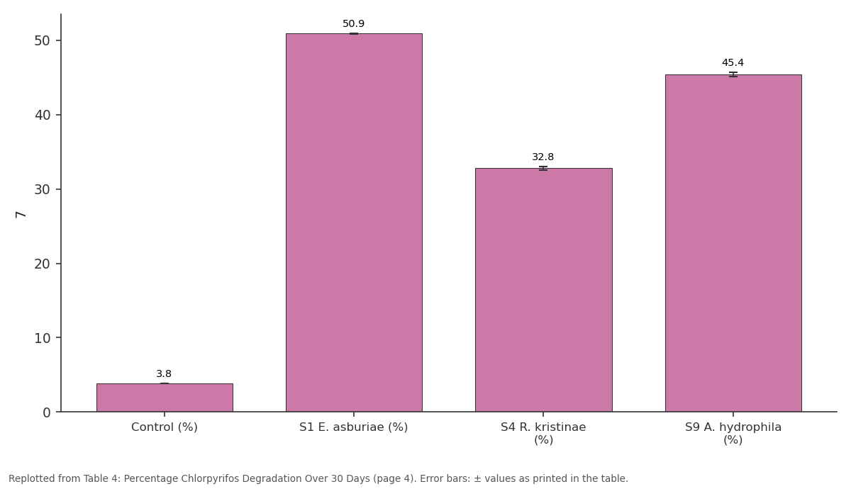
7: Control (%) vs S1 E. asburiae (%) vs S4 R. kristinae (%) vs S9 A. hydrophila (%)
Replotted from Table 4 (page 4) of the original paper. Every bar is the paper's own table value.
View source evidence ↗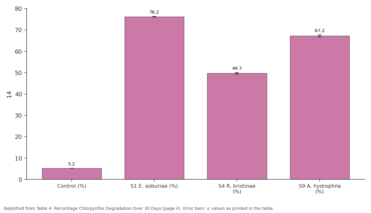
14: Control (%) vs S1 E. asburiae (%) vs S4 R. kristinae (%) vs S9 A. hydrophila (%)
Replotted from Table 4 (page 4) of the original paper. Every bar is the paper's own table value.
View source evidence ↗Figures & tables
From the original paper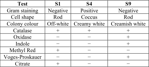 Table 1
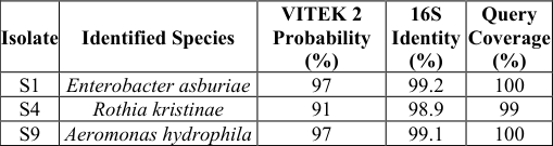 Table 2
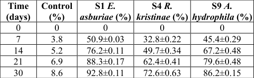 Table 4
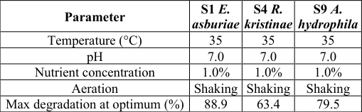 Table 3
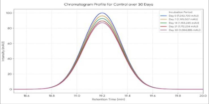 Figure 1
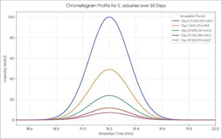 Figure 2
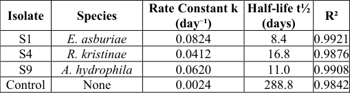 Table 5
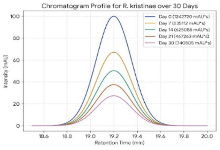 Figure 3
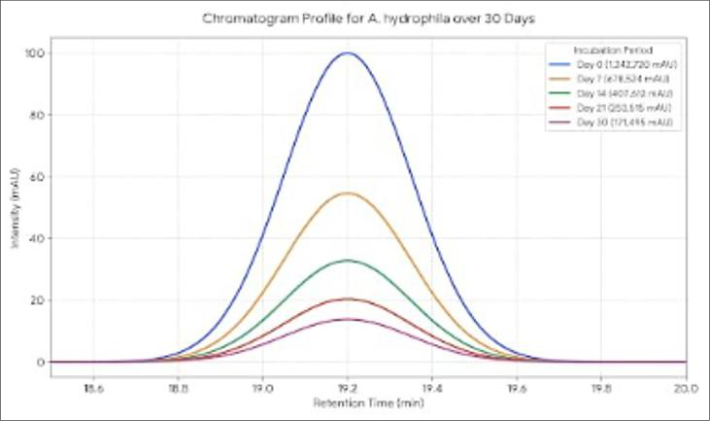 Figure 4
Introduction
Pesticides are extensively applied in modern agriculture to protect crops from insect pests and to maximize yields for the growing global population. Organophosphorus (OP) compounds account for more than 50% of all insecticides used worldwide, and their global market has been growing at a compound annual growth rate of approximately 5.5% (Aktar et al., 2009; Foong et al., 2020) [1,7] . Among the most widely used OP insecticides, chlorpyrifos [O,O-diethyl O-(3,5,6-trichloro-2-pyridyl) phosphorothioate] holds particular significance. First introduced in the United States in 1965 as a replacement for DDT, it is now applied across more than 100 countries for the control of soil-borne and foliar pests on crops including maize, cotton, citrus, and vegetables (John & Shaike, 2015; Foong et al., 2020) [11,7] . Despite its agronomic utility, the indiscriminate and repeated application of chlorpyrifos has led to widespread contamination of agricultural soils and water resources. Chlorpyrifos exhibits moderate to significant environmental persistence, with a soil half-life ranging from 60 to 120 days, and occasionally up to 365 days depending on temperature, pH, and microbial activity (Foong et al., 2020) [7] . Its primary hydrolytic metabolite, 3,5,6-trichloro-2-pyridinol (TCP), is of even greater concern TCP has a soil half-life of up to 360 days and high water solubility, enabling it to leach into groundwater and accumulate in the food chain (Deng et al., 2016; Žabar et al., 2016) [4,24] . The presence of three chlorine atoms on its aromatic ring renders TCP highly resistant to microbial breakdown, compounding the risk of environmental contamination (Rayu et al., 2017) [16] . The toxicological profile of chlorpyrifos is well established. It acts primarily through irreversible inhibition of acetylcholinesterase (AChE), the enzyme responsible for hydrolysis of the neurotransmitter acetylcholine at cholinergic synapses, leading to accumulation of acetylcholine and prolonged nerve stimulation (Eaton et al., 2008; Burke et al., 2017) [5,3] .
Acute exposure produces classical cholinergic symptoms including salivation, lacrimation, tremors, and respiratory distress, while chronic or prenatal exposure has been associated with neurodevelopmental disorders, endocrine disruption, and reproductive toxicity (Rauh et al., 2011; Testai et al., 2010) [15,19] . The World Health Organization classifies chlorpyrifos as a Class II moderately hazardous compound, with an LD₅₀ of 135-163 mg/kg in mammals (WHO, 2009). Given the scale of contamination and associated ecological and human health risks, there is an urgent need for effective remediation strategies. Conventional physicochemical methods such as adsorption, chemical oxidation, and incineration are often costly, non-selective, and may generate harmful secondary products. Bioremediation particularly the use of indigenous soil bacteria capable of metabolizing chlorpyrifos is recognized as a cost-effective and environmentally sustainable alternative (Singh et al., 2004; Huang et al., 2021) [17,9] . Microorganisms residing in pesticide-contaminated soils are known to develop specialized enzymatic systems, particularly organophosphate hydrolases (OPH), that cleave the P-O bond of chlorpyrifos, enabling its use as a carbon and/or phosphorus source (Singh et al., 2004; Anwar et al., 2009) [17, 2] . Numerous bacterial genera with chlorpyrifos-degrading capabilities have been documented from contaminated agricultural soils worldwide. Enterobacter strain B-14, isolated from Australian soil, was among the earliest reported chlorpyrifos degraders capable of utilizing the compound as a sole carbon and phosphorus source (Singh et al., 2004) [17] . Subsequently, degraders belonging to Pseudomonas, Bacillus, Sphingomonas, Stenotrophomonas, Achromobacter, Paracoccus, Klebsiella, Arthrobacter, and Rhizobium have been isolated and characterized (Yang et al., 2006; Anwar et al., 2009; Lakshmi et al., 2009; Xu et al., 2008; Farhan et al., 2021) [23,2,12,22,6] . Molecular identification via 16S rRNA gene sequencing has become the standard approach for accurate taxonomic assignment of these isolates, with sequence similarities of 96-100% to GenBank reference sequences reliably confirming genusand species-level identity (Madhuri & Rangaswamy, 2019; Farhan et al., 2021) [14,6] . Indigenous strains isolated from locally contaminated soils are particularly valuable because of their pre-adaptation to site-specific conditions of pH, temperature, and co-contaminants, making them superior candidates for in situ bioremediation (Wepukhulu et al., 2024; Ishag et al., 2016) [21,10] . Despite a growing body of literature, comprehensive isolation and molecular characterization studies from region-specific contaminated agricultural soils remain limited, particularly in areas of intensive pesticide use in developing countries. The present study was therefore undertaken to isolate, identify, and characterize chlorpyrifos-degrading bacteria from pesticidecontaminated agricultural soil using enrichment culture technique, standard biochemical characterization, and 16S rRNA gene sequencing, with the aim of identifying potent bacterial candidates for bioremediation of chlorpyrifoscontaminated environments.
Materials and Methods
Materials and Methods
Collection and Preparation of Soil Samples
Soil samples were collected from agricultural fields of Okra village, Balrampur district, Chhattisgarh, India, with a documented history of repeated chlorpyrifos application. Samples were collected aseptically from a depth of 10-15 cm using sterile spatulas, pooled to obtain a representative composite sample, transferred into sterile polythene bags, and transported to the laboratory in an ice box. Samples were stored at 4°C until further analysis. Prior to processing, soil was passed through a ≤2 mm sieve to remove debris, and moisture content was maintained at 40% by spraying with distilled water to support aerobic microbial activity (Gong et al., 2016) [8] .
Serial Dilution and Isolation of Bacteria
Serial dilutions were prepared from 1 g of processed soil suspended in 9 mL of sterile distilled water, up to 10⁻⁷, under aseptic conditions. Bacterial isolation was performed by the pour plate method using Nutrient Agar (beef extract 3.0 g, peptone 5.0 g, NaCl 5.0 g, agar 15.0 g per 1000 mL, pH 7.0), sterilized at 121°C and 15 psi for 15 minutes. One mL aliquots from dilutions 10⁻³ to 10⁻⁶ were plated and incubated at 37±1°C for 24-48 hours. Morphologically distinct colonies were subcultured repeatedly on fresh Nutrient Agar to obtain pure cultures, which were stored on Nutrient Agar slants at 4°C.
Screening for Chlorpyrifos Degradation
Pure bacterial isolates were screened for chlorpyrifos degradation using Minimal Salt Medium (MSM) containing KH₂PO₄ 1.2 g, K₂HPO₄ 4.8 g, NH₄NO₃ 1.0 g, MgSO₄•7H₂O 0.2 g, Ca(NO₃)₂•4H₂O 0.04 g, and FeSO₄ 0.001 g per liter, pH 7.0, with chlorpyrifos as the sole carbon source. Isolates were inoculated into MSM-chlorpyrifos flasks and incubated at 30°C under shaking at 120 rpm for 5-7 days. Turbidity development, biomass formation, and clearance zone formation were taken as indicators of degradation activity.
Morphological and Biochemical Characterization
Colony morphology was recorded after incubation on Nutrient Agar at 37±1°C for 24-48 hours. Microscopic characteristics were studied by Gram staining under oil immersion objective. Standard biochemical tests including catalase, oxidase, and IMViC tests (indole, methyl red, Voges-Proskauer, and citrate utilization) were performed following standard procedures.
Identification by VITEK 2 and 16S rRNA Gene Sequencing
Pure bacterial isolates were identified using the VITEK 2 Compact automated identification system at Ganga Diagnostic and Medical Research Centre, Raipur. Bacterial suspensions were prepared in 0.45% NaCl saline, adjusted to 0.5 McFarland standard, and loaded into appropriate VITEK identification cards. The system automatically analyzed biochemical reaction patterns and compared them against its reference database to generate species identification with probability percentage. For molecular identification, genomic DNA was extracted using the CTAB method (Sambrook and Russell, 2001) [18] and purity was confirmed by NanoDrop spectrophotometry (A260/A280 ratio 1.8-2.0). The 16S rRNA gene was amplified using universal primers 27F (5'-AGAGTTTGATCCTGGCTCAG-3') and 1492R (5'-TACGGTTACCTTGTTACGACTT-3') under the following thermal cycling conditions: initial denaturation at 95°C for 5 minutes, 30 cycles of 94°C for 30 seconds, 55°C for 30 seconds, and 72°C for 1 minute, followed by final extension at 72°C for 7 minutes (Lane, 1991) [13] . PCR products were verified by 1.5% agarose gel electrophoresis, purified, and submitted for Sanger sequencing. Sequences were analyzed using BLASTn against the NCBI GenBank database, and isolates showing ≥97% sequence identity were identified to species level. Phylogenetic analysis was performed using the Neighbor-Joining method with 1000 bootstrap replicates in MEGA version 11.0 (Tamura et al., 2021) [20] .
Optimization of Biodegradation Conditions
The effect of temperature (20-45°C), pH (4-9), nutrient concentration (0.5-2.0% yeast extract and peptone), aeration (shaking vs. static), and NaCl concentration (0.5-2.5%) on chlorpyrifos degradation was evaluated by incubating selected isolates in MSM-chlorpyrifos broth under each condition while keeping all other parameters constant. Each variable was tested independently and degradation efficiency was measured after incubation to determine the optimum condition for each parameter.
HPLC Analysis of Chlorpyrifos Degradation
Chlorpyrifos degradation was quantified by HPLC at 7, 14, 21, and 30 day intervals (Singh et al., 2004) [17] . Culture aliquots of 20 mL were centrifuged at 7000 rpm for 10 minutes, and the supernatant was extracted with dichloromethane. The organic layer was dried, dissolved in HPLC grade acetonitrile, and filtered through a 0.22 µm nylon syringe filter before injection. Analysis was performed on a C18 reverse phase column (250 × 4.6 mm, 5 µm) with acetonitrile:water (75:25 v/v) acidified with 1% phosphoric acid as the mobile phase, at a flow rate of 0.8-1.0 mL/min, injection volume of 10 µL, detection wavelength of 230 nm, and column temperature of 40°C. A standard calibration curve was constructed from chlorpyrifos standards at 10-100 mg/L. Degradation percentage was calculated as:
Degradation kinetics were analyzed using the first-order model:
Statistical Analysis
All experiments were performed in triplicate and results expressed as mean ± Standard deviation (SD). One-way ANOVA followed by Tukey's HSD post-hoc test was used to compare degradation efficiency among isolates. A pvalue of <0.05 was considered statistically significant. Statistical analysis was performed using GraphPad Prism.
Results
Results
Microbial Isolation and Colony Count
Soil samples collected from chlorpyrifos-contaminated agricultural fields of Okra village, Balrampur district, Chhattisgarh yielded a viable bacterial population of 6.8 × 10⁵ CFU/g of soil, calculated from countable plates at 10⁻⁴ dilution. A total of 28 bacterial colonies were observed on Nutrient Agar plates, from which 12 morphologically distinct colonies were picked and 9 pure cultures were successfully obtained after repeated subculturing.
Screening for Chlorpyrifos Degradation
All nine bacterial isolates were screened on Minimal Salt Medium (MSM) supplemented with chlorpyrifos as the sole carbon source. Three isolates S1, S4, and S9 demonstrated positive growth, visible turbidity, and clearance zone formation in MSM, indicating active chlorpyrifos utilization. These three isolates were selected for further characterization and degradation studies.
Morphological and Biochemical Characterization
Morphological examination on Nutrient Agar and standard biochemical tests were performed for all three selected isolates. S1 appeared as Gram-negative rods with smooth, opaque, off-white colonies. S4 appeared as Gram-positive cocci with creamy white, smooth colonies of 1-2 mm diameter. S9 appeared as Gram-negative rods with smooth, moist, glistening creamish white colonies. Biochemical profiles of all three isolates are summarized in Table 1.
| Test | S1 | S4 | S9 |
|---|---|---|---|
| Gram staining | Negative | Positive | Negative |
| Cell shape | Rod | Coccus | Rod |
| Colony colour | Off-white | Creamy white | Creamish white |
| Catalase | + | + | + |
| Oxidase | − | − | + |
| Indole | − | − | + |
| Methyl Red | + | − | − |
| Voges-Proskauer | − | − | + |
| Citrate | + | − | − |
Table text extracted from the original PDF; superscript letters are shown inline. The original table image is below.
View original table image
Identification by VITEK 2 and 16S rRNA Gene Sequencing
All three isolates were confirmed by VITEK 2 Compact automated identification system at Ganga Diagnostic and Medical Research Centre, Raipur. S1 was identified as Enterobacter asburiae (97% probability), S4 as Rothia kristinae (91% probability), and S9 as Aeromonas hydrophila/punctata (97% probability). Molecular identification by 16S rRNA gene sequencing using universal primers 27F/1492R and BLASTn analysis against NCBI GenBank confirmed all three identifications with sequence identities of 99.2%, 98.9%, and 99.1% respectively. DNA purity was acceptable for all isolates with A260/A280 ratios between 1.83 and 1.89. Identification results are summarized in Table 2.
Optimization of Biodegradation Conditions
Process optimization experiments revealed that all three bacterial isolates shared common optimal conditions: temperature of 35°C, pH 7.0, nutrient concentration of 1.0%, and aerobic shaking conditions. Under shaking conditions, degradation was approximately double that observed under static conditions across all isolates, confirming the strictly aerobic nature of chlorpyrifos degradation. Salt tolerance experiments showed that degradation declined sharply beyond 1.5% NaCl for all isolates. Optimum conditions for each isolate are summarized in Table 3.
| Parameter | S1 E. asburiae | S4 R. kristinae | S9 A. hydrophila |
|---|---|---|---|
| Temperature (°C) | 35 | 35 | 35 |
| pH | 7.0 | 7.0 | 7.0 |
| Nutrient concentration | 1.0% | 1.0% | 1.0% |
| Aeration | Shaking | Shaking | Shaking |
| Max degradation at optimum (%) | 88.9 | 63.4 | 79.5 |
Table text extracted from the original PDF; superscript letters are shown inline. The original table image is below.
View original table image
HPLC Analysis and Degradation Kinetics
Chlorpyrifos degradation was quantified by HPLC over 30 days at a retention time of 19.2 minutes and detection wavelength of 230 nm. All three isolates showed progressive and substantial degradation over the study period far exceeding the uninoculated control which retained 91.4% of chlorpyrifos at day 30, confirming that observed degradation was entirely microbial in origin.
Enterobacter asburiae (S1) achieved the highest degradation of 92.8% by day 30, followed by Aeromonas hydrophila (S9) at 86.2% and Rothia kristinae (S4) at 72.6%. Degradation was fastest between days 7 and 21, after which the rate slowed as residual chlorpyrifos became limited. Percentage degradation over time is presented in Table 4. Chlorpyrifos was detected by HPLC with a retention time of 19.2 minutes at a detection wavelength of 230 nm. This retention time served as the reference for identifying and quantifying chlorpyrifos in the degradation samples. Chromatograms for all three bacterial isolates and the uninoculated control were recorded at days 0, 7, 14, 21, and 30 as shown in the figure 1 to 4. Chlorpyrifos degradation followed first-order kinetics for all three isolates with R² values of 0.98-0.99, confirming that degradation rate was proportional to residual substrate concentration. E. asburiae showed the highest rate constant (k = 0.0824 day⁻¹) and shortest half-life of 8.4 days, while R. kristinae was the slowest with a half-life of 16.8 days. The control half-life of 288.8 days confirms negligible abiotic degradation. Kinetic parameters are presented in Table 5.
| Isolate | Species | Rate Constant k (day⁻¹) | Half-life t½ (days) | R² |
|---|---|---|---|---|
| S1 | E. asburiae | 0.0824 | 8.4 | 0.9921 |
| S4 | R. kristinae | 0.0412 | 16.8 | 0.9876 |
| S9 | A. hydrophila | 0.0620 | 11.0 | 0.9908 |
| Control | None | 0.0024 | 288.8 | 0.9842 |
Table text extracted from the original PDF; superscript letters are shown inline. The original table image is below.
View original table image
| Time (days) | Control (%) | S1 E. asburiae (%) | S4 R. kristinae (%) | S9 A. hydrophila (%) |
|---|---|---|---|---|
| 0 | 0 | 0 | 0 | 0 |
| 7 | 3.8 | 50.9±0.03 | 32.8±0.22 | 45.4±0.29 |
| 14 | 5.2 | 76.2±0.11 | 49.7±0.34 | 67.2±0.48 |
| 21 | 6.9 | 88.3±0.17 | 62.4±0.41 | 79.6±0.48 |
| 30 | 8.6 | 92.8±0.11 | 72.6±0.63 | 86.2±0.15 |
Table text extracted from the original PDF; superscript letters are shown inline. The original table image is below.
View original table image
Discussion
A population of 6.8 10 CFU/g bacterial isolated from chlorpyrifos-contaminated agricultural soil of Okra village, Balrampur district has revealed a functional microbial population. Microbial population similar to the ones previously identified in the soils under pesticide stress has also been observed in this study, and is indicative of the general understanding that application of pesticide over time stimulates selection of microbes tolerant to pesticide as well as degrading microorganisms. The formation of visible colonies of bacteria up to 10 dilution shows that the application of chlorpyrifos for long period has not eliminated the native microflora from the soil but altered to the selection of microorganisms capable to metabolize chlorpyrifos.
The screen was performed with MSM with chlorpyrifos as the only carbon source, and three bacterial isolates, S1, S4 and S9 out of nine screened showed growth and were further evaluated. This screening procedure is methodologically adequate since only those bacteria which are able to effectively mineralise or co-mineralise chlorpyrifos will be able to grow, and thus the observation of formation of turbidity and clearance zone reflect genuine degradation rather than mere tolerance. HPLC analysis later confirmed the results obtained from the screen. The optimum conditions for maximum phosphotriesterase production were observed at a temperature of 35°C, pH 7.0, a substrate concentration of 1.0%, and under aerobic shaking conditions, respectively. This optimal temperature of 35°C reflects the mesophilic environment in which the organisms thrive and is representative of the thermal stability range for bacterial phosphotriesterases. The universal pH optima of 7.0 also coincide with the typical operating range for hydrolytic enzymes that participate in pesticide breakdown and represent an agricultural pH value that, in turn has practical relevance to any potential field application. The dramatic differences observed in degradation efficiency between aerobic shaking and nonshaking conditions clearly indicate the strictly aerobic conditions that limit the rate of chlorpyrifos breakdown by the bacteria. The degradation data were well-fitted with the first-order kinetics and R-values ranged from 0.98 to 0.99 for all isolates. This demonstrates that the rate of degradation was directly correlated to the amount of residual chlorpyrifos during the investigation. The half-life decreased from 288.8 days (uninoculated control) to 8.4 days (E. Asburiae) and thus increased the removal rate of chlorpyrifos over 34 times. Because there was virtually no abiotic degradation found in the control study it is evident that chlorpyrifos removals were entirely contributed by the microbials. Thus, our study reveals that E. Asburiae, A. Hydrophila, and R. Kristinae from chlorpyrifos contaminated agricultural fields in Chhattisgarh are good chlorpyrifos degrading bacteria with high prospects for its bioremediation in the area. It is however, imperative to reiterate that this study was carried out under laboratory conditions and environmental factors like presence of soil organic matter, antagonistic bacteria and the soil physiochemical characteristics would surely have a bearing on these degradation values under field conditions. This study has also failed to achieve a full metabolite spectrum for its degradation products. The fate of tcp and other degradation intermediates remains elusive. This research opens the pathway for undertaking research on metabolites profiling of tcp, studies on biochemical pathways and enzymatic studies on chlorpyrifos degrading enzymes, in addition to, undertaking soil microcosmos studies under typical field conditions to implement it as an environment friendly tool in field bioremediation.
Conclusion
Three indigenous chlorpyrifos-degrading bacterial isolates, Enterobacter asburiae (S1), Rothia kristinae (S4), and Aeromonas hydrophila (S9), were successfully isolated and identified from pesticide-contaminated agricultural soils of Balrampur district. All isolates efficiently utilized chlorpyrifos as the sole carbon source under aerobic conditions. Optimum degradation was achieved at 35°C, pH 7.0, 1.0% substrate concentration, and under shaking conditions. HPLC analysis demonstrated that Enterobacter asburiae exhibited the highest degradation efficiency (92.8%; t½ = 8.4 days), followed by Aeromonas hydrophila (86.2%) and Rothia kristinae (72.6%). In contrast, the uninoculated control showed only 8.6% degradation after 30 days (t½ = 288.8 days), confirming that chlorpyrifos removal was predominantly due to microbial activity. Degradation by all isolates followed first-order kinetics (R² = 0.98-0.99), indicating predictable degradation behavior. The isolation of these efficient degraders from native agricultural soils highlights their potential as environmentally sustainable agents for the bioremediation of chlorpyrifos-contaminated environments. Among the isolates, Enterobacter asburiae demonstrated the greatest biodegradation potential and represents a promising candidate for field-scale bioremediation. Future studies should focus on metabolite identification using GC-MS or LC-MS, molecular characterization of degradation pathways, and validation under soil microcosm and field conditions to facilitate practical application.
Figures and tables from the paper
References
24 in the original paper- Aktar MW, Sengupta D, Chowdhury A. Impact of pesticides use in agriculture: their benefits and hazards. Interdisciplinary Toxicology. 2009;2(1):1-12.
- Anwar S, Liaquat F, Khan QM, Khalid ZM, Iqbal S. Biodegradation of chlorpyrifos and its hydrolysis product 3,5,6-trichloro-2-pyridinol by Bacillus pumilus strain C2A1. Journal of Hazardous Materials. 2009;168(1):400-405.
- Burke RD, Todd S. W, Lumsden E, Trask JAM, Batten SR, Bhatt P, et al. Developmental neurotoxicity of the organophosphorus insecticide chlorpyrifos: from clinical findings to preclinical models and potential mechanisms. Journal of Neurochemistry. 2017;142(S2):162-177.
- Deng W, Lin D, Yao K, Yuan H, Wang Z, Li J, et al. Characterization of a novel 𝛽-cypermethrin-degrading Aspergillus niger YAT strain and the biochemical degradation pathway of 𝛽-cypermethrin. Applied Microbiology and Biotechnology. 2015;99:8187-8198.
- Eaton DL, Daroff RB, Autrup H, Bridges J, Buffler P, Costa LG, et al. Review of the toxicology of chlorpyrifos with an emphasis on human exposure and neurodevelopment. Critical Reviews in Toxicology. 2008;38(S2):1-125.
- Farhan M, Ahmad M, Kanwal A, Butt ZA, Khan QF, Raza SA, et al. Biodegradation of chlorpyrifos using isolates from contaminated agricultural soil, its kinetic studies. Scientific Reports. 2021;11:10320.
- Foong SY, Ma NL, Lam SS, Peng W, Low F, Lee BHK, et al. A recent global review of hazardous chlorpyrifos pesticide in fruit and vegetables: prevalence, remediation and actions needed. Journal of Hazardous Materials. 2020;400:123006.
- Gong T, Liu R, Che Y, Xu X, Zhao F, Yu H, et al. Engineering Pseudomonas putida KT2440 for simultaneous degradation of carbofuran and chlorpyrifos. Microbial Biotechnology. 2016;9(6):792800.
- Huang Y, Zhang W, Pang S, Chen J, Bhatt P, Mishra S, et al. Insights into the microbial degradation and catalytic mechanisms of chlorpyrifos. Environmental Research. 2021;194:110660.
- Ishag SA, Abdelbagi AO, Hammad AMA, Elsheikh EAE, Elsaid OE, Hur JH, et al. Biodegradation of chlorpyrifos, malathion, and dimethoate by three strains of bacteria isolated from pesticide-polluted soils in Sudan. Journal of Agricultural and Food Chemistry. 2016;64(45):8491-8498.
- John EM, Shaike JM. Chlorpyrifos: pollution and remediation. Environmental Chemistry Letters. 2015;13:269-291.
- Lakshmi CV, Kumar M, Khanna S. Biotransformation of chlorpyrifos and bioremediation of contaminated soil. International Biodeterioration & Biodegradation. 2009;62(2):204-209.
- Lane DJ. 16S/23S rRNA sequencing. In: Stackebrandt E, Goodfellow M, editors. Nucleic acid techniques in bacterial systematics. New York (NY): Wiley; 1991. p. 115-175.
- Madhuri RJ, Rangaswamy V. Chlorpyrifos biodegradation in relation to metabolic attributes and 16S rRNA gene phylogeny of bacteria in a tropical vertisol. Discover Applied Sciences. 2019;1:161.
- Rauh VA, Garfinkel R, Perera FP, Andrews HF, Hoepner L, Barr DB, et al. Impact of prenatal chlorpyrifos exposure on neurodevelopment in the first 3 years of life among inner-city children. Pediatrics. 2011;118(6):e1845-e1859.
- Rayu S, Nielsen UN, Nazaries L, Singh BK. Isolation and molecular characterization of novel chlorpyrifos and 3,5,6-trichloro-2-pyridinol-degrading bacteria from sugarcane farm soils. Frontiers in Microbiology. 2017;8:518.
- Singh BK, Walker A, Morgan JAW, Wright DJ. Biodegradation of chlorpyrifos by Enterobacter strain B-14 and its use in bioremediation of contaminated soils. Applied and Environmental Microbiology. 2004;70(8):4855-4863.
- Sambrook J, Russell DW. Molecular cloning: A laboratory manual. 3rd ed. Cold Spring Harbor (NY): Cold Spring Harbor Laboratory Press; 2001.
- Testai E, Buratti FM, Di Consiglio E. Chlorpyrifos. In: Encyclopedia of Environmental Health. Amsterdam: Elsevier; 2010. p. 617-628.
- Tamura K, Stecher G, Kumar S. MEGA11: Molecular evolutionary genetics analysis version 11. Molecular Biology and Evolution. 2021;38(7):3022-3027.
- Wepukhulu M. Optimization of growth conditions for chlorpyrifos-degrading bacteria in farm soils in Nakuru County, Kenya. BioMed Research International. 2024;2024:1611871.
- Xu G, Zheng W, Li Y, Wang S, Zhang J, Yan Y. Biodegradation of chlorpyrifos and 3,5,6-trichloro-2pyridinol by a newly isolated Paracoccus sp. strain TRP. International Biodeterioration & Biodegradation. 2008;62(1):51-56.
- Yang C, Liu N, Guo X, Qiao C. Cloning of mpd gene from a chlorpyrifos-degrading bacterium and use of this strain in bioremediation of contaminated soil. FEMS Microbiology Letters. 2006;265(1):118-125.
- Žabar R, Komel T, Fabjan J, Kramberger MB, Trebše P. Photocatalytic degradation with immobilized TiO₂ of three selected neonicotinoid insecticides: imidacloprid, thiamethoxam and clothianidin. Chemosphere. 2016;112, 192-199.