
Research article · 2026 · Full text
International Journal of Biology SciencesPathological spectrum of avian colibacillosis in commercial broiler chickens reared in Northern Kashmir
Volume 8(7) · Pages 10–17 · DOI: 10.33545/26649926.2026.v8.i7a.892 ↗
Research areasEscherichia coli research studiesMicrobial infections and disease researchAnimal Virus Infections Studies
Abstract
Author-writtenThis study was conducted to investigate the pathomorphological alterations associated with naturally occurring colibacillosis in broiler chickens from February to July 2025 in commercial poultry farms of the Srinagar & Ganderbal district of Kashmir. A total of 10 outbreaks of colibacillosis were recorded across different farms and dead birds were submitted to the Division of Veterinary Pathology for comprehensive postmortem examination. Although the disease occurred across various age groups, mortality was most pronounced in 3-4-week-old birds and confirmation was based on typical lesions, with key pathomorphological changes such as fibrinous pericarditis, myocarditis, fibrinous perihepatitis, and airsacculitis observed in different organs. Colibacillosis continues to pose a significant economic burden on the poultry industry due to increased morbidity and mortality. The findings of this study indicate that pathogenic Escherichia coli can induce systemic lesions and cause immunosuppression in broiler chickens, as demonstrated by lymphoid depletion in lymphoid tissues.
Summary
Source-checkedPlain-language reading of the full paper, prepared with AI and checked against the text. Each point links to the authors' own words.
Why it matters
- Poultry farming is a critical part of global food security because it provides an affordable source of high-quality protein. View evidence ↗
- Avian colibacillosis, caused by avian pathogenic Escherichia coli (APEC), leads to significant economic losses through increased bird mortality and reduced growth. View evidence ↗
- Factors such as overcrowding, poor ventilation, and environmental stress can weaken a bird's immune system, making it more susceptible to colibacillosis. View evidence ↗
The question
- The study aimed to evaluate the pathological changes in the organs of broiler chickens infected with colibacillosis due to a lack of information on the disease in Kashmir. View evidence ↗
What they did
- The research was conducted at the Division of Veterinary Pathology, SKUAST-Kashmir, between February and July 2025. View evidence ↗
- The study analyzed 100 dead birds from 10 different colibacillosis outbreaks across various poultry farms. View evidence ↗
- Tissues from the liver, heart, air sacs, kidney, spleen, bursa, and thymus were fixed in 10% formalin and stained with Haematoxylin and Eosin for histopathological analysis. View evidence ↗
What they found
- While colibacillosis affected various ages, the highest mortality was observed in birds aged 3 to 4 weeks. View evidence ↗
- The liver showed fibrinous perihepatitis, which ranged from a thin layer in moderate cases to a thick exudate creating a 'bread and butter' appearance in severe cases. View evidence ↗
- Fibrinous pericarditis and congestion were consistently found in the hearts of most affected birds. View evidence ↗
- Air sacs exhibited inflammation characterized by cloudiness, thickening, and the presence of fibrinous exudate. View evidence ↗
- Most affected birds showed an enlarged, inflamed, and edematous bursa of Fabricius. View evidence ↗
- Histopathological analysis of the spleen revealed significant lymphoid depletion, specifically a reduction of lymphocytes in the white pulp. View evidence ↗
- The bursa of Fabricius exhibited severe lymphoid depletion and a marked reduction in the lymphocyte population within its follicles. View evidence ↗
- The thymus showed marked lymphoid depletion, which led to reduced cellularity in the thymic lobules. View evidence ↗
What it means
- The authors conclude that the observed lymphoid depletion and resulting immunosuppression highlight the severity of colibacillosis and its negative impact on poultry productivity. View evidence ↗
- The study identifies colibacillosis as a major cause of mortality and a substantial economic challenge for the poultry industry and small-scale farmers in Kashmir. View evidence ↗
Visual summary
1 visualsIllustrations explain the paper; they do not show study data.
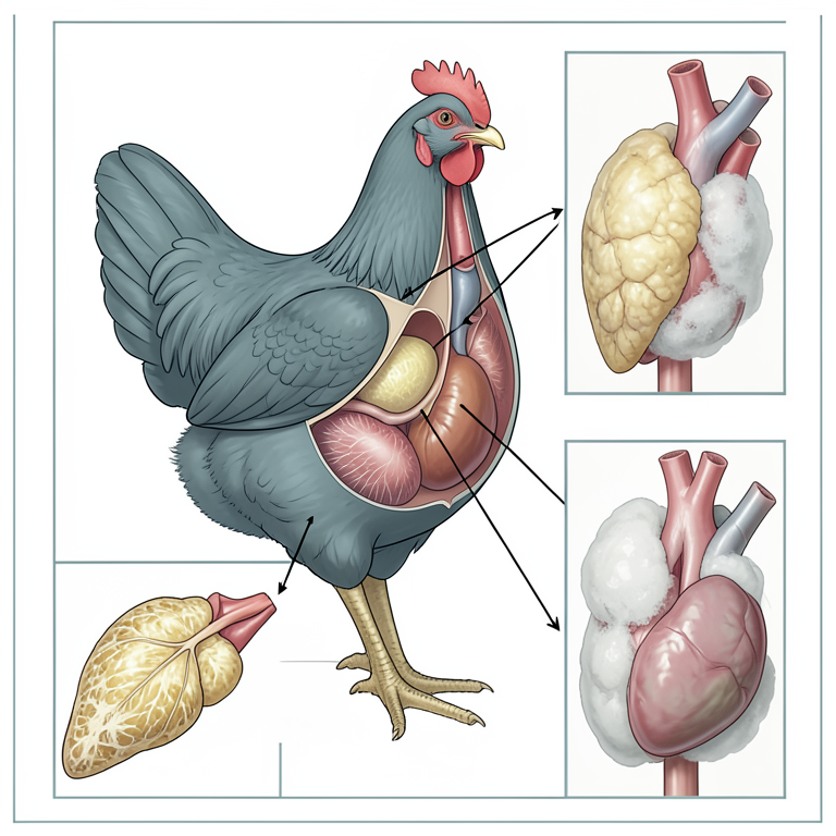
Systemic Pathological Effects
AI-generated conceptual illustration (commercial broiler chickens). It explains the idea and does not show study data.
View source evidence ↗Figures & tables
From the original paper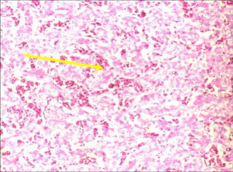 Figure 1
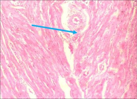 Figure 2
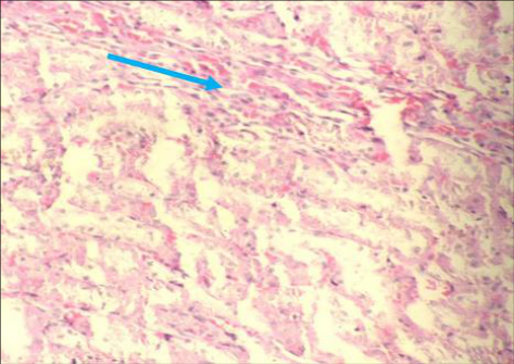 Figure 3
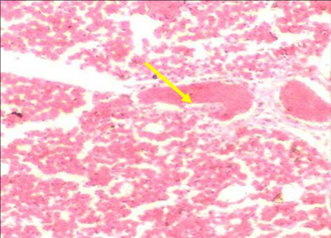 Figure 4
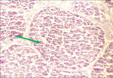 Figure 5
Introduction
Agriculture refers to the organized and methodical practice of growing crops, raising animals and utilizing biological resources to produce food, fibre, medicinal products and other essential materials that support and improve human life. It includes a broad spectrum of activities such as farming, animal husbandry, forestry and fisheries and plays a fundamental role in ensuring food security, promoting economic growth and enhancing societal wellbeing. Livestock farming is a major component of agriculture, involving the rearing of animals for products like meat, milk, eggs, wool and other by-products. The common livestock species include cattle, pigs, sheep, goats, and poultry such as chickens, turkeys and ducks. The efficient livestock production depends on proper nutrition, suitable housing, effective disease management and well-planned breeding strategies to ensure optimal productivity and animal health. Poultry farming is commonly known as aviculture, which involves the domestication and rearing of birds such as chickens, turkeys, ducks and geese for the production of meat, eggs and feathers. It represents one of the most rapidly expanding sectors within agriculture and contributes significantly to global food security by providing an economical source of high-quality protein. Moreover, poultry production plays a vital role in fulfilling the increasing protein requirements of the growing human population and serves as an important source of income worldwide (Kumar et al., 2013) [7] . Based on production objectives, poultry farming is mainly divided into layer farming and broiler farming. Layer farming is aimed at egg production under controlled conditions that maximize laying efficiency, whereas broiler farming focuses on the production of meat, with birds bred for rapid growth and typically reaching market weight within 6-7 weeks under intensive rearing systems (Yadav et al., 2024) [10] . A modern broiler production systems prioritize optimal environmental management, including temperature control, ventilation and balanced feeding, to enhance growth performance and feed conversion efficiency.
Disease control is an essential aspect of poultry production, as it directly influences the health status of birds, productivity and economic profitability. Poultry diseases may be caused by infectious agents such as viruses, bacteria, fungi and parasites, as well as non-infectious factors like nutritional deficiencies. The effective disease management strategies include strict implementation of biosecurity measures, vaccination programs, sanitation, hygiene, regular health monitoring, quarantine protocols, quality feed management and veterinary care. Biosecurity remains a key preventive measure that limits the introduction and spread of pathogens through controlled access, disinfection practices and maintenance of hygienic conditions (Ewers et al., 2004) [3] . The recent findings have further reinforced the role of biosecurity in minimizing bacterial infections such as colibacillosis and improving production efficiency in broiler systems (Tilli et al., 2024) [18] . Vaccination is widely practiced to safeguard poultry against major infectious diseases, including Newcastle disease, infectious bronchitis, infectious bursal disease and Marek's disease. In addition, maintaining proper sanitation, efficient waste disposal and continuous monitoring of flock health are essential for early detection and control of disease outbreaks. Adequate nutrition and access to clean drinking water are equally important in strengthening the immune system of birds, while veterinary guidance ensures timely diagnosis, treatment and prevention of diseases. Colibacillosis is also referred to as avian colibacillosis or colisepticemia, which is a significant bacterial disease in poultry caused by avian pathogenic Escherichia coli (APEC). It is responsible for considerable economic losses in the poultry industry due to increased mortality, decreased growth performance and carcass condemnation. The disease can present in various forms, including respiratory infections, septicemia and localized lesions in multiple organs. Recent research has demonstrated that APEC strains possess a wide range of virulence factors that facilitate colonization, systemic spread and persistence within the host (Joseph et al., 2023) [6] . Moreover, colibacillosis remains a major global concern due to its complex pathogenesis and its potential implications for food safety and zoonotic transmission (Abdelhamid et al., 2024) [1] . The transmission of E. coli occurs through contaminated feed, water, environment and carrier birds, with evidence also supporting vertical transmission via eggs (Abdelhamid et al., 2024) [1] . Some predisposing factors, including overcrowding, poor ventilation, environmental stress, transportation and concurrent infections, can compromise the immune system of birds and increase their susceptibility to infection. In addition, poor hygiene and inadequate sanitation facilitate the survival and spread of pathogenic bacteria within poultry farms. Clinically, colibacillosis exhibits a wide range of manifestations depending on the severity and type of infection. The respiratory signs include coughing, sneezing, nasal discharge, difficulty in breathing, swollen sinuses and abnormal respiratory sounds. The clinical manifestations are characterized by depression, reduced feed intake, diarrhoea, dehydration, increased mortality and poor growth performance. The localized infections may affect various organs, resulting in conditions such as arthritis, peritonitis and salpingitis in laying hens. The recent studies have also emphasized that colibacillosis induces significant immunological disturbances and systemic pathological alterations in affected birds, thereby contributing to substantial production losses (Ghahramani et al., 2024) [5] . Keeping in view the paucity of information regarding Colibacilosis in broiler chickens in Kashmir, this study was undertaken to evaluate the pathological alterations on the structure and functionality of different organs of infected broiler chickens. [2] and all observable lesions were systematically recorded. The representative tissue samples, including those from the liver, heart, air sacs, kidney, spleen, bursa, thymus and other organs exhibiting typical gross lesions, were collected for histopathological analysis. The samples were fixed in 10% formalin, processed by using routine histological techniques and stained with Haematoxylin and Eosin as per the method described by Lillie (1954) [8] .
Materials and Methods
Materials and Methods
Results:
The pathological findings of this investigation are systematically described under clinical, gross and microscopic observations recorded in poultry infected with Avian Pathogenic E. coli.
Clinical Signs:
The affected birds generally exhibited signs of weakness, depression and decreased activity. A marked reduction in feed intake was commonly observed. Diarrhoea was a frequent finding and frequently resulted in dehydration, which was evident by sunken eyes, dryness of the skin and increased thirst. In some cases, respiratory involvement was noted, characterized by coughing, sneezing, nasal discharge and dyspnea. In laying birds, infection was associated with a decline in egg production and the laying of abnormal or misshapen eggs. The musculoskeletal involvement was occasionally evident, with swollen joints leading to lameness and impaired locomotion. Some severe cases were often accompanied by high mortality, particularly among younger birds. The infected birds also showed poor weight gain or progressive weight loss, along with increased water consumption and a rough, ruffled feather appearance. The reproductive performance in breeding flocks was adversely affected, resulting in reduced fertility or infertility. Neurological manifestations such as tremors, convulsions, paralysis and incoordination were observed in some cases. The affected birds frequently exhibited a hunched posture, drooping wings and a tendency to huddle together. The mucous membranes appeared pale and thin. Some cases revealed skeletal abnormalities including tibial dyschondroplasia, bending of tibial bones, enlarged hock joints and sternal recumbency. Some additional clinical signs included lacrimation, excessive salivation, restlessness and ataxia were also observed in the affected birds. The emaciation was evident in severely affected birds, with a prominent keel bone. A consistent rise in body temperature was recorded from the onset of infection until the end of the observation period. Birds that survived for a few days displayed stunted growth, persistent diarrhoea, muscle weakness, staggering gait and diminished response to external stimuli prior to death. The affected birds often remained motionless for prolonged periods with puffed feathers and closed eyes. The respiratory distress was prominent, with repeated sneezing and accumulation of fluid in the oral cavity. Death was generally attributed to respiratory failure, often preceded by sudden collapse, uneasiness, paddling movements of the limbs, and loss of consciousness.
Pathomorphology:
Colibacillosis in broilers is characterized by pronounced, multisystemic gross pathological alterations observed during post-mortem examination. The carcasses are typically emaciated and dehydrated, exhibiting ruffled feathers and a generally poor body condition. The detailed observations of gross lesions across different organs are outlined below
Gross Pathology
• Liver: In cases of colibacillosis, the liver exhibited prominent gross alterations depending upon the severity of infection. In moderate cases, the hepatic surface was covered with a thin, delicate fibrinous layer, whereas in severe cases, a dense and thick fibrinous exudate enveloped the organ, imparting the characteristic "bread and butter" appearance. The liver was markedly enlarged with rounded margins and appeared congested and friable upon manipulation. The colour varied from dark reddish to pale discolored areas, indicating varying degrees of vascular disturbance and parenchymal damage. There are numerous multifocal necrotic foci, ranging from pinpoint to pinhead size, that were distributed throughout the hepatic parenchyma. In advanced stages, these lesions coalesced to form larger areas of necrosis accompanied by extensive hemorrhages. The hepatic capsule was noticeably thickened and often adhered firmly to adjacent visceral organs, suggesting chronic inflammatory changes. In some cases, diffuse necrosis with petechial hemorrhages was observed, reflecting severe septicemic involvement of the liver. • Heart: The heart revealed varying degrees of congestion along with fibrinous pericarditis, which was a consistent finding in most of the affected birds. In mild cases, a thin fibrinous layer was observed on the pericardial surface, however, in severe cases, a thick, opaque fibrinous exudate completely covered the heart.
Cardiomegaly was frequently noted, with dilation and engorgement of one or more cardiac chambers. The myocardium appeared congested and the coronary blood vessels were prominently distended with blood, imparting a bluish or dark discoloration to the cardiac tissue. The edema of the myocardial and pericardial tissues contributed to a swollen appearance of the heart.
In some cases, the pericardial sac contained serofibrinous fluid, further indicating severe inflammatory involvement. • Kidneys: The kidneys were consistently enlarged, swollen and oedematous, with a congested appearance. The external surface showed the presence of petechial and ecchymotic hemorrhages along with multiple necrotic foci. In early stages, the kidneys appeared hyperemic and smooth, whereas in advanced stages, they exhibited a mottled appearance due to the presence of multifocal necrotic lesions. The renal surface often appeared uneven and irregular, reflecting underlying pathological changes. In severe cases, the kidneys were pale in some regions due to necrosis and ischemia, while other areas showed intense congestion. • Spleen: The spleen in affected birds was markedly enlarged and congested, often appearing dark in colour due to excessive blood accumulation. The organ frequently exhibited scattered necrotic foci on its surface. The lymphoid depletion resulted in a reduction in the size and number of splenic follicles, leading to shrinkage and atrophy in chronic cases. The splenic capsule was sometimes tense due to congestion and the parenchyma appeared soft and friable. • Trachea: The trachea showed pronounced congestion and hyperemia of the mucosal lining, accompanied by swelling. In severe cases, the lumen contained fibrinous exudates forming greyish, adherent membranes along the tracheal wall. These pseudomembranes partially obstructed the airway and indicated severe inflammatory changes. The mucosal surface appeared thickened and inflamed, with occasional accumulation of mucus. • Air Sacs: The air sacs were cloudy, thickened and opaque, indicating inflammation. They were coated with fibrinous exudate, which in mild cases appeared as a thin film, while in severe cases formed dense fibrin layers. Caseous deposits were frequently observed, particularly in thoracic air sacs, giving them a cheesy appearance. In advanced cases, the air sac membranes were markedly thickened and lost their normal transparency. Necrotic lesions occasionally extended to adjacent lung surfaces, indicating spread of infection. • Lungs: The lungs exhibited marked congestion, edema and consolidation. The affected areas ranged from focal patches to diffuse involvement of the entire lung tissue.
In severe cases, the lungs appeared heavy, firm and dark due to extensive inflammatory exudation and vascular congestion. The cut surface often exuded fluid, indicating edema. The consolidated areas were firm and failed to collapse, reflecting severe pneumonia. • Bursa of Fabricius: The bursa of Fabricius was enlarged, oedematous and inflamed in most of the affected birds. The mucosal surface showed petechial and ecchymotic hemorrhages, along with accumulation of fibrinous and caseous exudates. In severe cases, the mucosa appeared thickened, discoloured and friable. The lumen often contained gelatinous or cheesy material. In chronic cases, the bursa appeared atrophied due to lymphoid depletion, with reduced size and loss of normal architecture. • Thymus: The thymus was reduced in size due to lymphoid depletion and atrophy. In severe cases, the organ exhibited congestion and edema and the tissue appeared soft, fragile and sometimes pale. The lobules of the thymus were less distinct, indicating degenerative changes. In chronic cases, the thymus showed marked shrinkage due to prolonged immune suppression.
• Proventriculus:
The proventriculus appeared congested, haemorrhagic and necrotic in affected birds. The mucosa showed areas of discoloration and damage due to vascular compromise and inflammatory processes. The lumen contained serous, fibrinous and necrotic debris. In severe cases, the organ appeared edematous and swollen, with accumulation of exudate. The glandular regions were often indistinct due to necrosis and inflammation.
• Intestine: The intestine exhibited congestion, haemorrhage and edema of the mucosal and submucosal layers. The intestinal wall was thickened, giving a swollen appearance. The necrotic lesions and ulcerations were frequently observed, particularly in severe cases. The excessive mucus accumulation was evident, forming a slimy coating over the mucosal surface. Fibrinous plaques consisting of inflammatory exudate and cellular debris were also present, giving the mucosa a rough and irregular appearance. In some cases, narrowing of the intestinal lumen due to inflammation and fibrosis was observed in affected intestines.
Gross Pathology
Gross Pathology
Histopathology
• Liver: Microscopically, the liver exhibited pronounced hepatocellular degeneration characterized by cellular swelling, vacuolation and loss of normal architecture. Distortion and disorganization of hepatic cords were evident, accompanied by marked congestion of central veins and sinusoids. Hepatitis was a consistent finding, with infiltration of inflammatory cells, predominantly heterophils and lymphocytes, within the hepatic parenchyma. In severe cases, there was marked thickening of the hepatic capsule due to fibrin deposition and intense heterophilic infiltration. Kupffer cell hyperplasia was prominent, reflecting enhanced phagocytic activity. Additional changes included cytoplasmic vacuolation, nuclear degeneration such as karyolysis and multifocal areas of coagulative necrosis. Sinusoidal congestion and hemorrhage were frequently observed, along with dense leukocytic infiltration around portal triads. In advanced stages, extensive disruption of hepatic architecture and widespread necrosis were evident. • Heart: Histologically, the heart showed lesions consistent with fibrinous pericarditis, characterized by deposition of fibrin on the pericardial surface along with infiltration of heterophils and other inflammatory cells. In severe cases, the pericardium was markedly thickened due to accumulation of fibrinous exudate and inflammatory debris. The myocardium exhibited degenerative changes, including vacuolation, swelling and loss of cross-striations in myocardial fibers.
Fragmentation and degeneration of muscle fibers were also evident. In advanced stages, areas of coagulative necrosis and interstitial hemorrhage were observed, indicating severe myocardial damage. The inflammatory infiltration extended into myocardial tissue, further compromising cardiac integrity and function.
• Kidneys: Microscopic examination of the kidneys revealed interstitial and glomerular nephritis, characterized by congestion of blood vessels and hemorrhages within the renal parenchyma. The renal tubules exhibited degenerative changes such as epithelial swelling, vacuolation and sloughing into the tubular lumen. Tubular necrosis and luminal obstruction were commonly observed. The glomerular changes included atrophy, hypocellularity and widening of Bowman's space. The interstitium showed infiltration by inflammatory cells, including heterophils and mononuclear cells. The progressive nephrosis, along with structural disorganization of renal tissue, was evident in severe cases. Persistent vascular congestion and hemorrhage further exacerbated renal damage. • Spleen: The spleen showed marked lymphoid depletion, characterized by a significant reduction in lymphocyte population within the white pulp. Follicular atrophy and disorganization of splenic architecture were prominent features. Small areas of necrosis and splenitis, with infiltration of inflammatory cells, were frequently observed. In advanced cases, secondary germinal centers were formed as a response to antigenic stimulation. The perivascular infiltration of leukocytes and thickening of blood vessel walls were also evident, indicating chronic inflammatory changes and vascular involvement.
• Trachea: Microscopically, the trachea exhibited lesions of tracheitis, characterized by epithelial hyperplasia and infiltration of inflammatory cells, including heterophils and lymphocytes, within the mucosal and submucosal layers. Degenerative changes such as epithelial necrosis, desquamation and loss of cilia were commonly observed in the affected birds. The submucosal edema and vascular congestion contributed to thickening of the tracheal wall. Fibrin deposition within the mucosa and lumen further indicated severe inflammatory response and contributed to airway obstruction. • Air Sacs: Histopathological examination of the air sacs revealed thickening of the air sac membranes due to edema and inflammatory infiltration. The lining epithelium showed degenerative changes and the lumen contained abundant fibrinous exudate mixed with cellular debris. The infiltration by heterophils and mononuclear cells was prominent. In severe cases, the membranes appeared markedly thickened and opaque, reflecting chronic inflammatory changes and impaired respiratory function.
• Lungs: The microscopic examination of the lungs revealed lesions consistent with pneumonia, characterized by congestion, haemorrhage and infiltration of inflammatory cells within the pulmonary parenchyma. The interlobular septa and parabronchi showed hemorrhages and edema. Bronchioles and parabronchi contained exudates composed of heterophils, macrophages and cellular debris. In acute cases, bronchopneumonia was evident, while chronic cases showed fibrotic changes with excessive deposition of collagen fibers, leading to distortion of normal lung architecture. Degeneration and necrosis of bronchial epithelium, along with infiltration of inflammatory cells, further contributed to impaired respiratory function.
• Bursa of Fabricius: The bursa of Fabricius showed severe lymphoid depletion, along with marked reduction in lymphocyte population within the follicles. Follicular atrophy and disruption of normal architecture were prominent findings. Degenerative changes such as nuclear pyknosis, karyorrhexis and necrosis were evident within the lymphoid tissue. Interstitial edema and vascular congestion were also observed in the affected lymphoid organ. In advanced cases, the lymphoid tissue was replaced by fibrous connective tissue, indicating chronic immunosuppression and irreversible damage to the organ. • Thymus: Histologically, the thymus exhibited marked lymphoid depletion, resulting in reduced cellularity of thymic lobules. The inflammatory infiltration by heterophils, macrophages and lymphocytes was observed within the interstitium. Small areas of necrosis were present, reflecting severe tissue damage. The cortical and medullary regions appeared poorly defined due to loss of lymphoid cells. These changes indicated significant impairment of the immune system and reduced capacity to mount an effective immune response.
• Intestine: Microscopically, the intestinal mucosa exhibited degeneration, necrosis and denudation of the epithelial lining. The villi were blunted, shortened and in many cases fused together, resulting in loss of absorptive surface area. The inflammatory infiltration within the lamina propria, consisting of heterophils and lymphocytes, was prominent in the intestinal tissue section. Glandular atrophy and disruption of crypt architecture were also evident. Hemorrhage and fibrin deposition were frequently observed within the mucosa and submucosa. In severe cases, complete destruction of mucosal architecture was noted. • Proventriculus: The proventriculus showed epithelial degeneration and necrosis of glandular tissue. Edema and vascular congestion were prominent features in the affected proventriculus. The lumen contained serous and necrotic debris, reflecting severe inflammatory changes. The infiltration of inflammatory cells within the glandular tissue and surrounding stroma was also observed, leading to disruption of normal structure and function.
Histopathology
Histopathology
Discussion
Poultry farming refers to the rearing of domesticated avian species such as chickens, ducks, turkeys and geese for the production of meat, eggs and other by-products, represents a vital component of modern agriculture as reported early by Singh et al., 2018 [17] . With the passage of time, this sector has undergone substantial transformation owing to advancements in genetics, nutrition and management practices. Although early poultry production systems primarily emphasized egg production, contemporary systems have expanded significantly to include intensive broiler production for meat. The industry is characterized by high efficiency, rapid turnover and relatively lower land and capital requirements as compared to other livestock enterprises, making it accessible to both commercial and small-scale farmers. Consequently, poultry farming contributes significantly to employment generation, income enhancement and global food security. This investigation was undertaken to evaluate the clinical manifestations and pathomorphological alterations associated with naturally occurring colibacillosis in broiler chickens. The affected birds exhibited a spectrum of clinical signs, including depression, reluctance to move, reduced feed intake, watery diarrhea, dehydration, ruffled feathers, tremors, gasping and vent picking. These findings are in agreement with earlier reports by Shafi et al. (2016) [12] & Shah et al., (2019) [11] who documented similar clinical presentations in Escherichia coli infections. Gross pathological examination revealed that the liver was one of the most severely affected organs, showing congestion, multifocal necrosis, fibrinous exudation, adhesions and rounding of edges, along with distension of the gall bladder and accumulation of fluid in the abdominal cavity. Histopathological findings included fibrin deposition on the hepatic capsule, heterophilic infiltration and Kupffer cell hyperplasia, which are consistent with earlier reports by Kumar et al., (2013) [7] & Shafi et al. (2017) [13] . These observations are further supported by recent studies emphasizing hepatic involvement as a hallmark of septicemic Colibacillosis as revealed previously by Shafi et al. (2017) [13] & Joseph et al., (2023) [6] Renal lesions varied from congestion and edema to the presence of petechial hemorrhages in severe cases. Microscopically, kidneys exhibited congestion, haemorrhage, tubular degeneration and infiltration of both mononuclear cells and heterophils. These findings are in accordance with earlier reports by Shafi et al. (2018) [14] & Gangane (2016) [4] . Similar renal alterations have also been described in recent investigations, suggesting that nephritis and tubular damage are common consequences of systemic E. coli infection as published by Tilli et al., (2024) [18] . Cardiac lesions were characterized grossly by congestion, fibrinous pericarditis and adhesions between the heart and surrounding structures. Cardiomegaly involving one or more chambers was frequently observed. Histologically, the heart showed thickening of the pericardium, mononuclear cellular infiltration, and fibrinous exudation. These findings are in close agreement with previous studies reported by Nakamura et [9,4,7] . The recent literature also supports the occurrence of fibrinous pericarditis as a key diagnostic lesion in avian colibacillosis (Abdelhamid et al., 2024) [1] . Air sac lesions were consistently observed, with pathological findings including cloudiness and deposition of thin to thick fibrinous layers, particularly in thoracic air sacs. Advanced cases exhibited caseous exudates and thickened, opaque air sac membranes. Microscopically, there was marked thickening of air sac membranes with infiltration of heterophils and accumulation of fibrinous exudate. These findings are in agreement with earlier reports published by Gangane et al., (2016) & Kumar et al., (2013) [4, 7] . The recent studies have emphasized that airsacculitis is a major lesion contributing to respiratory compromise and economic losses in broiler production (Joseph et al., (2023) & Tilli et al., (2024) [6,18] . Pulmonary lesions ranged from mild congestion to severe edema and consolidation. In advanced cases, lungs exhibited diffuse consolidation and marked congestion. Histopathological examination revealed infiltration of heterophils and mononuclear cells within bronchioles and parabronchi, along with hemorrhage and congestion. These findings are consistent with earlier observations reported by Gangane et al. (2016), Tottori et al. (2013), and Kumar et al. (2013) [4,19,7] . Similar pulmonary changes have been reported in recent studies, emphasizing bronchopneumonia as a frequent manifestation of colibacillosis as reported earlier by Shafi et al. (2019) & Ghahramani et al., (2024) [15,5] . Lymphoid organs, including the bursa of Fabricius, spleen and thymus, exhibited marked atrophy and haemorrhagic changes. Histopathologically, severe lymphoid depletion was observed in all lymphoid tissues, indicating immunosuppression. These findings are in agreement with Shah et al. (2019) [11] , who reported similar depletion in affected birds. The spleen showed congestion and necrotic foci grossly, while microscopically, there was severe lymphoid depletion, vascular thickening and necrosis. These observations are consistent with earlier reports by Shafi et al. (2020) & Kumar et al., (2013) [16,7] and are further supported by recent studies indicating that APEC infections cause significant immunological impairment as reported earlier by Abdelhamid et al., (2024) & Ghahramani et al., (2024) [1,5] . Thus, the findings of this study corroborate earlier research and align with recent advancements in understanding the pathogenesis of avian colibacillosis. The disease is characterized by systemic involvement affecting multiple organs, with prominent lesions in the liver, heart, kidneys, lungs, air sacs and lymphoid tissues. The observed lymphoid depletion and associated immunosuppression further emphasize the severity of the disease and its impact on poultry health and productivity.
Conclusion
This study revealed significant histopathological alterations in various organs of E. coli infected broiler chickens. Among the observed changes, pronounced destruction of lymphoid follicles in the spleen and bursa of Fabricius was the most notable finding, indicating strong immunosuppressive and immunomodulatory effects of Escherichia coli infection. Moreover, colibacillosis was identified as a major etiological factor contributing to mortality in broiler chickens. This disease also plays a critical role in reducing productivity, thereby posing a substantial economic challenge to small-scale poultry farmers as well as the poultry industry in Kashmir.
Figures and tables from the paper
References
19 in the original paper- Abdelhamid K, Rehman HU, Liebhart D, Hess M, Paudel S. Recent advances in avian colibacillosis: Pathogenesis, diagnosis and control strategies. Poultry Science Journal. 2024;103(2):101-115.
- Calnek BW. Diseases of Poultry. 10th ed. Ames (IA): Iowa State University Press; 1997.
- Ewers C, Janssen T, Kiessling S, Philipp HC, Wieler LH. Molecular epidemiology of avian pathogenic Escherichia coli (APEC). International Journal of Medical Microbiology. 2004;294(6-7):357-369.
- Gangane G. Pathological studies of colibacillosis in broiler chickens. Indian Journal of Veterinary Pathology. 2016;30(2):141-145.
- Ghahramani A, Mosleh N, Shomali T, Nazifi S, Khodakaram-Tafti A. Pathological and immunological alterations in broiler chickens affected with colibacillosis. Veterinary Research Communications. 2024;48(1):55-68.
- Joseph S, Dziva F, Stevens MP, Gally DL. Virulence mechanisms and systemic spread of avian pathogenic Escherichia coli in poultry. Frontiers in Veterinary Science. 2023;10:118-130.
- Kumar V, Singh A, Sharma M, Mahajan NK. Pathological and microbiological studies on colibacillosis in poultry. Veterinary World. 2013;6(9):678-682.
- Lillie RD. Histopathologic Technique and Practical Histochemistry. New York (NY): McGraw-Hill Book Company; 1954.
- Nakamura K, Maeda M, Imada Y, Imada T, Sato K. Pathology of colibacillosis in broiler chickens. Avian Diseases. 2015;29(2):402-414.
- Yadav K, Shah SA, Shafi M, Kamil SA, Mir MS, Rather MA, et al. Etiological and histomorphological studies on early chick mortality in broiler chicken in Kashmir, India. Open Veterinary Journal. 2024;14(11):3037-3046.
- Shah MS, Khan JA, Khan MS, Aslam A, Rabbani M, Anjum AA. Clinicopathological studies on colibacillosis in broiler chickens. Journal of Entomology and Zoology Studies. 2019;7(3):123-127.
- Shafi M, Kamil S, Mir M, Shah S, Adil S. In vivo evaluation of ziram induced acute toxicity on pathomorphology of broiler chicken. Journal of Applied and Natural Science. 2016;8(4):2206-2211.
- Shafi M, Kamil S, Mir M, Darzi M, Shah S, Baba O. Gross and histopathological study of ziram induced subchronic toxicity in broiler chickens. Nature Environment and Pollution Technology. 2017;16(3):821-828.
- Shafi M, Shabia K. In vivo evaluation of diclofenac induced sub-acute toxicity in Japanese quails. Journal of Entomology and Zoology Studies. 2018;7(2):10481053.
- Shafi M, Shabia K. Immunosuppressive effect of ibuprofen in Japanese quails. Journal of Entomology and Zoology Studies. 2019;7(3):253-258.
- Shafi M, Shabia K. Pathomorphological studies of nimesulide induced subacute toxicity in Japanese quails. The Pharma Innovation Journal. 2020;9(1):383390.
- Singh R, Kumar P, Singh V, Kumar A, Kumar D. Poultry production and management practices: A review. International Journal of Livestock Research. 2018;8(3):1-10.
- Tilli A, Vougat Ngom R, Ferreira H, Apostolakos I, Paudel S, Piccirillo A, et al. Role of biosecurity in controlling bacterial infections in broiler production systems. Poultry Science. 2024;103(4):102345.
- Tottori J, Yamaguchi S, Murakawa Y, Sato S, Wada Y. Experimental infection of avian pathogenic Escherichia coli in chickens. Avian Diseases. 2013;41(2):223-230. International Journal of Biology Sciences https://www.biologyjournal.net