
Research article · 2026 · Full text
International Journal of Advanced Chemistry ResearchThe diagnostic value of neutrophil gelatinase-associated protein (NGAL) in serum and its relationship to renal function markers in women with urinary tract infections
Volume 8(7) · Pages 12–18 · DOI: 10.33545/26646781.2026.v8.i7a.693 ↗
Research areasUrinary Tract Infections ManagementAcute Kidney Injury ResearchPediatric Urology and Nephrology Studies
Abstract
Author-writtenUrinary tract infections are among the most common bacterial infections in women and may be associated with inflammatory responses and changes in kidney function. The aim of this study was to evaluate the level of neutrophil gelatinase-associated lipocalin (NGAL) in the serum of women with urinary tract infections and to examine its relationship with certain indicators of kidney function compared to healthy women. The study included a group of women with urinary tract infections and a control group of healthy women. Serum NGAL levels were measured along with urea, creatinine, estimated glomerular filtration rate (eGFR), sodium, and potassium. The results showed a statistically significant increase in NGAL levels in women with urinary tract infections compared to the control group (p<0.05). Significant changes were also observed in some markers of kidney function, including elevated urea levels and decreased estimated glomerular filtration rate (eGFR), sodium, and potassium levels in the patients, while creatinine levels did not show a significant difference between the two groups. Correlation analysis also showed no statistically significant relationship between NGAL levels and any of the renal parameters studied. ROC curve analysis demonstrated acceptable diagnostic performance of NGAL in distinguishing between women with urinary tract infections and healthy women, with an area under the curve (AUC = 0.744), a sensitivity of 61.1%, and a specificity of 83.3%. These results suggest that serum NGAL may be a useful biomarker reflecting the inflammatory response associated with urinary tract infections in women and may contribute to the diagnostic evaluation alongside other clinical and laboratory markers. In the general urine examination method, chemical and microscopic examination tapes were used to confirm the infection of urinary tract infections in women with clinical symptoms identical to the examination results.
Summary
Source-checkedPlain-language reading of the full paper, prepared with AI and checked against the text. Each point links to the authors' own words.
Why it matters
- Urinary tract infections (UTIs) are a significant health issue and are among the most frequent bacterial infections occurring in women. View evidence ↗
- UTIs are concerning because complicated or recurrent infections can damage the kidneys, affecting electrolyte balance and renal tubule function. View evidence ↗
- Neutrophil gelatinase-associated lipocalin (NGAL) is considered an early marker for renal tubular injury, potentially increasing before creatinine levels change. View evidence ↗
The question
- The study aimed to compare serum NGAL levels and several renal function markers (urea, creatinine, eGFR, sodium, and potassium) between women with UTIs and a healthy control group. View evidence ↗
- The researchers also sought to determine if NGAL correlates with kidney function indicators and to assess its diagnostic accuracy using ROC analysis. View evidence ↗
What they did
- This case-control study was conducted from December 2025 to February 2026, involving 90 women (aged 18-50), consisting of 60 UTI patients and 30 healthy controls. View evidence ↗
- Participants were excluded if they had diabetes, chronic kidney/liver disease, autoimmune disorders, malignancies, or had used anti-inflammatory/antioxidant medications in the previous three months. View evidence ↗
- Serum NGAL was measured via ELISA, while urea, creatinine, sodium, and potassium were measured using standard laboratory methods; eGFR was calculated from creatinine levels. View evidence ↗
What they found
- Serum NGAL levels were significantly higher in women with UTIs (230.52±42.25 pg/ml) compared to healthy women (188.01±33.20 pg/ml). View evidence ↗
- Urea levels were significantly increased in UTI patients (25.080±5.951 mg/dL) compared to the control group (22.510±6.522 mg/dL), with p < 0.05. View evidence ↗
- There was no statistically significant difference in serum creatinine levels between UTI patients (0.564±0.091 mg/dL) and healthy women (0.549±0.096 mg/dL). View evidence ↗
- Both potassium (3.439±0.546 vs 3.724±0.201 mmol/L, p < 0.001) and sodium (138.740±2.588 vs 140.540±3.245 mmol/L, p < 0.01) levels were significantly lower in women with UTIs. View evidence ↗
- Correlation analysis revealed no statistically significant relationship between serum NGAL levels and any of the studied renal function or electrolyte markers. View evidence ↗
- ROC analysis showed that serum NGAL has acceptable diagnostic performance (AUC = 0.744, p = 0.004), with a sensitivity of 61.1% and specificity of 83.3% at a cut-off of >214 pg/ml. View evidence ↗
What it means
- The authors conclude that serum NGAL is a useful supportive blood biomarker for evaluating kidney function and inflammatory changes in women with UTIs. View evidence ↗
- The authors recommend future research with larger cohorts, a distinction between lower UTIs and pyelonephritis, and a more detailed classification of infection severity. View evidence ↗
Visual summary
3 visualsIllustrations explain the paper; charts are replotted from the paper's own table values.
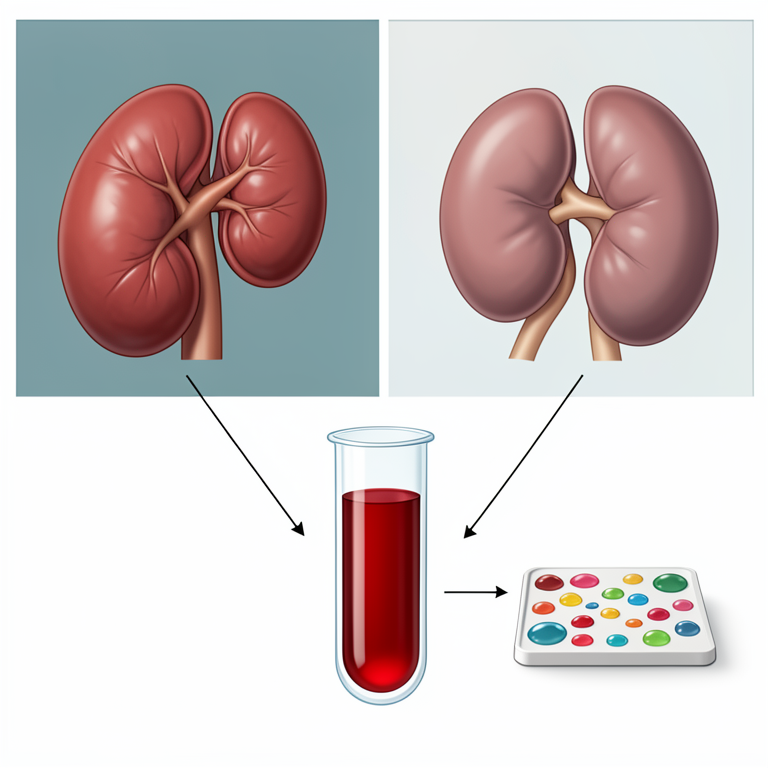
Study Workflow
AI-generated conceptual illustration (Human female kidneys and blood serum). It explains the idea and does not show study data.
View source evidence ↗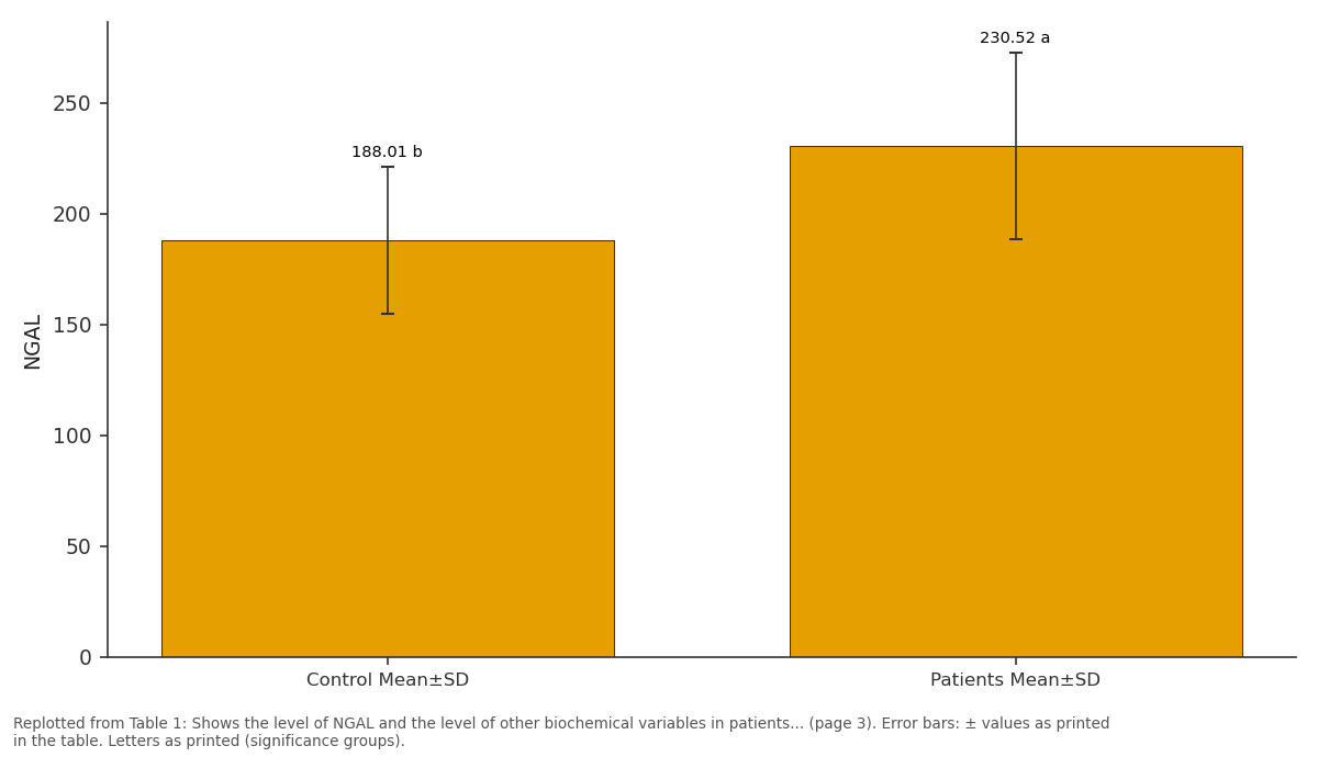
NGAL: Control Mean±SD vs Patients Mean±SD
Replotted from Table 1 (page 3) of the original paper. Every bar is the paper's own table value.
View source evidence ↗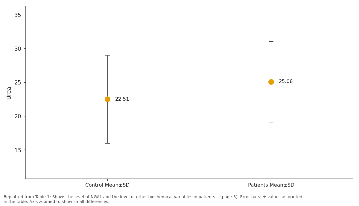
Urea: Control Mean±SD vs Patients Mean±SD
Replotted from Table 1 (page 3) of the original paper. Every bar is the paper's own table value.
View source evidence ↗Figures & tables
From the original paper Table 1
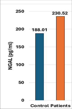 Figure 1
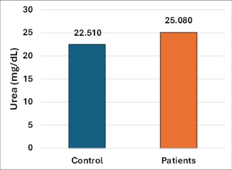 Figure 2
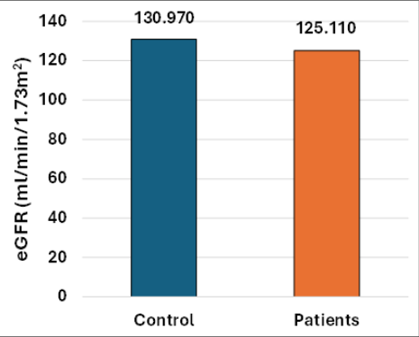 Figure 3
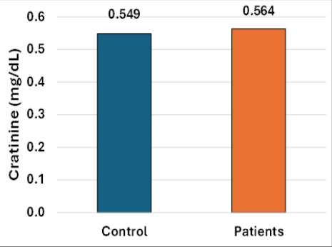 Figure 4
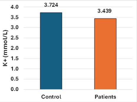 Figure 5
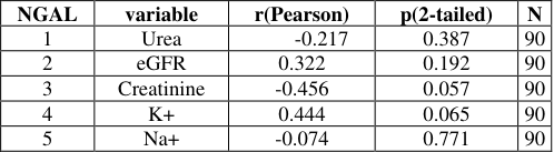 Table 2
 Table 3
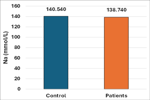 Figure 6
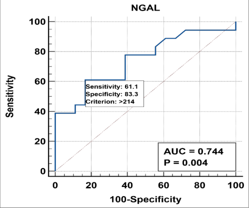 Figure 7
Introduction
Urinary tract infection (UTI) is one of the most common bacterial infections of the urinary system and represents an important health problem, especially in women [1,2] . The urinary system consists of the kidneys, ureters, bladder, and urethra. The infection may affect the lower part of the urinary system, such as cystitis, or the upper part, such as pyelonephritis, which may be associated with negative effects on kidney function [1,3,5,22] . Infection rates are higher in women than in men due to anatomical and physiological factors, most notably the shortness of the female urethra and its proximity to the vagina and rectum, which facilitates the transmission of bacteria to the bladder [1,2] . Recurrent urinary tract infections are also a common problem and are usually defined as more than two symptomatic episodes within six months or more than three episodes within one year [2,6] . The most common pathogens of urinary tract infections include Escherichia coli, Klebsiella pneumoniae, Proteus mirabilis, Pseudomonas aeruginosa, Staphylococcus aureus, Enterococcus faecalis, Staphylococcus saprophyticus, and Candida spp. [1,2,[31][32][33]48] .
Patterns of bacterial isolates may vary depending on population characteristics, clinical conditions, and laboratory methods used [30][31][32][33]46] . The infection usually begins with colonization of the area surrounding the urethra and then the rise of pathogens into the bladder, and in some cases may extend to the kidneys, causing pyelonephritis [1,2,22] . The likelihood of recurrent or complicated infections is also increased in the presence of risk factors such as urinary tract obstruction, kidney stones, urinary catheterization, diabetes, pregnancy, vesicoureteral reflux, neurogenic bladder, immunosuppression, kidney transplantation, and chronic kidney disease [5,16,26,[28][29][30]40] . Kidney injury is of particular importance in cases of urinary tract infections, as recurrent, ascending, or complicated infections may affect glomerular filtration rate, renal tubule function, and regulation of electrolyte balance [5,22] . Previous studies have shown changes in kidney function indicators, including creatinine, urea, sodium, and potassium, in patients with severe urinary tract infections or pyelonephritis, reflecting the possibility that kidney function may be affected during infection [19,21,22] . Neutrophil gelatinase-associated lipocalin (NGAL), also known as lipocalin-2, is a protein belonging to the lipocalin family and is secreted from neutrophils, renal tubular epithelial cells, and a number of other immune cells in response to inflammation or tissue injury [7][8][9][10][11] . This protein plays an important role in innate immunity by binding to bacterial siderophores and reducing the availability of iron necessary for bacterial growth [7,8,10,17] . In addition, NGAL is an early biomarker of renal tubular injury, as it may rise before obvious changes in blood creatinine levels appear [11,12,20] . It has been evaluated in many pathological conditions, including acute renal injury, chronic kidney disease, diabetic nephropathy, kidney transplantation, vesicoureteral reflux, and urinary tract infections [6][7][8][9][10][11][12][13][14][15][16] . Although most previous studies have focused on measuring NGAL in urine [6,7,14,16,40] , measuring it in serum may provide additional information about the systemic inflammatory response and renal tubular stress [8,11,13,15] . Therefore, assessing serum NGAL levels in conjunction with conventional renal function indices, including urea, creatinine, estimated glomerular filtration rate (eGFR), sodium, and potassium, may contribute to a more comprehensive picture of the inflammatory and renal changes associated with urinary tract infections. Accordingly, this study aimed to evaluate serum NGAL levels and some renal function parameters, including urea, creatinine, estimated glomerular filtration rate (eGFR), sodium, and potassium, in women with urinary tract infections compared to a healthy group. The study also aimed to determine the relationship between NGAL and kidney function indicators and evaluate the diagnostic performance of NGAL in blood serum using receptor operational properties curve (ROC) analysis.
Materials and working methods 2.1 Study design and participants
This case-type study -witness (Case-Control) was conducted during the period from December 2025 to February 2026. The study included 90 women aged 18-50 years, with 60 patients diagnosed with urinary tract infections and 30 healthy women as a control group. The patients were selected from the consulting clinics at Al Amal Hospital for Women and Children, and the diagnosis was confirmed by specialist doctors. The control group was matched to the patient group in terms of age.
Inclusion and exclusion criteria
The study included women with clinically confirmed urinary tract infections. Women with diabetes, chronic kidney or liver disease, autoimmune disorders, acute inflammatory conditions, or malignancies, as well as smokers who had used anti-inflammatory or antioxidant medications during the three months prior to the study, were excluded.
Sample collection
5 ml of venous blood was collected from all participants after a night fast. Samples were left to coagulate at room temperature and then centrifuged at 3000 rpm for 10 minutes. The serum was separated and stored at -32°m3 for analysis.
Biochemical measurements
Serum concentration of neutrophil gelatinase-associated lipocalin (NGAL) was measured using enzyme-linked immunoassay (ELISA) technology according to the manufacturer's instructions Human NGAL ELISA Kit, Sunlong Biotech Co., Ltd., China. Concentrations of urea, creatinine, sodium, and potassium were also measured using standard laboratory methods, and the estimated glomerular filtration rate (eGFR) was calculated depending on the serum creatinine level [19-22, 24, 25] .
Statistical analysis
Statistical analysis was performed using SPSS software, and the results were presented in mean±standard deviation format. The comparison test between the two groups was used to evaluate statistical differences at a significance level (P ≤ 0.05). Pearson's correlation coefficient was also used to study the relationship between NGAL level and renal function indices, and receiver operating characteristic curve (ROC) analysis was performed to evaluate the diagnostic value of NGAL, calculating area under the curve (AUC), sensitivity, specificity, and best cut-off point.
Results and discussion
Results and discussion
Serum NGAL level in women with UTIs who are healthy
The serum NGAL level as shown in Table (1) and Figure (1) was significantly higher in women with UTIs compared to healthy women. The average serum concentration of NGAL was (230.52±42.25 pg/ml) in women with UTIs versus (188.01±33.20 pg/ml) in healthy women. Different letters in the statistical comparison indicate a significant difference between the two groups. This increase may reflect activation of inflammatory and innate immune pathways during a UTI. NGAL protein is produced by neutrophils and epithelial cells during infection and is also secreted from renal tubule cells in stressful situations [7][8][9][10] . Its ability to bind to bacterial siderophores and restrict bacterial iron absorption is attributed to its role in antibacterial defense [8,10,17] . Therefore, elevated serum NGAL levels in women with urinary tract infections may reflect systemic inflammatory activation and possibly infection-related renal tubule stress. This finding is consistent with previous studies that have shown elevated NGAL levels in urinary tract infections, pyelonephritis, recurrent urinary tract infections, and kidney injury [6,7,11,16] . Liu and colleagues reported that persistently elevated urinary NGAL levels after antibiotic treatment may predict recurrence or persistence of urinary tract infections in women [6] . Pamuk and colleagues also reported that NGAL levels were higher in children with urinary tract infections and pyelonephritis and may help differentiate between upper and lower urinary tract infections [7] . Although these studies mainly evaluated the level of NGAL in urine or plasma, the current study supports the benefit of serum NGAL as a hematological biomarker in women with UTIs. Urea is a traditional indicator of kidney function as it reflects the kidneys' excretion of nitrogenous waste, protein metabolism, and hydration status [19,24,25] . A slight, albeit statistically significant, rise in urea levels in women with urinary tract infections may indicate renal functional stress, impaired processing of nitrogenous waste, or infectionrelated changes in hydration and metabolism. Previous studies have indicated that renal dysfunction and chronic renal failure are associated with high levels of urea and creatinine in the blood [24,25] . Hussein and colleagues also reported higher urea levels in patients with urinary tract infections and autoimmune thyroid diseases compared to control groups, suggesting that urinary tract infections may contribute to impaired kidney function when combined with systemic diseases [25] . However, the increase in urea levels in this study was slight and the mean remained within the limited clinical range. This suggests that women with urinary tract infections in this study may have experienced early or minor renal functional changes rather than advanced renal failure.
Estimated glomerular filtration rate (eGFR) in women with urinary tract infections who are healthy
The estimated glomerular filtration rate as shown in Table (1) and Figure (2) (eGFR) was significantly lower in women with urinary tract infections compared to healthy women.
The estimated average glomerular filtration rate was 125, 110±10, 949 ml/min/1.73 m² in women with urinary tract infections versus 130, 970±8, 302 ml/min/1.73 m² in healthy people with a p value < 0.01. Although the estimated average glomerular filtration rate remained within the normal range, the decrease was statistically significant and may indicate an early renal functional response to infection. Previous studies have reported that severe urinary tract infections, urinary tract infections requiring hospitalization, and pyelonephritis may be associated with kidney dysfunction or renal failure [5,22] . Experimental ascending pyelonephritis has been associated with decreased creatinine clearance, supporting the possibility that upper urinary tract infection may affect renal filtration [22] . In this study, a slight decrease in estimated glomerular filtration rate (eGFR) may reflect subclinical renal functional stress rather than renal failure.
Serum creatinine in healthy, upper urinary tract infections
Serum creatinine as shown in Table (1) and Figure ( 4) did not show a statistically significant difference between women with upper urinary tract infections and healthy women. The average creatinine level was 0.564±0.091 mg/dL in women with upper urinary tract infections and 0.549±0.096 mg/dL in healthy people with a p value > 0.05. Creatinine is widely used to evaluate kidney function and estimate estimated glomerular filtration rate (eGFR) [20] . Serum creatinine levels may remain unchanged during early stages or minor renal stress and may be affected by muscle mass, diet, age, gender, and medications [20] . Studies examining biomarkers of acute kidney injury confirm that NGAL levels may be elevated before creatinine and may better reflect renal tubule injury or renal inflammatory stress [9, 11, 12] . Therefore, a marked increase in serum NGAL levels despite no significant difference in creatinine levels may indicate that NGAL is more responsive to inflammation or early tubular stress than creatinine in women with urinary tract infections.
serum potassium in healthy women with urinary tract infections
The serum potassium level as shown in Table (1) and Figure (5) was significantly lower in women with urinary tract infections compared to healthy women. The average potassium level was 3.439±0.546 mmol/L in women with urinary tract infections and 3.724±0.201 mmol/L in healthy people with a p value < 0.001. Potassium is an essential intracellular electrolyte and is involved in membrane potential, neuromuscular activity, cardiac excitability, acidity and alkalinity balance, and fluid and electrolyte homeostasis [21] . The kidneys play a key role in regulating potassium through filtration, reabsorption, and excretion [21,36,37] . Low potassium levels in women with urinary tract infections may be associated with changes in renal tubule processing, hydration status, diet, inflammatory response, or physiological changes associated with infection. Experimental research on pyelonephritis has shown a change in potassium processing during renal infection although the direction and severity of potassium change may vary depending on the severity of the infection and the extent to which the kidneys are affected [22] . Chronic kidney disease guidelines also emphasize that the interpretation of potassium levels depends on kidney function, diet, medications, and compensation mechanisms [19] .
serum sodium in healthy women with urinary tract infections
The serum sodium level as shown in Table (1) and Figure (6) was significantly lower in women with urinary tract infections compared to healthy women. The average sodium level was 138, 740±2, 588 mmol/L in women with urinary tract infections and 140, 540±3, 245 mmol/L in healthy people with a p value < 0.01. Sodium is the main extracellular cation and plays an essential role in extracellular fluid balance, osmotic regulation, and blood pressure control [19] . The kidneys are the two main organs responsible for sodium homeostasis. Low sodium levels in women with urinary tract infections may be associated with an inflammatory response, hydration status, water balance, or a slight disturbance in renal tubule function. An experimental study of pyelonephritis showed a decrease in blood sodium levels and an increase in its excretion, suggesting that renal infection may affect sodium homeostasis [22] . However, a small study did not show any statistically significant association between low blood sodium levels and the severity of UTI, suggesting that sodium changes in UTI may be variable and contextdependent [23] . In this study, sodium levels remained within the physiological range, indicating a slight, statistically significant but clinically slight decrease.
Relationship between serum NGAL and renal function indices
Pearson correlation analysis as in Table (2) and Figure (2) showed no statistically significant relationship between serum NGAL and the kidney or electrolyte function indicators studied. NGAL showed a weak negative correlation with urea r = -0.217 p = 0.387 and a weak positive correlation with estimated glomerular filtration rate (eGFR) r = 0.322 p = 0.192 and a moderate negative correlation with creatinine r = -0.456 p = 0.057 and a moderate positive correlation with potassium r = 0.444 p = 0.065 and a very weak negative correlation with sodium r = -0.074 p = 0.771. These results suggest that the high serum NGAL level in this study may be more closely related to activation of inflammation or early renal tubular stress than to known changes in conventional renal function indicators. This explanation is consistent with the biological role of NGAL as a marker associated with inflammation and renal tubule injury [7][8][9][10][11][12] . The absence of statistically significant associations with relatively low creatinine values and estimated glomerular filtration rate (eGFR) in the women included in the study can also be explained.
ROC curve analysis of serum NGAL level
Analysis of the ROC curve as in Table (3) and Figure (7) showed that serum NGAL level has acceptable diagnostic performance in distinguishing between women with urinary tract infections and healthy women. The area under the curve was 0.744 with a value of p = 0.004. At a cut-off value >214 pg/ml, serum NGAL levels showed a sensitivity of 61.1% and a specificity of 83.3%. This area under the curve value indicates average diagnostic accuracy. The relatively high specificity suggests that serum NGAL may be useful as an adjunctive marker for identifying inflammatory changes associated with UTI in women. However, moderate sensitivity suggests that serum NGAL should not be used as the sole diagnostic marker. Rather, it should be interpreted in conjunction with clinical findings, urine culture or urinalysis to confirm the diagnosis of UTI, and serum renal function parameters. Previous studies also support NGAL as a useful biomarker but emphasize the need to interpret it in a clinical context because it may be elevated in infections, inflammation, acute renal failure, chronic kidney disease, diabetes, organ transplantation, and other renal or systemic disorders [8, 11, 1316] .
General interpretation
The current study showed a significant increase in serum NGAL levels in women with UTIs, accompanied by elevated urea levels and decreased estimated glomerular filtration rate, sodium, and potassium. Creatinine levels did not differ significantly between groups. This pattern suggests that UTIs in women may be associated with inflammatory activation and subtle changes in kidney or electrolyte function without a significant increase in creatinine levels. The results are consistent with previous studies suggesting that NGAL may respond faster than creatinine and may reverse inflammation or renal tubular stress during infection [9,11,12] . The results also support the importance of traditional kidney function tests and electrolyte analyses in assessing the systemic and renal effects of UTI [19-22, 24, 25] . Although many studies on NGAL have focused on urinary NGAL, this study differs because it evaluated serum NGAL. This distinction is important because serum NGAL may reflect systemic inflammation and renal tubular stress while urinary NGAL may directly reflect secretions from the urinary tract epithelium or renal tubules [8,11,13,14] . Serum NGAL may therefore be useful as a supportive blood biomarker in UTI research in women especially when combined with urea, creatinine, estimated glomerular filtration rate, sodium, and potassium.
Conclusion
This study showed that women with UTIs had significantly higher serum NGAL levels than healthy women. Urea levels increased significantly while estimated glomerular filtration rate (eGFR), potassium, and sodium decreased significantly in women with urinary tract infections. Blood creatinine levels did not show a statistically significant difference between the two groups. The absence of statistically significant associations between blood NGAL and conventional renal function parameters suggests that NGAL may reflect inflammatory activation or early renal tubule stress rather than proven renal dysfunction. ROC curve analysis showed that serum NGAL had acceptable diagnostic performance with a subcurve area (AUC) of 0.744, sensitivity of 61.1%, specificity of 83.3%, and a cutoff value of >214 pg/ml. Since all variables in this study were measured using blood/serum samples and not urine samples, serum NGAL can be considered a useful bloodbased supportive biomarker for assessing inflammatory changes and kidney function associated with urinary tract infections in women. Further studies in larger groups of women, a clear separation between lower urinary tract infection and pyelonephritis, and a detailed clinical classification of the severity of infection are recommended.
Figures and tables from the paper
| Parameters | Control Mean±SD | Patients Mean±SD | P-value | Significance |
|---|---|---|---|---|
| NGAL | 188.01±33.20 b | 230.52±42.25 a | ||
| Urea | 22.510±6.522 | 25.080±5.951 | < 0.05 | * |
| eGFR | 130.970±8.302 | 125.110±10.949 | < 0.01 | ** |
| Creatinine | 0.549±0.096 | 0.564±0.091 | > 0.05 | NS |
| K+ | 3.724±0.201 | 3.439±0.546 | < 0.001 | ** |
| Na+ | 140.540±3.245 | 138.740±2.588 | < 0.01 | ** |
Table text extracted from the original PDF; superscript letters are shown inline. The original table image is below.
View original table image
| NGAL | variable | r(Pearson) | p(2-tailed) | N |
|---|---|---|---|---|
| 1 | Urea | -0.217 | 0.387 | 90 |
| 2 | eGFR | 0.322 | 0.192 | 90 |
| 3 | Creatinine | -0.456 | 0.057 | 90 |
| 4 | K+ | 0.444 | 0.065 | 90 |
| 5 | Na+ | -0.074 | 0.771 | 90 |
Table text extracted from the original PDF; superscript letters are shown inline. The original table image is below.
View original table image
References
45 in the original paper- Sujith S, Solomon AP, Rayappan JBB. Comprehensive insights into UTIs: from pathophysiology to precision diagnosis and management. Front Cell Infect Microbiol. 2024;14:1402941. DOI:10.3389/fcimb.2024.1402941
- Sheetal, Yadav S, Ranawat S. A review on urinary tract infection. Int J Multidiscip Res. 2025;7(3):IJFMR250346917. DOI:10.36948/ijfmr.2025.v07i03.46917
- Bonkat G, Wagenlehner F, Cai T, Geerlings S, MedinaPolo J, Köves B, et al. Classification of urinary tract infections in 2025: moving beyond uncomplicated and complicated. Eur Urol Open Sci. 2025;75:44-47. DOI:10.1016/j.euros.2025.03.010
- Dadzie I, Quansah E, Dakorah MP, Abiade V, TakyiAmuah E, Adusei R. The effectiveness of dipstick for the detection of urinary tract infection. Can J Infect Dis Med Microbiol. 2019;2019:8642628. DOI:10.1155/2019/8642628
- Sako A, Yasunaga H, Matsui H, Fushimi K, Yanai H, Gu Y, et al. Hospitalization for urinary tract infections in Japan, 2010-2015: a retrospective study using a national inpatient database. BMC Infect Dis. 2021;21:1048. DOI:10.1186/s12879-021-06735-y
- Liu MC, Jiang YH, Jhang JF, Chang TL, Yang CC, Kuo HC. Persistent elevation in urinary neutrophil gelatinase-associated lipocalin levels can be a predictor of urinary tract infection recurrence or persistence in women. Int J Mol Sci. 2024;25(23):12670. DOI:10.3390/ijms252312670
- Pamuk U, Kalman S, Tascilar ME, Sertoglu E. Neutrophil gelatinase-associated lipocalin as a biomarker for the diagnosis of urinary tract infection in children. Medicine Science. 2022;11(1):70-74. DOI:10.5455/medscience.2021.06.202
- Romejko K, Markowska M, Niemczyk S. The review of current knowledge on neutrophil gelatinase-associated lipocalin (NGAL). Int J Mol Sci. 2023;24(13):10470. DOI:10.3390/ijms241310470
- Jasotani K, Dahiya K, Ahlawat R, Gupta M, Kumar S, Dhankhar R, et al. NGAL: an upcoming biomarker of interest. Indian J Med Biochem. 2022;26(1):26-30. DOI:10.5005/jp-journals-10054-0206
- Guardado S, Ojeda-Juárez D, Kaul M, Nordgren TM. Comprehensive review of lipocalin 2-mediated effects in lung inflammation. Am J Physiol Lung Cell Mol Physiol. 2021;321(5):L726-L733. DOI:10.1152/ajplung.00080.2021
- Virzì GM, Morisi N, Oliveira Paulo C, Clementi A, Ronco C, Zanella M. Neutrophil gelatinase-associated lipocalin: biological aspects and potential diagnostic use in acute kidney injury. J Clin Med. 2025;14(5):1570. DOI:10.3390/jcm14051570
- Jana S, Mitra P, Roy S. Proficient novel biomarkers guide early detection of acute kidney injury: a review. Diseases. 2023;11(1):8. DOI:10.3390/diseases11010008
- Greco M, Chiefari E, Mirabelli M, Salatino A, Tocci V, Cianfrone P, et al. Plasma or urine neutrophil gelatinase-associated lipocalin (NGAL): which is better at detecting chronic kidney damage in type 2 diabetes? Endocrines. 2022;3(2):175-186. DOI:10.3390/endocrines3020016
- Li A, Yi B, Liu Y, Wang J, Dai Q, Huang Y, et al. Urinary NGAL and RBP are biomarkers of normoalbuminuric renal insufficiency in type 2 diabetes mellitus. J Immunol Res. 2019;2019:5063089. DOI:10.1155/2019/5063089
- Mărunțelu I, Șerboi DV, Tudor TA, Necșeriu GC, Baciu MR, Ovidiu ML, et al. The importance of neutrophil gelatinase-associated lipocalin (NGAL) in monitoring patients with renal transplantation. Ann Acad Rom Sci Ser Med. 2025;6(1):22-29. DOI:10.56082/annalsarscimed.2025.1.22
- Gavrilovici C, Dusa CP, Iliescu Halitchi C, Lupu VV, Spoiala EL, Bogos RA, et al. The role of urinary NGAL in the management of primary vesicoureteral reflux in children. Int J Mol Sci. 2023;24(9):7904. DOI:10.3390/ijms24097904
- Mercado-Evans V, Branthoover H, Chew C, Serchejian C, Saltzman AB, Mejia ME, et al. TammHorsfall protein augments neutrophil NETosis during urinary tract infection. JCI Insight. 2025;10(1):e180024. DOI:10.1172/jci.insight.180024 18. Darwish B, Chamaa F, Awada B, Lawand N, Saadé NE, Abou Fayad AG, et al. Urinary tract infections impair adult hippocampal neurogenesis. Biology (Basel). 2022;11(6):891. DOI:10.3390/biology11060891
- Ikizler TA, Burrowes JD, Byham-Gray LD, Campbell KL, Carrero JJ, Chan W, et al. KDOQI clinical practice guideline for nutrition in CKD: 2020 update. Am J Kidney Dis. 2020;76(3 Suppl 1):S1-S107. DOI:10.1053/j.ajkd.2020.05.006
- Kashani K, Rosner MH, Ostermann M. Creatinine: from physiology to clinical application. Eur J Intern Med. 2020;72:9-14. DOI:10.1016/j.ejim.2019.10.025
- Toft U, Riis NL, Jula A. Potassium: a scoping review for Nordic Nutrition Recommendations 2023. Food Nutr Res. 2024;68:10365. DOI:10.29219/fnr.v68.10365
- Skowron B, Juszczak K, Baranowska A, Machowska A, Strus M, Więcek G, et al. Laboratory diagnostics of renal function in an experimental model of ascending pyelonephritis with high-virulent Escherichia coli. Folia Med Cracov. 2012;52(1-2):25-37.
- Sankar A, Madhuri B, Umashankar, Surya A. Association of low serum sodium levels with urinary tract infections: a case series. J Adv Zool. 2023;44(S3):306-309. DOI:10.53555/jaz.v44iS3.600
- Khudhair HI, Abood ZH. Evaluation of IL-6 and IL-30 serum level in addition to some biochemical markers in Iraqi patients with chronic renal failure. Iraqi J Biotechnol. 2025;24(Special Issue 2):184-191.
- Hussien RA, Zwain GF, Abood ES. Study of專 UTI effect on renal function for the patient with thyroid autoimmune disease. Hilla Univ Coll J Med Sci. 2024;2(1):40-45. DOI:10.62445/2958-4515.1013
- Zaidi SMJ, Kaneez M, Almas T, Fatima L, Safian HA, Jamal AM, et al. Gauging the risk factors for asymptomatic bacteriuria in type-2 diabetic women: a case-control study. Cureus. 2020;12(7):e9069. DOI:10.7759/cureus.9069
- Hernández C, Simó R. Albumin excretion rate is not affected by asymptomatic urinary tract infection: a prospective study. Diabetes Care. 2004;27(7):15651569. DOI:10.2337/diacare.27.7.1565
- Kocur SB, Noppenberg M, Sowińska I, Gniadek A. Selected risk factors for urinary tract infections. Nurs Probl. 2023;31(3):128-132. DOI:10.5114/ppiel.2023.133337
- Hernández González E, Alberti Amador E, Rivero Ojeda J. Clinical and microbiological characterization of the urinary infections in patients with neurogenic bladder. Acta Sci Microbiol. 2021;4(4):16-22. DOI:10.31080/ASMI.2021.04.0800
- Saeed KM, Yaseen EM. The occurrence of asymptomatic bacteriuria in pregnant women in Tikrit city. Sci Res J Med Sci. 2024;4(2):1-6. DOI:10.47310/srjms.2024.v0i402.009
- Mahesh S, Carmelin DS, Muthusamy R. Bacterial flora and treatment strategies in women with Escherichia coli urinary tract infections. Cureus. 2024;16(3):e56552. DOI:10.7759/cureus.56552
- Hamzah DAAH. Study of the prevalence of Staphylococcus aureus in urine among students of Wasit University. J Wasit Sci Med. 2026;19(1):11-19. DOI:10.31185/jwsm.598
- Adnan M, Aziz IH, Al-Deresawi MS. Molecular detection of Proteus mirabilis using PCR technique among urinary tract infection patients. Iraqi J Biotechnol. 2014;13(2):35-47.
- Bazira PJ. Anatomy of the kidney and ureter. Surgery (Oxford). 2022;40(8):481-488. DOI:10.1016/j.mpsur.2022.05.008
- Al Lawati H, Blair BM, Larnard J. Urinary tract infections: core curriculum 2024. Am J Kidney Dis. 2024;83(1):90-100. DOI:10.1053/j.ajkd.2023.08.009
- Inker LA, Eneanya ND, Coresh J, Tighiouart H, Wang D, Sang Y, et al. New creatinine-and cystatin Cbased equations to estimate GFR without race. N Engl J Med. 2021;385(19):1737-1749. DOI:10.1056/NEJMoa2102953
- Mohammed Raouf LL, Qadir SH, Alzbeede AA, Ghafoor DD. Evaluation of some physiological kidney function tests in athletes underuse of protein supplementation: a pilot study. Iraq Med J. 2024;8(3):74-78. DOI:10.22317/imj.v8i3.1287
- Bae S, Chung H. The urobiome and its role in overactive bladder. Int Neurourol J. 2022;26(3):190200. DOI:10.5213/inj.2244016.008
- Koukourikis P, Papaioannou M, Papanikolaou D, Apostolidis A. Urine biomarkers in the management of adult neurogenic lower urinary tract dysfunction: a systematic review. Diagnostics (Basel). 2023;13(3):468. DOI:10.3390/diagnostics13030468
- Jiang YH, Jhang JF, Wang JH, Wu YH, Kuo HC. A decision tree model using urine inflammatory and oxidative stress biomarkers for predicting lower urinary tract dysfunction in females. Int J Mol Sci. 2024;25(23):12857. DOI:10.3390/ijms252312857
- Yu WR, Jiang YH, Jhang JF, Kuo HC. Urine biomarker could be a useful tool for differential diagnosis of a lower urinary tract dysfunction. Tzu Chi Med J. 2024;36(2):110-119. DOI:10.4103/tcmj.tcmj_221_23
- Jiang YH, Jhang JF, Kuo HC. Urinary oxidative stress biomarker levels might be useful in identifying functional bladder disorders in women with frequency and urgency syndrome. J Clin Med. 2023;12(6):2336. DOI:10.3390/jcm12062336
- Rada MP, Ciortea R, Măluţan AM, Doumouchtsis SK, Bucuri CE, Clim A, et al. The profile of urinary biomarkers in overactive bladder. Neurourol Urodyn. 2020;39(8):2305-2313. DOI:10.1002/nau.24487
- Ünal K, Çöllüoğlu ML, Erdem E, Özbaş C, Özdemir Ö. The relationship of neurotrophin levels with stressinduced urinary incontinence in multiparous premenopausal women. Qatar Med J. 2025;2025(1):3. DOI:10.5339/qmj.2025.3
- Saleh MA. Bacteriological, biochemical study of urinary tract infections in some urine samples and comparison of the results of analyses between Bani Waleed laboratories. J Bani Waleed Univ Humanit Appl Sci. 2023;(29):513-533.
- ElNahal ASM, Ibrahim A, Abdelhalim E, et al. Serum neutrophil gelatinase-associated lipocalin as a novel biomarker for diagnosing acute pyelonephritis in adults. Sci Rep. 2025;15:19651. DOI:10.1038/s41598-025-03646-9