
Research article · 2026 · Full text
International Journal of Advanced Chemistry ResearchHistopathological changes in resistant and susceptible guava (Psidium gujava) varieties due to Root knot nematode M. enterolobi
Volume 8(6) · Pages 68–72 · DOI: 10.33545/26646781.2026.v8.i6a.690 ↗
Research areasNematode management and characterization studiesPlant Disease Management TechniquesInsect-Plant Interactions and Control
Abstract
Author-writtenThe root-knot nematode Meloidogyne enterolobii is an emerging and highly virulent pathogen causing severe damage in Guava (Psidium guajava), particularly overcoming host resistance in many commercial cultivars. This study investigates the histopathological alterations in resistant and susceptible guava genotypes following nematode infection to elucidate host-pathogen interactions and mechanisms of resistance. Root samples were collected at different stages post-inoculation and processed for microscopic examination. In susceptible guava plants, nematode infection induced extensive cellular modifications, including the formation of well-developed giant cells characterized by hypertrophy, dense cytoplasm, multinucleation, and dissolution of cell walls in the vascular cylinder. These changes facilitated the establishment of permanent feeding sites, leading to gall formation and disruption of normal vascular function. In contrast, resistant guava genotypes exhibited restricted nematode development, with limited or poorly formed giant cells, increased deposition of phenolic compounds, and localized cell necrosis around infection sites. The vascular tissues in resistant plants remained largely intact, indicating an effective defense response. Comparative histological analysis revealed that resistance in guava is associated with early defence activation, structural barriers, and inhibition of giant cell formation, thereby impairing nematode establishment and reproduction. These findings provide critical insights into the anatomical and cellular basis of resistance against M. enterolobii and contribute to the development of resistant cultivars and integrated nematode management strategies.
Summary
Source-checkedPlain-language reading of the full paper, prepared with AI and checked against the text. Each point links to the authors' own words.
Why it matters
- The root-knot nematode M. enterolobii is a highly virulent pathogen that causes severe damage to guava and can overcome the resistance of many commercial varieties. View evidence ↗
- Understanding the compatibility between the host plant and the nematode during root penetration and feeding site creation is essential for developing resistant plants for breeding programs. View evidence ↗
The question
- The study aims to investigate the anatomical changes in the root tissues of both susceptible and resistant guava species when infected by M. enterolobii. View evidence ↗
What they did
- The researchers used P. cattleianum and P. gujava seeds planted in heat-sterilized sandy loamy soil within a net house. View evidence ↗
- Plants were inoculated with M. enterolobii juveniles hatched from egg masses previously cultivated in tomato plants. View evidence ↗
- Root segments (0.5-1.0 cm) were collected 30 days after inoculation and fixed using a formalin-aceto alcohol solution. View evidence ↗
- Transverse cross sections of the roots were cut to a thickness of 10 μm using a rotary microtome. View evidence ↗
- Root sections were stained with toluidine blue for light microscopy, and acid fuchsin was used to verify the presence of egg masses and females. View evidence ↗
What they found
- In P. gujava, the presence of female nematodes and egg masses caused disruption and rupture of the root cortex. View evidence ↗
- Giant cells in P. cattleianum were small with thin walls and lacked cytoplasmic material, which failed to support the development of female nematodes. View evidence ↗
- Infested roots of P. cattleianum showed an increase in vacuoles, suggesting a host resistance response. View evidence ↗
- Acid fuchsin staining revealed that egg masses did not develop in the roots of P. cattleianum. View evidence ↗
What it means
- The authors conclude that guava resistance to M. enterolobii is linked to early defense activation and structural barriers that inhibit the formation of giant cells, preventing nematode reproduction. View evidence ↗
- The study concludes that P. cattleianum exhibits resistance compared to P. gujava due to the disrupted development of fewer and less cytoplasmic giant cells. View evidence ↗
Visual summary
1 visualsIllustrations explain the paper; they do not show study data.
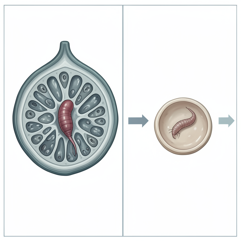
Comparison of Giant Cell Development
AI-generated conceptual illustration (Guava (Psidium guajava) root tissue and Meloidogyne enterolobi nematodes). It explains the idea and does not show study data.
View source evidence ↗Figures & tables
From the original paper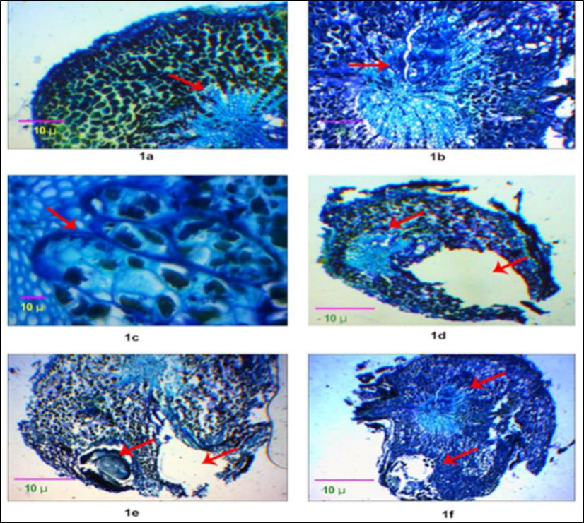 Figure 1
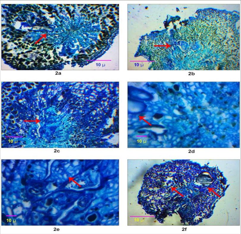 Figure 2
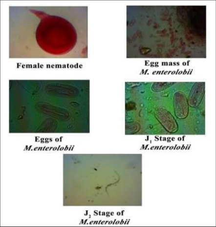 Figure 3
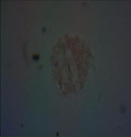 Figure 4
Introduction
The fruit which is known as the "poor man's fruit," guava (Psidium guajava L.) is a significant commercial fruit in India. After citrus, bananas, and mangos, it is the fourth most significant fruit. Although guava is native to South America and the West Indies, it is also produced in other tropical and subtropical regions, such as India. India's guava cultivation area is 261 MHa, producing 3961 MT annually with a yield of 13.9 MT/Ha (NHB Data Base 2017-18). Similarly, Tamil Nadu's guava production is 77.41MT and its area is 9.78Mha. A new issue with guava is Meloidogyne enterolobii, which has been documented from (Poornima et al., 2016).Meloidogyne mayaguensis is a synonym for M. enterolobii, a species of root knot nematode that may have spread from other nations through saplings. Phaseolus vulgaris (bean), Coffea arabica (coffee), Gossypium hirsutum L. (cotton), Carica papaya L. (papaya), Psidium guajava (guava), Solanum quitoense (naranjilla), Solanum tuberosum (potato), Glycine max (soybean), Ipomoea batatas (sweet potato), Nicotiana tobacum (tobacco), Lycopersicon esculentum (tomato), and Citrullus lanatus (watermelon) are the main hosts (Rammah and Hirschmann, 1988;Brito et al., 2007;Bitencourt and Silva, 2010). Garlic, grape fruit, and sour oranges are a few examples of non-hosts. In order to effectively employ resistant plants in breeding programs, the host plant and nematode must be compatible during root penetration and feeding site creation for the establishment of a successful host-parasite relationship. The goal of the current study is to understand the anatomical alterations in the root tissues of both vulnerable and resistant guava species.
Materials and methods
P. cattleianum and P. gujava seeds were acquired from IIHR Bangalore. Pre-germinated seeds were planted in tumbler pots with heat-sterilized sandy loamy soil and cultivated in a net house. From a pure culture of M. enterolobii cultivated in tomato plants, the egg mass was extracted and allowed to hatch in tap water at room temperature. Each tumbler pot received an inoculation of M. enterolobii juveniles. Roots from each treatment were plucked and gently cleaned of any adhering soil 30 days following inoculation. Formalin-aceto alcohol solution (FAA) was used to fix two to six root galled root segments (0.5-1.0 cm) p plant. The root segments were then infiltrated and immersed in paraffin wax (50-55 °C) after being dehydrated in a graded series of ethanol (35, 50, 75, and 100%, 1 hour in each series of concentration). Using a rotary microtome (Weswox optic MT1090), serial transverse cross sections with a thickness of 10 μm were cut, placed on glass slides, and allowed to dry for two minutes. The sections were soaked in xylene to remove paraffin wax, and then they were cleaned with ethanol and distilled water (Hoppert, 2003). Sections were examined under a light microscope with a digital camera (Medline ML105) after being stained with toluidine blue (Parker et al., 1982). Acid fuchsin staining of the roots verified the existence of females and egg mass. The roots were examined under a stereomicroscope following staining.
Perineal pattern identification Perineal pattern analysis of adult females:
The method created by Hartman.K et.al. 1985 was used to prepare the perineal patterns of nematode females. Ten adult females were chosen from each district's water convolvulus roots, and they were placed under a stereomicroscope on a transparent plastic sheet after the anterior and posterior portions were separated using a razor. In order to remove cuticles and clean the pattern, the perineal patterns were placed in a drop of 45% lactic acid. The perineal pattern was then moved to a drop of 4% glycerine that was placed on a glass slide, sealed with nail varnish, and covered with a coverslip. After labeling the slide, it was examined under a compound microscope to determine its morphology. Measurements for J2 and males: Using Sienhorst's approach, males taken from infected roots and J2 hatched from the single egg mass were fixed on glass slides. Under a compound microscope, the males and J2 of each isolate were examined and captured on camera.
Results and discussion
Results and discussion
Histopathology of healthy roots
The cortex of healthy guava roots has thin walls, several layers, and circular or polygonal parenchyma cells with no intercellular gaps, according to histopathology. This is mostly employed to move salts and water from the root hairs to the root's centre (Abbasi et al., 2013) [19] . The vascular cylinder encircles the cortex, endodermis, and pericycle (Fig. 1a). The outermost layer of the steel is called the pericycle, and it is made up of a single layer of parenchymatous cells with no intervals between them. Aly Khan et al. (2017) also noted that secondary xylem and phloem tissue are arranged in a linear chain.
Histopathology of p. Gujava roots infected with m. enterolobii
The absence of complex galls and group feeding of nematodes were shown by anatomical alterations brought on by the second stage juveniles of M. enterolobii (Fig. 1b). The maximum number of large cells is just four or five (Fig. 1c). The xylem and phloem cells are dislocated by giant cells, impeding the movement of nutrients and water (Fig. 1d). In the large cells (Fig. 1c), thickened cell walls with granular cytoplasm and four to ten hypertrophied nuclei and nucleoli were also noted; this is consistent with Vovlas's (2004) findings about olive root knot nematodes. The displacement of xylem and phloem arteries caused by large cells was also documented by Hussey and Williamson (1997). It was discovered that females and egg masses disrupted the cortical area of the roots (Fig. 1e).
According to Teliz et al. (2007), they also deform the root surface, which causes the root cortex to rupture (Fig. 1f).
According to Fawoe (1988), the root knot nematode's large cell development caused the xylem tissues in tomatoes to distort.
Histopathology of P. cattleianum roots infected with M. enterolobii Giant cell development was observed in P. cattleianum (Fig. 2c). This demonstrates that there was no host reaction to nematode penetration in this species (Fig. 2a). Nematode group feeding and complicated gall development are not seen. The number of large cells was found to be lower, ranging from two to three (Fig. 2d). Alain Denis de Souse et al. (2016) found that several Psidium species had poor feeding site development and degraded giant cells (Fig. 2c). More vacuoles were seen in M. enterolobii-infested roots (Fig. 2f), which may indicate a host resistance response. Eissa et al. (1988) noted a similar finding in a resistant host. Giant cells have thin walls and are tiny, with no cytoplasmic material (Fig. 2e). Female development was not supported by these large cell deformities (Fig. 2b). [13] , nematodes in resistant roots appear shrunken in comparison to those in susceptible roots because the large cells are unable to give nourishment. However, the vulnerable roots have healthy giant complexes with abundant cytoplasm, and the nematode matures into a female for more infection.This study concludes that, in comparison to P. gujava, the large cells of P. cattlieianum develop normally with disruption, having less thick cytoplasm and fewer in number. Giant cells of P. gujava had abundant cytoplasm, multinucleation, and thick walls. P. cattleianum did not exhibit complex galls or nematode group feeding. Nematode development was minimal, however P. gujava speci
Figures and tables from the paper
References
21 in the original paper- Sreekavya G, Muthuvel I, Rajangam J, Prabhu S. Comparative study on histopathological changes caused by Meloidogyne enterolobii in Psidium guajava and Psidium cattleianum guava species. International Journal of Current Microbiology and Applied Sciences. 2019;8(5):21982203. https://doi.org/10.20546/ijcmas.2019.805.259
- Poornima K, Ashokkumar N, Kalaiarasan P, Kavino M. Screening and histological characterization of guava (Psidium guajava L.) cultivars against root-knot nematode, Meloidogyne enterolobii. 2019.
- Nagachandrabose S, Rajendran P, Ganeshan S, Arunachalam A, Prabhu S, Muthuvel I, Shanmugasundaram KA. Resistance to Meloidogyne enterolobii in guava: Screening of cultivated and wild types, resistance principles, and graft compatibility. Scientia Horticulturae. 2024; 338:113825. https://doi.org/10.1016/j.scienta.2024.113825
- Costa SNO, da Paschoa RP, Alexandrino CR, Cremonez PM, Ribeiro JM, da Cunha e Castro JM, et al. Psidium defenses against Meloidogyne enterolobii: Proteomic and microscopic analysis of this plant-predator association. Proteomics. 2025;25(16):70015.
- Khan MR, Poornima K, Somvanshi VS, Walia RK. Meloidogyne enterolobii: A threat to guava (Psidium guajava) cultivation. Archives of Phytopathology and Plant Protection. 2022;55(4):1-37. https://doi.org/10.1080/03235408.2022.2132623
- Ghule TM, Phani V, Somvanshi VS, Patil M, Bhattacharyya S, Khan MR. Further observations on Meloidogyne enterolobii infecting guava (Psidium guajava) in India. Journal of Nematology. 2021;53: e2021120.
- Santos CAF, Costa SR, Boiteux LS, Grattapaglia D, SilvaJunior OB. Genetic associations with resistance to Meloidogyne enterolobii in guava (Psidium spp.). PLoS ONE. 2022;17(11): e0273959. https://doi.org/10.1371/journal.pone.0273959
- Carneiro RMDG, Almeida MRA, Braga RS, Almeida CA, Gomes ACM. First record of Meloidogyne mayaguensis on guava in Brazil. 2007.
- Hussey RS, Williamson VM. Physiological and molecular aspects of nematode parasitism. In: Plant and Nematode Interactions. 1998:87-108.
- Moens M, Perry RN, Starr JL. Root-knot nematodes. CAB International; 2009.
- Jones JT, Haegeman A, Danchin EGJ, Gaur HS, Helder J, Jones MGK, et al. Top 10 plant-parasitic nematodes in molecular plant pathology. Molecular Plant Pathology. 2013;14(9):946-961.
- Williamson VM, Kumar A. Nematode resistance in plants: The battle underground. Trends in Genetics. 2006;22(7):396-403.
- Das S, DeMason DA, Ehlers JD, Close TJ, Roberts PA. Histological characterization of root-knot nematode resistance in cowpea and its relation to reactive oxygen species modulation. Journal of Experimental Botany. 2008;59(6):1305-1313.
- Khan MR, Jain RK, Ghule TM, Pal S. Root-knot nematodes in India: A comprehensive review. Indian Journal of Nematology. 2014;44(1):1-23.
- Karssen G. The plant-parasitic nematode genus Meloidogyne Göldi, 1892. Brill Academic Publishers; 2002.
- Byrd DW, Kirkpatrick T, Barker KR. An improved technique for clearing and staining plant tissues for detection of nematodes. Journal of Nematology. 1983;15(1):142-143.
- Schindler AF. A simple extraction method for nematodes from plant tissues. Plant Disease Reporter. 1961; 45:747748.
- Sayed SA, Abd-Elgawad MMM, El-Nagdi WMA. Histopathological responses of fruit tree roots infected with root-knot nematodes. 2008.
- Swarnsingh, Abbasi MW, Hisamuddin. Histopathological response of Lens culinaris roots towards root-knot nematode. Pakistan Journal of Biological Sciences. 2013;16(7):317-324.
- Téliz D, Landa BB, Rapoport HF, Pérez-Camacho F, Jiménez-Díaz RM, Castillo P. Susceptible reaction to rootknot nematodes of grapevine rootstocks reported as moderately resistant. Plant Disease. 2007;91(9):11471154.
- Hartman KM, Sasser JN. Identification of Meloidogyne species on the basis of differential host test and perinealpattern morphology. In: Barker KR, Carter CC, Sasser JN, editors. An Advanced Treatise on Meloidogyne, Volume II: Methodology. Raleigh (NC): North Carolina State University Graphics; 1985. p. 69-77.