
Research article · 2026 · Full text
International Journal of Biology SciencesPostnatal developmental morphology of the kidney in Black Bengal goats (Capra hircus) from birth to maturity
Volume 8(4) · Pages 160–163 · DOI: 10.33545/26649926.2026.v8.i4b.873 ↗
Research areasComparative Animal Anatomy StudiesVeterinary Medicine and SurgeryReproductive Physiology in Livestock
Abstract
Author-writtenBackground: Postnatal kidney development in goats follows a predictable growth trajectory influenced by genetic, nutritional, and environmental factors. Establishing age-specific kidney morphometric norms is critical for distinguishing pathological changes from normal developmental variation in clinical and forensic veterinary contexts. Objectives: This research investigated the postnatal developmental trajectory of kidney morphology in Black Bengal goats from birth through maturity at the University of Veterinary Science, Myanmar, during 2025-2026. Methods: Kidneys from 36 Black Bengal goats across six age groups (birth, 1, 3, 6, 12 months, and adult >24 months; n=6 per group, equal sex distribution) were subjected to gross morphometric analysis including weight, length, width, volume, cortex thickness, medulla thickness, and capsule thickness. Results: Kidney weight increased 8.3-fold from 5.8±0.4 g at birth to 47.9±3.6 g at maturity. The most rapid growth phase occurred between birth and 3 months, accounting for 47.4% of total weight gain. Kidney length showed a 2.8-fold increase from 2.1 to 5.9 cm. Cortex and medulla growth was proportional, maintaining a stable cortex-to-medulla ratio. Conclusion: Kidney development in Black Bengal goats follows a triphasic postnatal growth pattern. These age-specific morphometric baselines provide essential reference standards for clinical diagnosis and pathological evaluation.
Summary
Source-checkedPlain-language reading of the full paper, prepared with AI and checked against the text. Each point links to the authors' own words.
Why it matters
- The kidney is a critical organ for maintaining homeostasis, electrolyte balance, and waste elimination, undergoing significant maturation from birth to adulthood in small ruminants. View evidence ↗
- Defining age-specific kidney measurements is essential for veterinary clinicians and forensic experts to tell the difference between normal growth and disease. View evidence ↗
- The Black Bengal goat is a meat-type breed from South and Southeast Asia whose compact size and early maturity may affect how its internal organs develop. View evidence ↗
The question
- The study aimed to characterize the developmental path of kidney morphology in Black Bengal goats from birth until they reach maturity. View evidence ↗
What they did
- The researchers studied 36 Black Bengal goats divided equally by sex into six age groups: birth, 1 month, 3 months, 6 months, 12 months, and adults over 24 months. View evidence ↗
- The study measured kidney weight, length, width, thickness, and volume using digital balances, vernier calipers, and the water displacement method. View evidence ↗
- Mid-sagittal sections of the kidneys were used to measure the thickness of the cortex, medulla, and capsule, as well as the length of the hilus. View evidence ↗
- Data were analyzed using one-way ANOVA with Tukey HSD tests and nonlinear regression in GraphPad Prism 10, with a significance threshold of P<0.05. View evidence ↗
What they found
- Kidney weight increased 8.3-fold over the growth period, rising from 5.8±0.4 g at birth to 47.9±3.6 g in adults. View evidence ↗
- The fastest growth occurred between birth and 3 months, where weight increased from 5.8 g to 22.7 g, representing 47.4% of the total weight gain. View evidence ↗
- Kidney length increased 2.8-fold, growing from 2.1±0.2 cm at birth to 5.9±0.3 cm in adults. View evidence ↗
- Kidney width increased 2.8-fold (1.2 to 3.4 cm) and thickness increased 2.5-fold (0.9 to 2.3 cm) during postnatal development. View evidence ↗
- The length-to-width ratio stayed stable between 1.71 and 1.75, showing that the kidney grew proportionally in all dimensions. View evidence ↗
- The cortex thickness increased 2.0-fold (2.8 mm to 5.6 mm) and the medulla thickness increased 1.9-fold (4.1 mm to 7.9 mm) from birth to adulthood. View evidence ↗
- The kidney capsule thickness increased 1.8-fold, from 0.21±0.03 mm at birth to 0.38±0.04 mm at maturity. View evidence ↗
- Most kidney measurements showed no significant difference between 12-month-old goats and adults, indicating that kidneys reach near-mature size by one year. View evidence ↗
What it means
- The established morphometric baselines serve as essential reference standards for clinical diagnosis, pathology, and forensic age estimation in this breed. View evidence ↗
Visual summary
2 visualsIllustrations explain the paper; charts are replotted from the paper's own table values.
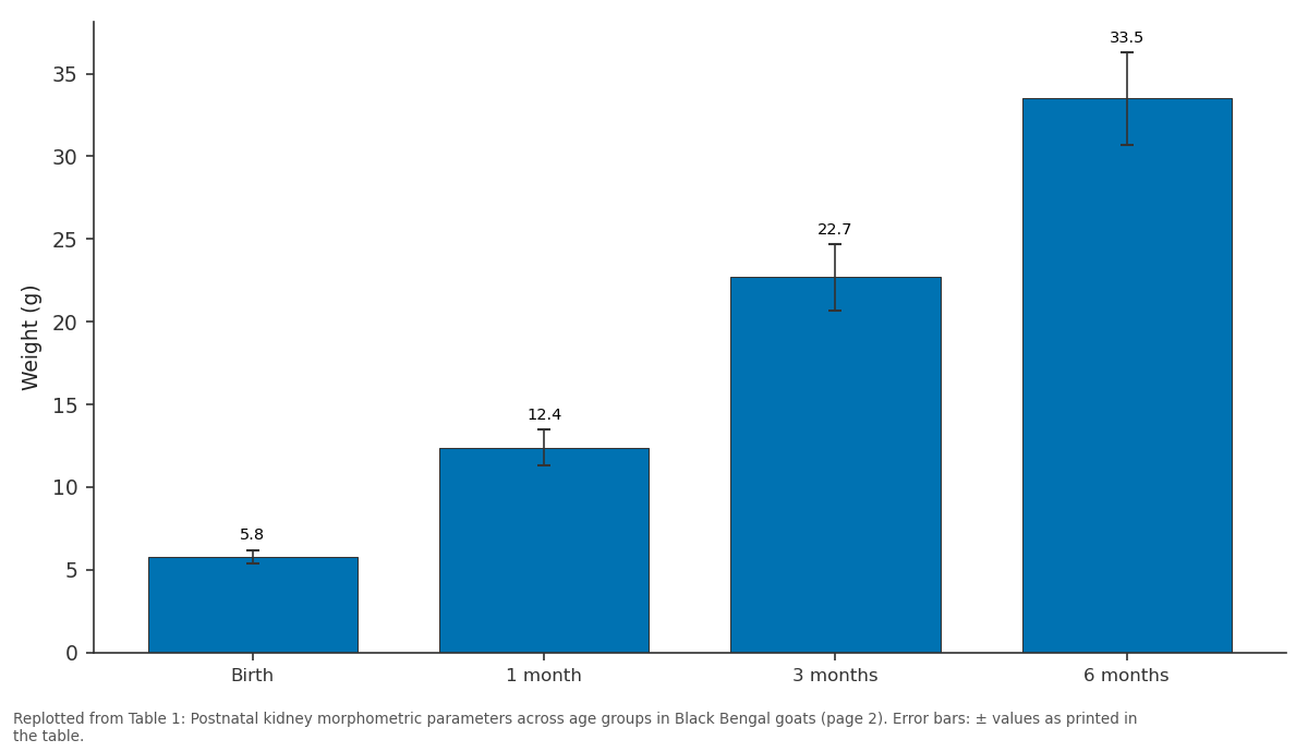
Weight (g): Birth vs 1 month vs 3 months vs 6 months
Replotted from Table 1 (page 2) of the original paper. Every bar is the paper's own table value.
View source evidence ↗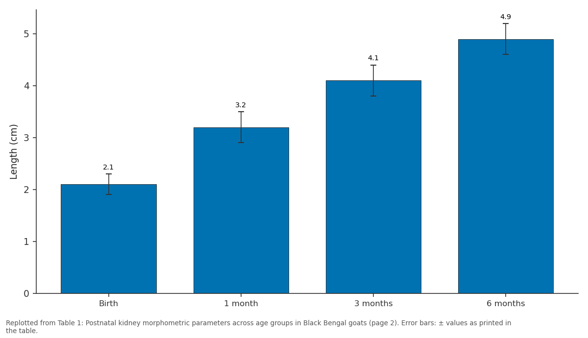
Length (cm): Birth vs 1 month vs 3 months vs 6 months
Replotted from Table 1 (page 2) of the original paper. Every bar is the paper's own table value.
View source evidence ↗Figures & tables
From the original paper Table 1
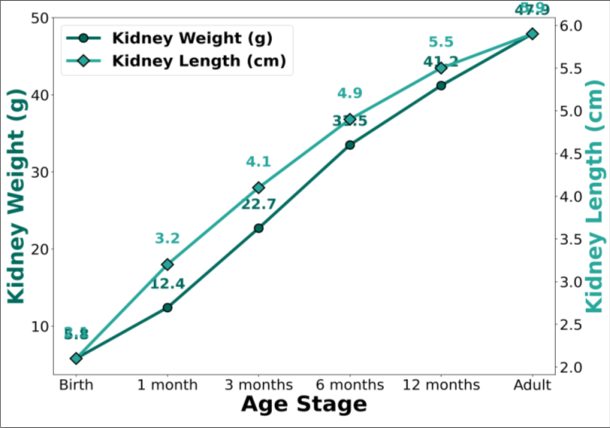 Figure 1
 Table 2
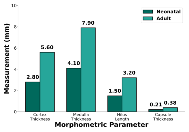 Figure 2
Introduction
Postnatal organ development in domestic animals follows predictable growth trajectories governed by genetic programming, nutritional adequacy, and environmental influences [1] . The kidney, as a vital organ responsible for homeostatic regulation, electrolyte balance, acidbase maintenance, and metabolic waste elimination, undergoes substantial morphological and functional maturation from birth through adulthood in small ruminants [2] . Gross morphological documentation of the Barbari goat kidney established important baseline parameters including organ dimensions, shape characteristics, and surface features in adult specimens [3] . However, the temporal progression of kidney development across postnatal life stages in goat breeds has received limited attention, particularly for the Black Bengal breed [4] . The Black Bengal, a prolific meat-type breed native to South and Southeast Asia, exhibits compact body size and early maturity characteristics that may influence the pattern and rate of visceral organ development. Understanding age-specific kidney morphometric norms is essential for veterinary practitioners to accurately distinguish pathological renal changes from normal developmental variation at different life stages [5] . This research was designed to characterise the complete postnatal developmental trajectory of kidney morphology in Black Bengal goats from birth through maturity, providing age-specific reference standards for clinical and forensic veterinary applications. Furthermore, age-specific reference standards enable forensic veterinary practitioners to estimate the approximate age of goat carcasses or isolated kidneys based on organ dimensions, which may be relevant in cases involving illegal slaughter, wildlife crime investigations, or insurance assessments. Systematic documentation of the complete postnatal developmental trajectory thus serves multiple practical and academic purposes across veterinary disciplines [6] .
Material and Methods
Material and Methods
Material
The research was conducted at the Department of Anatomy, University of Veterinary Science, Yezin, Myanmar, from January 2025 to March 2026. Animals were sourced from the university teaching and research farm where Black Bengal goats have been maintained under standardised management for over a decade. A total of 36 Black Bengal goats were assigned to six age groups with equal sex distribution: birth (day 0, n=6), 1 month (n=6), 3 months (n=6), 6 months (n=6), 12 months (n=6), and adult (>24 months, n=6). Animals were maintained under standard institutional management with access to natural grazing, concentrate supplementation, and clean water. All animals were clinically healthy and free from renal pathology at the time of examination.
Methods
Kidneys were harvested following humane slaughter conducted in accordance with institutional ethical guidelines, freed of perinephric fat, and processed within one hour of collection. Gross morphometric parameters measured included total weight (digital balance, 0.01 g accuracy), length, width, and thickness (digital vernier callipers, 0.01 mm accuracy), and volume (water displacement method).
Mid-sagittal sections were prepared for measurement of cortex thickness, medulla thickness, hilus length, and capsule thickness. Growth rate between consecutive age groups was calculated as percentage increase per parameter. One-way ANOVA with post-hoc Tukey HSD test was used for multiple age-group comparisons. Nonlinear regression was applied to fit growth curves. All analyses were performed using GraphPad Prism version 10 with significance at P<0.05.
Results
Kidney weight demonstrated a continuous progressive increase across all age groups, from 5.8±0.4 g at birth to 47.9±3.6 g at maturity, representing an 8.3-fold total increase over the postnatal growth period (Table 1). The most rapid growth phase occurred between birth and 3 months, with kidney weight increasing from 5.8 g to 22.7 g, a 3.9-fold increase accounting for 47.4% of total weight gain. The growth trajectory displayed three distinct phases: rapid growth from birth to 3 months, moderate growth from 3 to 12 months, and plateau from 12 months to adulthood (Figure 1). Kidney length followed a parallel trajectory with a 2.8-fold increase from 2.1±0.2 cm at birth to 5.9±0.3 cm in adults. Width and thickness followed parallel growth trajectories, with width increasing 2.8-fold (1.2 to 3.4 cm) and thickness 2.5-fold (0.9 to 2.3 cm) over the postnatal period. The length-to-width ratio remained relatively stable across all age groups (range 1.71-1.75), indicating isometric growth patterns with proportional expansion along all three anatomical axes. Regional morphometric analysis revealed differential but proportional growth patterns between cortex and medulla (Table 2, Figure 2). Cortex thickness increased 2.0-fold from 2.8 mm at birth to 5.6 mm in adults, while medulla thickness showed a 1.9-fold increase from 4.1 mm to 7.9 mm. The cortex-to-medulla thickness ratio remained relatively stable, ranging from 0.68 at birth to 0.71 at maturity. Capsule thickness increased from 0.21±0.03 mm at birth to 0.38±0.04 mm at maturity (1.8-fold increase). All morphometric parameters showed statistically significant increases between birth and 3-month age groups (P<0.001), with non-significant changes between 12-month and adult groups for most parameters (P>0.05), confirming attainment of near-mature kidney dimensions by one year of age.
| Parameter | Birth | 1 month | 3 months | 6 months | 12 months | Adult |
|---|---|---|---|---|---|---|
| Cortex thickness | 2.8±0.3 | 3.5±0.3 | 4.2±0.3 | 4.8±0.4 | 5.3±0.4 | 5.6±0.4 |
| Medulla thickness | 4.1±0.4 | 5.2±0.4 | 6.1±0.4 | 6.8±0.5 | 7.4±0.5 | 7.9±0.5 |
| Hilus length | 1.5±0.2 | 2.0±0.2 | 2.4±0.2 | 2.7±0.3 | 3.0±0.3 | 3.2±0.3 |
| Capsule thickness | 0.21±0.03 | 0.25±0.03 | 0.28±0.03 | 0.32±0.04 | 0.35±0.04 | 0.38±0.04 |
Table text extracted from the original PDF; superscript letters are shown inline. The original table image is below.
View original table image
| Parameter | Birth | 1 month | 3 months | 6 months | 12 months | Adult |
|---|---|---|---|---|---|---|
| Weight (g) | 5.8±0.4 | 12.4±1.1 | 22.7±2.0 | 33.5±2.8 | 41.2±3.2 | 47.9±3.6 |
| Length (cm) | 2.1±0.2 | 3.2±0.3 | 4.1±0.3 | 4.9±0.3 | 5.5±0.3 | 5.9±0.3 |
| Width (cm) | 1.2±0.1 | 1.9±0.2 | 2.5±0.2 | 2.9±0.2 | 3.2±0.2 | 3.4±0.2 |
| Volume (mL) | 5.1±0.4 | 11.2±1.0 | 20.5±1.8 | 30.1±2.5 | 37.8±3.0 | 43.5±3.4 |
Table text extracted from the original PDF; superscript letters are shown inline. The original table image is below.
View original table image
Discussion
The postnatal kidney growth pattern observed in Black Bengal goats follows the characteristic sigmoidal trajectory documented in other domestic ruminant species, with rapid early growth reflecting the intensive functional demands placed on developing kidneys during the transition from colostral to solid feed nutrition [6] . The concentration of 47.4% of total kidney weight gain within the first three months of life corresponds to the period of most rapid whole-body growth and increasing metabolic demands on renal excretory capacity.
The proportional growth of cortex and medulla, maintaining a stable cortex-to-medulla ratio throughout postnatal development, suggests coordinated developmental programming of both functional regions [7] . This finding has practical significance for veterinary pathologists, as deviations from this ratio at any given age may indicate pathological changes in cortical or medullary tissue. The plateau in kidney growth beyond 12 months coincides with the attainment of physiological maturity in Black Bengal goats [8] , suggesting that kidney maturation tracks closely with overall somatic growth cessation. These agespecific morphometric data fill an important gap in the comparative veterinary anatomy literature and provide clinicians with essential reference values for evaluating renal morphology across the lifespan of Black Bengal goats. From a clinical standpoint, the attainment of near-mature kidney dimensions by 12 months carries practical implications for veterinary diagnostic imaging. Ultrasonographic reference values established in adult animals may be applied with reasonable confidence to animals older than 12 months, but significant adjustment is necessary for younger age groups, particularly those under three months of age where kidney dimensions are less than half of adult values [9] . The growth curve equations derived from this research provide a quantitative framework for such age-specific adjustments in clinical practice.
Conclusion
Kidney development in Black Bengal goats follows a triphasic postnatal growth pattern characterised by rapid growth from birth to 3 months, moderate growth through 12 months, and subsequent plateau toward adult dimensions. The 8.3-fold increase in kidney weight with proportional cortical and medullary expansion demonstrates coordinated organ maturation aligned with whole-body growth. These age-specific morphometric baselines provide essential reference standards for clinical diagnosis, pathological evaluation, and forensic age estimation of renal tissue in Black Bengal goats, and contribute to the comparative developmental anatomy database for Capra hircus breeds.
References
12 in the original paper- McGeady TA, Quinn PJ, FitzPatrick ES, Ryan MT. Veterinary embryology: postnatal organ development in domestic species. 2nd ed. Wiley-Blackwell; 2025.
- Bacha WJ, Bacha LM. Color atlas of veterinary histology: renal morphogenesis in small ruminants. 3rd ed. Wiley-Liss; 2025.
- Gautam VK, Singh KN, Kumar M, Singh N, Gond SP, Kumar A. Gross morphological investigations on the kidney of Barbari goat (Capra hircus). Int J Vet Sci Anim Husb. 2024;9(3):191-194.
- Islam MR, Mia MM, Bhuiyan AKFH. Morphological characterization and production performance of Black Bengal goats. J Bangladesh Agric Univ. 2025;23(1):4558.
- Husain SS, Amin MR, Islam ABMM. Productive performance of Black Bengal goats under village management conditions. Asian-Australas J Anim Sci. 2025;38(5):872-885.
- Ferrell CL, Garrett WN, Hinman N. Growth, development and composition of organ systems during postnatal growth. J Anim Sci. 2025;103(2):1523-1538.
- Nickel R, Schummer A, Seiferle E. The viscera of the domestic mammals: renal development and functional maturation. 3rd ed. Verlag Paul Parey; 2025.
- Ahmed K, Hasan MN, Akter S. Age-related changes in organ proportions of Black Bengal goats under semiintensive management. J Livest Sci. 2026;17(2):112124.
- Dyce KM, Sack WO, Wensing CJG. Textbook of veterinary anatomy: comparative renal anatomy in ruminants. 5th ed. Elsevier; 2025.
- Abdalla MA, Abdalla AA. Morphometric investigation of the kidney in domestic ruminants: clinical implications. Anat Histol Embryol. 2025;54(3):312325.
- Ranjhan SK, Pathak NN. Allometric scaling of organ weights in small ruminants. Small Rumin Res. 2025;231:107-118.
- Sonfada ML, Shehu SA, Umar AA. Postnatal development of visceral organs in Nigerian goat breeds. J Vet Anat. 2025;18(2):85-96.