
Research article · 2026 · Full text
International Journal of Advanced Chemistry ResearchSynthesis, Structural Characterization, and Bioactivity Evaluation of Copper (II) oxide Nanoparticles Using X ray Diffraction and UV–Visible Spectroscopy
Volume 8(6) · Pages 16–21 · DOI: 10.33545/26646781.2026.v8.i6a.683 ↗
Research areasCopper-based nanomaterials and applicationsNanoparticles: synthesis and applicationsLaser-Ablation Synthesis of Nanoparticles
Abstract
Author-writtenRecently, copper oxide nanoparticles (CuONPs) have caught the attention of researchers due to their unique structural, optical, and catalytic properties. Copper oxide nanoparticles (CuO) were studied using a UV-Vis spectroscopy & the X-ray Diffraction (XRD) technique [6]. The synthesized nanoparticles were examined for their optical behaviour by measuring their UV–Vis absorption spectrum by using wavelength ranging from 200–800 nm. The formation of CuO nanoparticles and their optical semiconductor behaviour were confirmed by the characteristic appearance of an absorption band in the UV region. The nanoparticle size was found to be nearly 94 nm via Haiss Equation. Using the Tauc relation, the optical band gap energy was estimated. X-ray diffraction was used to study the crystalline structure and phase purity of nanoparticles. The XRD pattern, which features many diffraction peaks matching the monoclinic structure of copper oxide, demonstrates the successful creation of crystalline CuO nanoparticle. By applying Scherrer’s equation, the average crystallite size was found to be in nano range as the particle size was ~28 nm. Combining the results of UV-Vis and XRD studies leads to a better understanding of the structural and optical characteristics of copper oxide nanoparticles. Uses of CuO Nanoparticles.the antibacterial action of copper oxide nanoparticles were studied against S. golden and e. E. coli bacteria. As concentration increases the inhibition zone was wider.CuO nanoparticles can be used as antimicrobial agents for environmental and biomedical application as revealed the results.
Summary
Source-checkedPlain-language reading of the full paper, prepared with AI and checked against the text. Each point links to the authors' own words.
Why it matters
- Copper oxide is a p-type semiconductor with a small band gap (1.2 to 2.1 eV), making it useful for applications like wastewater treatment, sensors, and antimicrobial agents. View evidence ↗
- Nanoscale copper oxide is highly valued in material science due to its high surface area, catalytic activity, and tunable optical properties. View evidence ↗
- The functional properties of nanoparticles are primarily determined by their crystallinity, shape, surface characteristics, and size. View evidence ↗
The question
- The study aimed to synthesize copper oxide nanoparticles and evaluate their structural and optical properties using XRD and UV-Vis spectroscopy. View evidence ↗
- The research also sought to evaluate the antibacterial effectiveness of the synthesized nanoparticles against S. aureus and E. coli. View evidence ↗
What they did
- Copper oxide nanoparticles were synthesized via chemical precipitation using copper (II) nitrate trihydrate as the precursor and sodium hydroxide as the agent. View evidence ↗
- Antibacterial activity was tested using concentrations of 50, 150, 250, and 350 µg/mL of nanoparticles against bacterial strains. View evidence ↗
What they found
- XRD analysis using the Debye-Scherrer equation determined the average crystallite size to be 30.5 nm. View evidence ↗
- The particle size calculated via the Haiss equation using UV-Vis spectroscopy was 94 nm. View evidence ↗
- The optical band gap of the synthesized nanoparticles was estimated to be approximately 2.1–2.2 eV. View evidence ↗
- The nanoparticles were confirmed to be phase-pure with a monoclinic structure, matching the JCPDS No. 45-0937 standard. View evidence ↗
- Antibacterial activity increased with concentration; for S. aureus, the inhibition zone grew from 13.25 mm at 50 µg/mL to 25.5 mm at 350 µg/mL. View evidence ↗
- S. aureus (Gram-positive) showed higher sensitivity to the nanoparticles than E. coli (Gram-negative) across all tested concentrations. View evidence ↗
What it means
- The authors conclude that CuO nanoparticles are potent antibacterial agents suitable for medical and environmental applications. View evidence ↗
- The study confirms the successful synthesis of phase-pure CuO nanoparticles with potential for various nanotechnology uses. View evidence ↗
Visual summary
2 visualsIllustrations explain the paper; charts are replotted from the paper's own table values.
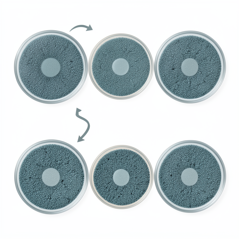
Concentration-Dependent Antibacterial Effect
AI-generated conceptual illustration (Petri dishes with bacterial lawns and copper oxide nanoparticle disks). It explains the idea and does not show study data.
View source evidence ↗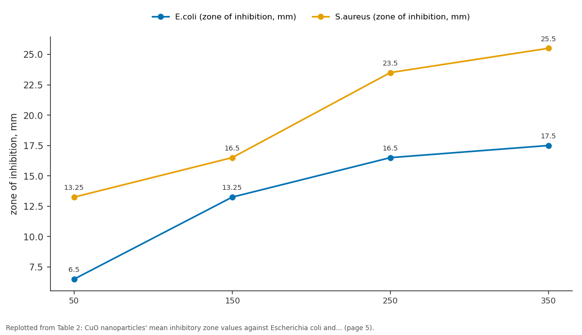
E.coli (zone of inhibition, mm) and S.aureus (zone of inhibition, mm): 50 vs 150 vs 250 vs 350
Replotted from Table 2 (page 5) of the original paper. Every bar is the paper's own table value.
View source evidence ↗Figures & tables
From the original paper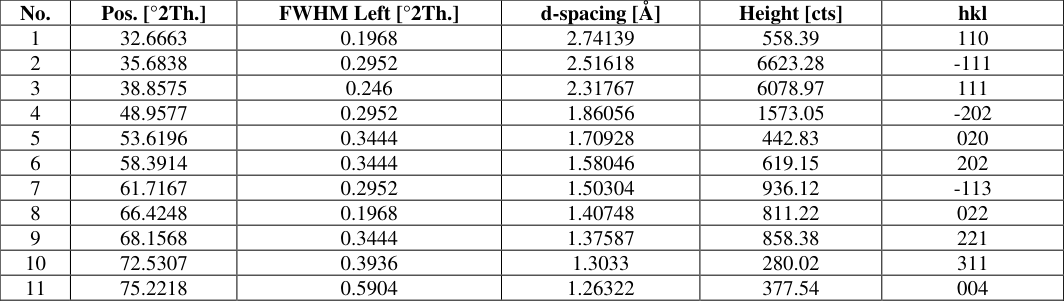 Table 1
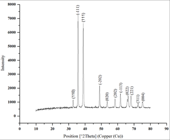 Figure 1
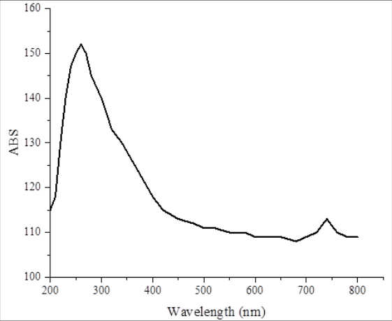 Figure 2
 Table 2
Introduction
Copper oxide nanoparticles' (CuO NPs) properties are different from bulk copper oxide in terms of physicochemical, optical and catalytic which is gaining serious attention of researchers [1] . The copper oxide is a transition metal oxide and is a p-type semiconductor [2] . Due to its small band gap, typically ranging from 1.2 to 2.1 eV, it can be used in a variety of technological and environmental applications including wastewater treatment, photocatalyst, sensor, solar cell and antimicrobial agent. Copper oxide nanoparticles are highly catalytic and have tunable optical properties and high surface area [3] . These properties make copper oxide nanoparticles a necessary element in the field of nanotechnology and material science. Recently, a lot of studies have been performed on the synthesis and characterisation of CuO nanoparticles by using various chemical, physical and biological methods. The nanoparticles' properties are essentially determined by their particle size, shape, crystallinity and surface characteristics and features [4] . Hence, Cabal et al.151 discussed the potential of CuO nanomaterials in diverse applications including as nano-adsorbents for the treatment of wastewater [5] . X-ray diffraction (XRD) and ultraviolet-visible (UV-Vis) spectroscopy are two well-known characterization methods that recognize the optical structural characteristics of nanomaterials. Analysis by X-ray diffraction is a valuable criterion for crystallite size, lattice parameters, spatial homogeneity, and crystal structure etc of nanoparticles [6] .
The presence of numerous diffraction peaks of monoclinic crystal structure of CuO confirms synthesis and crystallinity is depicted in the XRD spectra for copper oxide nanoparticles [7] . Ultraviolet spectroscopy or UV-Visible Spectrometry is an important technique for studying the optical and electrical transitions of semiconductor nanoparticles. The light interaction with the nanomaterial and optical band gap energy was acquired from the absorption spectra obtained by UV-Vis analysis. Quantum confinement effects and changes in the size of nanoparticles usually result in a modification of the absorption edge and band gap energy [8,9] . Material instability and limitations of titanium dioxide can be avoided by growing copper oxide nanoparticles due to their excellent properties. The antibacterial activity of such compounds is generally concentration-dependent, with enhanced inhibition zones associated with higher concentrations. Proposed mechanisms involved in this activity are generation of ROS and release of damaging Cu2+ ions that can hinder bacterial membrane and metabolism as well. As a result, CuO nanoparticles are prospective candidates for antibacterial applications along with their technological and environmental applications. [10] According to UV-Vis Spectra with XRD and UV-Vis spectra, structural and optical properties of copper oxide nanoparticles can be get with reasonable accuracy. During the study, the techniques were used to investigate the crystalline structure, phase evolution, and optical properties of copper oxide nanoparticles. The findings obtained establish a relationship between the optical properties and structural properties of CuO nanoparticles. The potential applications of these materials in technological uses, environmental applications, medical uses such as antimicrobial agents, drug delivery systems, and biomedical sensing.
Synthesis of Copper Oxide Nanoparticles (CuO NPs)
The chemical precipitation technique is simple, costeffective and more efficient for nanostructured materials. Accordingly, the chemical precipitation method has been used to prepare copper oxide nanoparticles (CuO). Sodium hydroxide (NaOH) was the agent and copper precursor was copper (II) nitrate trihydrate. A typical synthesis procedure involves dissolving a suitable weight of Cu(NO3)2⋅3H2O in distilled water which stirring. Through this process, a dark blue solid of Copper hydroxide (Cu(OH)2) formed. The solution was stirred for half an hour to one hour for complete precipitation. After filtration, the precipitate was washed with ethanol and distilled water several times to remove the excess ions. The precipitate was then washed and oven-heated at 80-100 °C for about 5 hours. The muffle furnace was employed to calcine the dried product for three hours at 600 °C. As per the following reaction, a thermal decomposition of copper hydroxide occurred followed by the formation of copper oxide nanoparticles:
In order to produce fine copper oxide nanoparticles appropriate for additional analysis, the resulting black powder was ultimately pulverized using an agate mortar. The optical and structural characteristics of the produced CuO nanoparticles were next examined using X-ray diffraction (XRD), UV-visible spectroscopy and and antibacterial.
Antibacterial Activity of CuO NPs
Using the liquid medium approach, the CuO NPs' antibacterial activity was initially evaluated against the gram-positive bacteria Staphylococcus aureus and the gramnegative bacterium Escherichia coli. 0.5 McFarland turbidity standard (5×10⁷ cell mL -1 ) tubes were used to regulate the bacterial suspension. On nutritional broth, each strain of bacteria was subcultured. The broth was inoculated with 0.2 mL of bacterial strains and subsequently 0.5 mL of CuO NPs at concentrations of 50, 150, 250, and 350 µg mL -1 with antibiotics in order to examine the inhibition rate of the CuO NPs. For a whole day, the tubes were incubated at 37°C. Each test's triple reading was used to get the mean inhibition values. The control was a broth tube that contained solely bacterial strains and no NPs.
Result and discussion
The produced copper oxide nanoparticles (CuO NPs) show distinct diffraction peaks in their XRD pattern, which suggests that a crystalline structure (monoclinic phase) has formed. The crystallographic planes (110), (-111), ( 111), (-202), ( 020), ( 202), (-113), ( 022), (221), and (311) are represented by the significant peaks seen at 2θ values of roughly 32.8°, 35.9°, 38.8°, 48.7°, 53.5°, 58.3°, 61.5°, 66.3°, 68.0°, and 75.2°, respectively. The effective fabrication of phase-pure CuO nanoparticles free of detectable impurities is confirmed by these peaks' good match to the standard JCPDS card for monoclinic CuO (JCPDS No. 45-0937). The diffraction peaks' sharpness and intensity indicate that the produced nanoparticles have a high degree of crystallinity. The Debye-Scherrer equation can be used to determine the average crystallite size based on the most intense peak seen at the (-111) plane:
Where λ is the X-ray wavelength (1.5406 Å for Cu Kα), D is the crystallite size, K is the form factor (usually 0.9), β is the peak's full width at half maximum (FWHM) in radians, and θ is the Bragg angle. Using this formula, the average crystallite size of the CuO nanoparticles is calculated to be 30.5 nm, which is in line with the typical nanoscale CuO documented in the literature. Furthermore, as shown in figure 1 and table 1, the lack of secondary peaks attests to the great purity and homogeneity of the produced nanoparticles.
| No. | Pos. [°2Th.] | FWHM Left [°2Th.] | d-spacing [Å] | Height [cts] | hkl |
|---|---|---|---|---|---|
| 1 | 32.6663 | 0.1968 | 2.74139 | 558.39 | 110 |
| 2 | 35.6838 | 0.2952 | 2.51618 | 6623.28 | -111 |
| 3 | 38.8575 | 0.246 | 2.31767 | 6078.97 | 111 |
| 4 | 48.9577 | 0.2952 | 1.86056 | 1573.05 | -202 |
| 5 | 53.6196 | 0.3444 | 1.70928 | 442.83 | 020 |
| 6 | 58.3914 | 0.3444 | 1.58046 | 619.15 | 202 |
| 7 | 61.7167 | 0.2952 | 1.50304 | 936.12 | -113 |
| 8 | 66.4248 | 0.1968 | 1.40748 | 811.22 | 022 |
| 9 | 68.1568 | 0.3444 | 1.37587 | 858.38 | 221 |
| 10 | 72.5307 | 0.3936 | 1.3033 | 280.02 | 311 |
| 11 | 75.2218 | 0.5904 | 1.26322 | 377.54 | 004 |
Table text extracted from the original PDF; superscript letters are shown inline. The original table image is below.
View original table image
UV-Visible (UV-Vis) Spectroscopy
The presence of nanoscale CuO particles with direct band gap transitions in the UV region about 280 nm. The electron excitation from CuO's valence band to its conduction band, which is typical of semiconductor behavior, is responsible for this absorption. prospective light-harvesting capacity and prospective uses in photocatalysis are suggested by the extensive absorption that extends towards 800 nm. The Tauc relation can be used to estimate the optical band gap (Eg):
(αhν) n = A(hν-Eg) Where α is the absorption coefficient, hv is the photon energy, Eg is the optical band gap, A is a material-dependent constant, and n denotes the type of electronic transition (n = 1/2 for a direct allowed transition and n = 2 for an indirect allowed transition). A band gap of roughly 2.1-2.2 eV can be inferred from the spectra by extrapolating the linear part of the Tauc figure. The quantum confinement effect seen in nanoparticles, where size reduction results in an increase in band gap energy, is compatible with this somewhat greater band gap (1.2-1.5 eV) compared to bulk CuO. Because of the phenomena of localized surface plasmon resonance (LSPR), metallic nanoparticles' optical characteristics can be utilized to assess their particle size. The dimension of spherical nanoparticles can be calculated from the UV-Vis absorption spectrum using the ratio between the absorbance at the plasmon peak and the absorbance at 450 nm, according to the empirical relation put out by Hai.
The Haiss equation is given as d = ln (λ SPR -λ°)/L1/L2 where: d is particle diameter (nm), λ SPR is absorbance at the surface plasmon resonance peak absorbance at 450 nm, L1 = 6.53 and L2 = 0.0 216. By Haiss equation the copper oxide nanoparticles is 94 nm, see figure 2.
XRD and UV-vis findings' correlation
The concomitant XRD and UV-Vis studies validate that the produced CuO particles are nanoscale. The blue-shifted absorption in the UV-Vis spectrum is a reflection of the quantum confinement effect, which is influenced by the small crystallite size seen via XRD. These CuO nanoparticles are attractive candidates for use in photocatalysis, sensors, and electronic devices because of their high crystallinity and nanoscale size, which improve the optical properties. The particle size determined by XRD (~30.5 nm) is less than that found by UV-Vis spectroscopy (~94 nm).This discrepancy is frequently seen in the characterisation of nanoparticles and can be attributed to a number of factors: Crystallite Size vs. Particle Size: The size of a single crystalline domain is measured via the Scherrer equation. However, a single nanoparticle may be made up of many crystallites together, so:
Particle size (UV-vis) > Crystallite size (XRD)
Particle Aggregation: UV-Vis spectroscopy is used to assess the optical response of whole particles, including surface layers (oxides or organic stabilizers). The apparent particle size rises as a result. Plasmon on the Surface Sensitivity: Because the plasmon resonance peak depends on particle form, the surrounding dielectric medium, and particleparticle interactions, particle size may be exaggerated when using optical models. XRD Instrumental Broadening: Peak broadening in XRD can be influenced by lattice strain, crystal structural flaws, and instrumental broadening, which may lead to somewhat underestimated crystallite sizes.
CuO Nanoparticles' Antibacterial Properties
The generated CuO nanoparticles were subjected to agar well diffusion process to test their bactericidal activity against varied bacteria. The study findings showed the presence of distinct inhibition zones surrounding the wells indicating that CuO nanoparticles have significant antibacterial activity against both Gm+ and Gm-bacteria.
As the concentration of CuO nanoparticles (B = 50, C = 150, D = 250 and E = 350 μg/mL) increased, so did the widening of the inhibitory zone. The reference antibiotic produced larger inhibition zones compared to CuO nanoparticles against bacterium strains mentioned in current results. As such, oxide effectiveness was re-evaluated and shown to be by using an ineffective reference (A = inactivated reference) (figure 2) [11] . The mechanism of metal oxide nanoparticle effectiveness as Antibacterial effectiveness is through indirect physicochemical mechanisms where as antibiotics are designed to interfere with or destroy the metabolic pathway of the specific bacteria. While the inhibition zones of the CuO nanoparticles were quite shorter compared to the antibiotic, they showed great activity. Many systems carry out this function. One of the primary causes is the creation of reactive oxygen species (ROS) like superoxide ions and hydroxyl radicals, which leads to oxidative stress within bacterial cells. ROS radicals harm essential macromolecules including proteins, lipids and DNA. Bacterial cells ultimately ends up dying. The second important mechanism is the liberating ions Cu2+ from the surface nanoparticles CuO. These ions affect enzymes and proteins and increase membrane permeability and interference with life processes. When bacteria malfunction, it ceases to grow and multiply.
If their insides leak out, they stop growing. Bacterial cell membrane disturbed. As stated by the source, there are different parameters which influence antibacterial activity of CuO nanoparticles [12] . This refers to size, shape, surface charge and method of synthesis. Small-sized nanoparticles typically display remarkable antibacterial activity due to their larger surface area and strong contact with bacterial cell walls. The differences in their antibacterial activities may depend on the differences in the cell wall architecture of bacteria. Gram-negative bacteria have an outer membrane made of lipopolysaccharides which could hinder the entry of nanoparticles. Gram positive bacteria,on other hand, are thick walled organism. In general, through various studies on CuO nanoparticles, it was found that they exhibit very good antibacterial properties which could play a crucial role as antimicrobial agents in the environmental and medical spheres [13] . Figure 2 and Table 2 show the results. Figure 2. CuO nanoparticles' antibacterial activity against E. coli and S. aureus at varying doses is displayed as a bar chart with zones of inhibition (mm) determined by the agar well diffusion method.
| CuO NPs concentration (μg/mL) | E.coli (zone of inhibition, mm) | S.aureus (zone of inhibition, mm) |
|---|---|---|
| 50 | 6.5 | 13.25 |
| 150 | 13.25 | 16.5 |
| 250 | 16.5 | 23.5 |
| 350 | 17.5 | 25.5 |
Table text extracted from the original PDF; superscript letters are shown inline. The original table image is below.
View original table image
Conclusion
The presence of nanoscale particles was confirmed by the UV-Vis spectrum, which showed a distinctive absorption band at about 270 nm. The Scherrer analysis of the XRD pattern produced a crystallite size of roughly 30.5 nm, whereas the particle size predicted using the Haiss equation was roughly 94 nm.The difference between the two numbers results from the fact that XRD measures the size of coherent crystalline domains within the nanoparticles, whereas UV-Vis spectroscopy assesses the entire particle size, including potential aggregation and surface effects. As a result, the particle size determined by optical measurements is usually larger than the crystallite size determined by XRD. XRD examination confirmed the remarkable crystallinity and nanoscale dimensions of the produced CuO nanoparticles. Because of quantum confinement effects, the UV-Vis spectroscopy data show semiconducting behavior with a wider band gap. Overall, the effective synthesis of phasepure CuO nanoparticles with potential for a range of nanotechnology applications is confirmed by the structural and optical characterizations. The crystalline nature of the produced nanoparticles was verified by the XRD examination. The (111) crystallographic plane was represented by the most intense of the diffraction pattern's multiple distinctive peaks. The crystallite size determined by XRD and the particle size determined by UV-Vis spectroscopy differed noticeably.
There are a number of reasons for this disparity, which is frequently noted in the characterisation of nanoparticles. First, UV-Vis spectroscopy shows the total particle size, which may comprise several crystallites within a single nanoparticle, while the Scherrer equation quantifies the size of coherent crystalline domains. Second, an apparent increase in the size determined by optical measurements may result from the aggregation or agglomeration of nanoparticles in solution. The surrounding medium, particle shape, and interparticle interactions all have an impact on the plasmon resonance peak and can also lead to a greater apparent size. The produced nanoparticles are therefore probably polycrystalline or partially aggregated, where many crystallites form a single nanoparticle structure, based on the smaller size derived by XRD compared with the UV-Vis estimation. Thus, a more thorough understanding of the optical particle size and the crystalline domain size of the produced nanomaterials can be obtained by combining UV-Vis spectroscopy with XRD. The antibacterial activity of copper oxide nanoparticles (CuO NPs) against several bacterial strains was successfully assessed in this investigation. The findings showed that CuO nanoparticles have strong antibacterial qualities, with the inhibitory zone growing with nanoparticle concentration. Because their cell walls differed, Gram-positive bacteria (S. aureus) were more sensitive than Gram-negative bacteria (E.coli). The CuO nanoparticles can therefore be regarded as potent antibacterial agents with possible uses in the environmental and medicinal domains.
References
10 in the original paper- Sivayogam D, Punithavathi IK, Jayakumar SJ, Mahendran N. Structural, optical and electrical properties of Cd doped CuO nanoparticles obtained by simple sol gel method. Mater Today Proc. 2022;49(7):2752-2757.
- Angayarkanni S, Neyvasagam K. Structural and optical studies of copper oxide nanoparticles synthesized by chemical precipitation method. Mater Today Proc. 2021;47(4):1149-1154.
- Khit SA. Eco-friendly synthesized of CuO nanoparticles using Anchusa strigosa L. flowers and study its adsorption activity. Baghdad Sci J. 2023;20(4):1322-1330.
- Shishir MKH, Alam MA, Bishwas RK, Karim MM, Alam SMN, Khan GMA. Crystallographic insights into copper oxide nanoparticles synthesized via a unique sol-gel method: antibacterial activity and photocatalytic evaluation. S Afr J Chem Eng. 2026;55:162-179.
- Ruvalcaba-Manzo SG, Ramírez-Bon R, Ochoa-Landín R, Castillo SJ. A comprehensive study of the optical, structural, and morphological properties of chemically deposited ZnO thin films. Inorganics. 2025;13(10):331.
- Maria KH, Bonet SGR, Bosco MV, Salam SRB, Dhar D, Nazzarro M, et al. Synthesis and characterization of CuO nanoparticles using heat treatment approach: annealing effects on crystallite size and band gap of CuO nanoparticles. ACS Omega. 2026;11:1114111154.
- Diachenko O, Kováč J Jr, Dobrozhan O, Novák P, Kováč J, Skriniarova J, et al. Structural and optical properties of CuO thin films synthesized using spray pyrolysis method. Coatings. 2021;11(11):1392.
- Rakshit S, Maji D, Mondal KG, Roy T, Jana PC, Kar BS, Datta R. Structural, optical and magnetic properties of chemically grown copper oxide nanoparticles: an insight into anticancer activities. Mater Chem Phys. 2024;325:129794.
- Gherasim C, Asandulesa M, Fifere N, Doroftei F, Tîmpu D, Airinei A. Structural, optical and dielectric properties of some nanocomposites derived from copper oxide nanoparticles embedded in poly(vinylpyrrolidone) matrix. Nanomaterials. 2024;14:759.
- Salem SS, Yousef A, Abd El Hamid ES, Ibrahim S, Elbawab RH. Evaluation of eco-friendly copper oxide nanoparticles for anticancer activity and antibacterial effects against Streptococcus mutans using molecular docking. Sci Rep. 2025;15:40921.