
Research article · 2026 · Full text
International Journal of Advanced Chemistry ResearchThe role of adhesion molecules and chemokines as factors associated with physiological, immunological, and chemical changes in Iraqi multiple sclerosis patients
Volume 8(6) · Pages 08–15 · DOI: 10.33545/26646781.2026.v8.i6a.682 ↗
Research areasMultiple Sclerosis Research StudiesNeuroinflammation and Neurodegeneration MechanismsCell Adhesion Molecules Research
Abstract
Author-writtenThe central nervous system is affected by Multiple Sclerosis (MS), a progressive, autoimmune, and inflammatory disease. Many physicians believe that MS is a terminal disease because it can only be treated by reducing the number of relapses and slowing the rate of physical and mental decline. About 4 million people are living with MS today, and the disease results from an acute inflammatory response to the breakdown of the barrier between the blood and the brain. The research was carried out in a specialized unit in Baghdad, at Dr. Saad Al-Watri Hospital for Neurosciences/ Baghdad (Neurological Diseases Unit) and in private clinics located in Baghdad Governorate during the period from 11/15/2024 to 4/15/2025, with an age range of participants between (30 years and 60 years); there were (30) samples from patients with multiple sclerosis, (20) male patients and (10) female patients; (30) samples will be taken from the control group (healthy) at the same range of ages; there will be (25) healthy males and (5) healthy females that have been free of all diseases. The blood samples were obtained by venipuncture (blood drawn through your arm) after disinfecting the area with a needle; each sample drawn will be a 5cc syringe. The syringe is (disposable) and will be discarded (immediately after use). Blood from each animal was collected in tubes containing a gelatinous material to allow both the clotted and serum portion of the blood to separate after being centrifuged at (3500 x G) for ten minutes. After centrifugation, the serum was removed and placed in dry plastic vials before being stored at 4°C until needed for the measurements carried out as part of the research bioassay (CCl2, CXCL10, PAF, P-Selectin, IL-6, and IL-12). When the separate male and female groups were compared, males exhibited statistically significant differences from females via a probability level of p≤0.05.
Summary
Source-checkedPlain-language reading of the full paper, prepared with AI and checked against the text. Each point links to the authors' own words.
Why it matters
- Multiple Sclerosis (MS) is a progressive inflammatory and autoimmune disease that affects the central nervous system. View evidence ↗
- The disease is caused by an acute inflammatory response triggered by the breakdown of the blood-brain barrier. View evidence ↗
- Chemokines, secreted by activated T lymphocytes, are critical for regulating the migration of monocytes to damaged areas of the nervous system during inflammation. View evidence ↗
The question
- The study aimed to investigate how adhesion molecules and chemokines act as factors associated with chemical and immunological changes in Iraqi patients with multiple sclerosis. View evidence ↗
What they did
- The researchers used enzyme-linked immunosorbent assay (ELISA) to measure levels of CCl2, CXCl10, PAF, P-Selectin, IL-6, and IL-12 in blood serum. View evidence ↗
- Statistical variance between the MS and control groups was analyzed using a t-test with a significance level of p≤0.05. View evidence ↗
What they found
- Serum CCl2 levels were significantly higher in MS patients (10.66±12.02 pg/ml) than in the control group (1.36±0.97 pg/ml). View evidence ↗
- Serum CXCl10 levels were significantly increased in MS patients (60.16±92.11 pg/ml) compared to controls (8.36±4.12 pg/ml). View evidence ↗
- Platelet-activating factor (PAF) levels were significantly higher in MS patients (35.16±15.41 ng/ml) than in controls (15.16±13.01 ng/ml). View evidence ↗
- P-Selectin levels were significantly elevated in MS patients (16.13±5.11 ng/ml) compared to the control group (4.58±2.62 ng/ml). View evidence ↗
- IL-6 levels were significantly higher in MS patients (14.07±0.43 pg/ml) than in the control group (5.01±0.24 pg/ml). View evidence ↗
- IL-12 levels were significantly increased in MS patients (476.02±66.18 pg/ml) compared to controls (167.04±29.85 pg/ml). View evidence ↗
- ROC analysis for CCl2 showed an area under the curve (AUC) of 1.000, with 100% sensitivity and specificity (p < 0.001). View evidence ↗
What it means
- The authors conclude that targeting platelets for the diagnosis and treatment of MS is a promising approach that warrants further research. View evidence ↗
- The authors state that chemokines and their receptors are highly significant in autoimmunity, inflammation, and the biological processes associated with multiple sclerosis. View evidence ↗
Visual summary
3 visualsIllustrations explain the paper; charts are replotted from the paper's own table values.
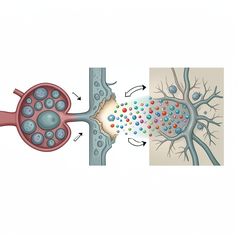
Mechanism of Neuroinflammation in MS
AI-generated conceptual illustration (Human blood-brain barrier and central nervous system tissue). It explains the idea and does not show study data.
View source evidence ↗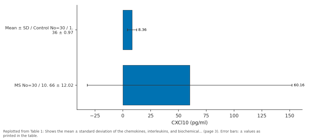
CXCl10 (pg/ml): Mean ± SD / Control... vs MS No=30 / 10. 66 ±...
Replotted from Table 1 (page 3) of the original paper. Every bar is the paper's own table value.
View source evidence ↗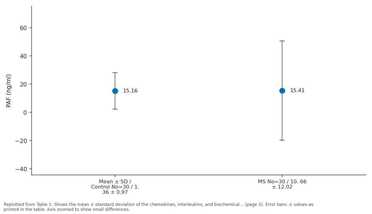
PAF (ng/ml): Mean ± SD / Control... vs MS No=30 / 10. 66 ±...
Replotted from Table 1 (page 3) of the original paper. Every bar is the paper's own table value.
View source evidence ↗Figures & tables
From the original paper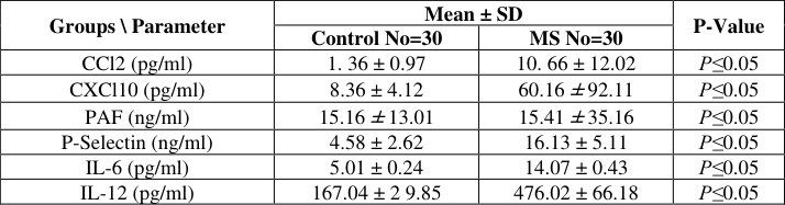 Table 1
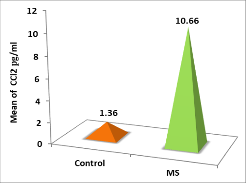 Figure 1
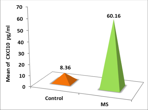 Figure 2
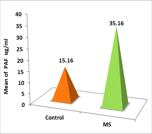 Figure 3
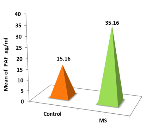 Figure 4
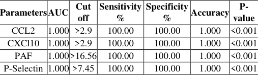 Table 2
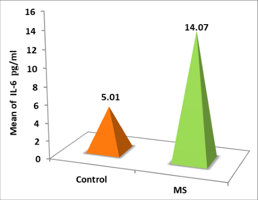 Figure 5
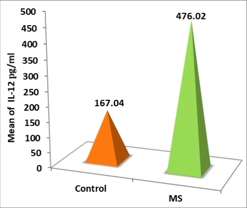 Figure 6
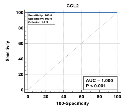 Figure 7
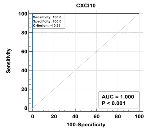 Figure 8
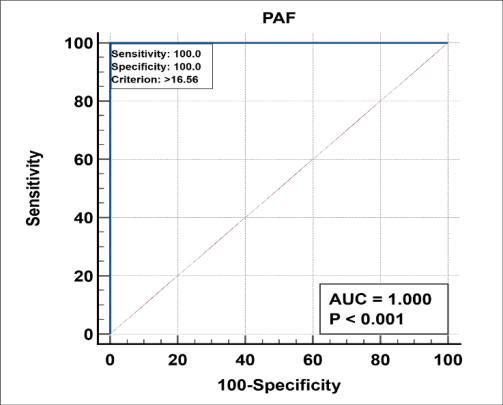 Figure 9
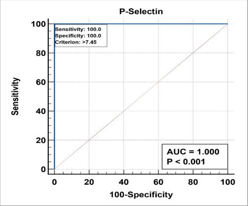 Figure 10
Introduction
Multiple Sclerosis (MS) is an inflammatory disease of the Central Nervous System (CNS) resulting from chronic inflammation that impairs the autoimmune system [1] . The hallmark symptoms of MS are inflammation and neurodegeneration, which occur together throughout the disease course [2] . Multiple sclerosis is characterized by demyelinating white matter lesions and neurodegeneration, and the disease usually presents with periods of clinical stability interspersed with acute relapses such as autoimmune encephalomyelitis. The ability to reduce clinical symptoms of brain lesions is a crucial factor influencing the course of the disease [3] . Multiple sclerosis is considered an incurable disease, and its treatment depends primarily on reducing relapses and slowing the process of motor and mental deterioration [4] . The incidence of multiple sclerosis may be somewhat hereditary, as immediate family members are more susceptible to this disease. Polymorphisms in the genes of some cytokines have been identified in the incidence of this disease, such as some types of interleukins IL-2, IL-6, and IL-13, as distinctive signs of multiple sclerosis [5,6] . In addition, the role played by B cells in the pathophysiology of multiple sclerosis has led to a shift from treatments that focus on T cells only, as well as the introduction of antibodies in the treatment of multiple sclerosis [7] . Biomarkers that reflect different aspects of the pathophysiology of the disease may be more accurate in characterizing individuals who develop this disease, because factors promoting chronic inflammation in MS are compartmentalized by the central nervous system within active lesions and are present diffusely within the meninges, choroid plexus, and follicles of the ectopic lymph nodes [8] . Inflammatory cytokines play a key role in the pathophysiology of MS by promoting blood-brain barrier (BBB) damage, activating the neuroinflammatory cascade in the CNS, and migration of innate and adaptive immune cells [9] . Peripheral and CNS-resident immune cells release cytokines, which interact with a large number of receptors expressed by various cell types, such as infiltrating lymphocytes, microglia, astrocytes, as well as neurons [10,11] . The organization of the complex functions of these molecules under these physiological conditions is regulated by basic regulatory principles, as these molecules form complex networks that work on the same cytokine, and they can perform multiple activities that depend on the state of the inflammatory environment [12] . Immune cells are activated by pro-inflammatory cytokines in the central nervous system, leading to demyelination, glial scarring, and axonal lesions of the affected parts. because the regulation of the immune response is done by the infiltration of cells into the central nervous system, which is done through chemokines and their receptors, which play an effective role between the natural internal balance, autoimmunity and inflammation, because chemokines and their receptors are effective therapeutic targets for multiple sclerosis [13] . Chemokines play an important role in inflammation, cell development and migration during immune surveillance, which are secreted by activated T lymphocytes and regulate the accumulation and migration of monocytes to damaged areas of the nervous system and promote their differentiation, thus maintaining the immune response process in the body [14,15] . CCL2 is a potent chemokine for the recruitment of macrophages, monocytes, and T lymphocytes to the CNS and is produced by endothelial cells, macrophages, and microglia [16] . Furthermore, CCL2 influx is mediated by ABC transporters of astrocytes, thus promoting the migration of immune cells across brain endothelial cells, which are involved in the neuroinflammatory process [17] . CXCL10 is one of the chemokines that has been used in flow cytometry to determine the immune phenotype of multiple sclerosis cases. CXCL10 is secreted from a variety of cells, such as leukocytes, activated neutrophils, eosinophils, epithelial cells, endothelial cells, and stromal cells [18] . Platelet activating factor (PAF) is a lipid molecule secreted by a number of cells including immune and endothelial cells. It is also a potent pro-inflammatory mediator and mediates a variety of chronic diseases, including some types of cancer, cerebrovascular disorders, and diseases of the central nervous system [19,20] . Platelet-activating factor has the ability to exert physiological pathological actions at low concentrations of up to 10-12 times, because it acts as a primary messenger between cells [21] . Through the increased concentration of chemokines and interleukins, the research aimed to investigate the role of adhesion molecules and chemokines as factors accompanying immune and chemical changes in Iraqi multiple sclerosis patients.
Collection of specimens
A total of thirty samples were collected from individuals suffering from multiple sclerosis. This sample consisted of twenty males and ten females. Thirty additional samples were also collected from a control group of healthy individuals within the same age bracket as those suffering from multiple sclerosis. The healthy sample consisted of twenty-five males and five females who have been determined to be free from any disease. A sample of whole blood was collected from a peripheral vein. The site of collection was sterilized prior to collecting the blood. Blood was collected into a disposable 5ml syringe (the syringes were immediately disposed of after use). The collected blood was placed in a tube containing a gelatinous substance so that, when subjected to centrifugation for 10 minutes at a speed of 3500 RCF, the serum could be separated from the coagulated portion of the blood. Serum was then removed from the tubes and transferred to sterile, dry plastic tubes for storage until later analysis (4°C). The bioassays performed on the blood samples analyzed in this study were: CCL2, CXCL10, PAF, P-Selectin, IL-6, and IL-12. The results were statistically different for males and females in both groups (p≤0.05).
Estimation of chemokines and interleukins in the samples under study
The immune parameters (CCl2, CXCl10, PAF, P-Selectin, IL-6, IL-12) were measured in patients and healthy controls using the enzyme-linked immunosorbent assay (ELISA) according to the manufacturers' recommendations. The assay was performed with a 96-well adsorption plate coated with antibodies to (CCl2, CXCl10, PAF, P-Selectin, IL-6, and IL-12). Antigens from the serum of patients and healthy controls were captured onto the wells by the presence of the captured antibodies and the enzyme conjugated to the detecting antibodies. This creates a noncompetative complex between the antigens and antibodies (a "sandwich" type complex). During the incubation period, an immunecomplex aggregation occurs in the titration tubes due to the high-affinity binding of the biotionylated antibody to streptavidin. After equilibrium and binding have occurred, the separated antibody fragments are washed free from unbound antigens. The enzyme activity in the bound antibody fragments is proportional to the concentration of the antigens as the enzyme activity is facilitated by horseradish peroxidase (HRP). Following addition, the wells will turn blue, then after adding the stopping solution, they will turn yellow. The OD will be measured at (450) nm. Each sample will be run in replicate, as an additional means of ensuring that there is accurate results.
Statistical Evaluation
The results of the study were evaluated using the Statistical Package for the Social Sciences (SPSS) statistical analysis program and using a completely randomized design (CRD) method. The t-test was used to analyze the variance between the study groups at a probability level of (p≤0.05).
Results
• Estimation of the concentration of chemokines, interleukins, and biochemical variables of the samples under examination 1 showed a significant increase in the level of CCl2 concentration in the blood serum of the group of patients with multiple sclerosis (10.66±12.02) pg/ml compared to the control group (1.36±0.97) pg/ml, at a probability level of p≤0.05, as in the following figures: Table (1) showed a significant increase in the level of P-Selectin concentration in the blood serum of the group of patients with multiple sclerosis (16.13±5.11) ng/ml compared to the control group (4.58±2.62) ng/ml, at a probability level of P≤0.05, as in the following figures: Table (1) showed a significant increase in the level of IL-6 concentration in the blood serum of the group of patients with multiple sclerosis (14.07±0.43) pg/ml compared to the control group (5.01±0.24) pg/ml, at a probability level of p≤0.05, as in the following figures: Table 1 showed a significant increase in the level of IL-12 concentration in the blood serum of the group of patients with multiple sclerosis (476.02±66.18) pg/ml compared to the control group (167.04±29.85) pg/ml, at a probability level of P≤0.05, as in the following figures:
| Groups \ Parameter | Mean ± SD | — | P-Value |
|---|---|---|---|
| Control No=30 | MS No=30 | ||
| CCl2 (pg/ml) | 1. 36 ± 0.97 | 10. 66 ± 12.02 | P≤0.05 |
| CXCl10 (pg/ml) | 8.36 ± 4.12 | 60.16 ± 92.11 | P≤0.05 |
| PAF (ng/ml) | 15.16 ± 13.01 | 15.41 ± 35.16 | P≤0.05 |
| P-Selectin (ng/ml) | 4.58 ± 2.62 | 16.13 ± 5.11 | P≤0.05 |
| IL-6 (pg/ml) | 5.01 ± 0.24 | 14.07 ± 0.43 | P≤0.05 |
| IL-12 (pg/ml) | 167.04 ± 2 9.85 | 476.02 ± 66.18 | P≤0.05 |
Table text extracted from the original PDF; superscript letters are shown inline. The original table image is below.
View original table image
ROC curve calculation -
The ROC curve was calculated for chemokines and other biochemical variables for patients with multiple sclerosis, as shown in Table 2. The results of chemokine (CCL2) showed that the value of the area under the curve (AUC) was (1), the (Cut off) was (>2.9), the sensitivity ratio was (100), while the specificity ratio was (100), and the accuracy reached ( 1) at a probability level of (<0.001), as in Figure (7). The results of the chemokine (CXCl10) showed that the area under the curve (AUC) value was (1), the (Cut off) was (>2.9), the sensitivity ratio was (100), while the specificity ratio was (100), and the accuracy reached ( 1) at a probability level of (<0.001), as in Figure (8). The results of (PAF) showed that the value of the area under the curve (AUC) was (1), the (Cut off) was (>16.56), the sensitivity ratio was (100), while the specificity ratio was (100), and the accuracy reached ( 1) at a probability level of (<0.001), as in Figure (9). The results of (P-Selectin) showed that the value of the area under the curve (AUC) was ( 1), (Cut off) was (>7.45), the sensitivity ratio was (100), while the specificity ratio was (100), and the accuracy reached ( 1) at a probability level of (<0.001), as in Figure (10).
Discussion
The mechanism of development of multiple sclerosis is still unclear and not well understood, as this disease is linked to several factors, including viral infections, genetic and environmental factors, as well as autoimmune inflammation, which plays a distinct and effective role in the development of this neurological disease [22] . Through the results we obtained, a close relationship between chemokines and multiple sclerosis was demonstrated, The result of increased CCl2 in the serum of patients with multiple sclerosis was consistent with the result of (Kurtuncu et al.) [23], as well as the result of CXCl10 compatibility, which was consistent with the result of (Comini-Frota et al.) [24] . The reason for the increased level of chemokines is likely due to the infiltration of lymphocytes, monocytes, and macrophages in the lesions and plaques affected by multiple sclerosis [25] . When the levels of chemokines (CCl2 and CXCl10) are high, they are able to infiltrate and continuously cross into immune cells, in order to attract white blood cells to sites of infection and inflammation via precise selective receptors because these chemokines are produced in various tissues of the body [26,27] . Chemokines can play an important and effective role in activating and stimulating leukocyte adhesion molecules, which in turn leads to a gradient of chemical concentration across endothelial cells. They can also increase cell adhesion and target migration across the blood-brain barrier by interacting with leukocytes, which promotes them in the central nervous system [28,29] . Such as autoreactive T cells that target myelin components, which are considered triggers of multiple sclerosis, and this step is vital for the transfer of inflammatory cells to the central nervous system of multiple sclerosis patients [30] . CCL2 can mediate its activity by binding to the chemokine receptor CCR2, this association is essential to elicit the clinical and histopathological characteristics of autoimmune encephalomyelitis in patients with multiple sclerosis, through the infiltration of immune cells through the activation of glial cells during autoimmune encephalomyelitis [31,32,33] . The result of the increase in platelet-activating factor (PAF) was consistent with the result of (Angela Midgley et al.) [34] and the result of (Leonardo et al.) [35] , while the adhesion molecule (P-Selectin) was consistent with the result of (Diego et al.) [36] . The results of the increase in both PAF and P-Selectin Hyperactivity of platelets in MS patients, regardless of their EDSS score, is indicated because elevated platelet levels are a developmental risk factor in MS patients, due to the ability of these platelets to cross the blood-brain barrier via damaged vascular basal lamina [37,38] . Another role of platelets is the use of P-selectin molecules; It has been proven that its levels increase in patients with multiple sclerosis, and these molecules are able to bind to lymphocytes, which leads to increased attachment of lymphocytes to the inner lining of the affected tissues [39,40] . Blood-brain barrier disruption and microvascular endothelial activation in MS result either, in an unclear way, from the effects of inflammatory cytokines on astrocytes and immune cells associated with the neurovascular unit, Or clearly for the cytokines secreted by myelin-stimulating T cells, as leaks in the blood-brain barrier lead to increased infiltration of immune cells [41,42] . In the end, it became clear to us that the influencing factors are still using different immune cell groups under study and research. Although some junctional proteins have been shown to remodel and influence migration pathways, often favoring either cell-tocell transport or transcellular migration [43,44] . The essential role of platelets in multiple sclerosis is important and promising, as this role includes the continuous appearance of platelet abnormalities and the release of their products in multiple sclerosis patients, in addition to the presence of those platelet products in postmortem multiple lesions. Evidence for this idea comes from the increased risk of cardiovascular events, including myocardial infarction, ischemic stroke, and thrombosis in MS patients, which is intrinsically linked to an abnormal coagulation cascade because premature platelet infiltration into CNS tissues causes neurodegeneration, Finally, targeting platelets to diagnose and treat patients with multiple sclerosis holds great promise and deserves further research and study on this topic [45,46] . The result of the increased level of interleukins, which included IL-6, was consistent with the result of (Omar et al.) [47], while the result of (IL-12) was consistent with the result of (Losy J et al.) [48] and (Matusevicius et al.) [49] . It is likely that the reason for the increased level of interleukin-6 is due to the activation of T cells in the group of patients, which is directly related to the increased binding of IL-6. because elevated IL-6 is involved in antibody-mediated demyelination pathways, it is well known to enhance the humoral immune response [50] . Furthermore, IL-6 plays an active role in immune modulation and demyelinating disorders because it is considered a pro-inflammatory activity [51] . IL-6 is the most important mediator of the immune response in the acute phase, as it is able to cross the blood-brain barrier and initiate the synthesis of prostaglandin E2 in the hypothalamus, which is secreted by T cells to stimulate the immune response to tissue damage, leading to chronic inflammation [52] . The reason for the increased concentration of IL-12 is due to the enhancement of the differentiation of T cells, because IL-12 enhances the migration of these cells to the central nervous system by increasing the regulation of adhesion molecules present in it [53] . Studies have shown that preventing the migration of T cells to the central nervous system is an effective and successful strategy to improve demyelination in patients with multiple sclerosis [54,55,56] .
Conclusions
Several inflammatory illnesses are closely associated with problems involving the immune system; these issues can frequently lead to either the death of adult stem cells, the myelin formation around axons within the nervous system, or other levels of damage to neurons. The central nervous system protects immune cells from damage with several types of pro-inflammatory cytokines. As a result, damage occurs through demyelination of axons, scar tissue formation by glial cells and damage to the axons themselves. Furthermore, chemokines and their respective receptors are highly significant in all aspects of autoimmunity, inflammation and many different biological processes, including the internal homeostasis that regulates lymphoid tissue growth, cellular trafficking, and importantly, multiple sclerosis.
Figures and tables from the paper
References
55 in the original paper- Thompson AJ, Baranzini SE, Geurts J, Hemmer B, Ciccarelli O. Multiple sclerosis. Lancet. 2018;391:1622-1636.
- Lassmann H. Multiple sclerosis pathology. Cold Spring Harbor Perspectives in Medicine. 2018;8.
- Kumar DP, Zanotto T, Cozart JS, Bruc AS. Association between frailty and sleep quality in people living with multiple sclerosis and obesity: An observational crosssectional study. Multiple Sclerosis and Related Disorders. 2024;81:105154.
- Di Meglio I, Vaz-Luis I. Systemic inflammation and cancer-related frailty: Shifting the paradigm toward precision survivorship medicine. ESMO Open. 2024;9(1):102205.
- Mouloud AF, Ahmed FF, Zeynel AA. Evaluation of some hormones and immunological variables in patients with polycystic ovary syndrome: A physiological study. International Research Journal of Pharmacy and Medical Sciences. 2025;8(6):1-8.
- Attfield KE, et al. The immunology of multiple sclerosis. Nature Reviews Immunology. 2022;23:83-20.
- Hauser SL, Cree BAC. Treatment of multiple sclerosis: A review. American Journal of Medicine. 2020;133:1380. doi:10.1016/j.amjmed.2020.05.049.
- Mado H, Adamczyk-Sowa M, Bartman W, Wierzbicki K, Tadeusiak B, Sowa P. Plasma interleukin-33 level in relapsing-remitting multiple sclerosis. Is it negatively correlated with central nervous system lesions in patients with mild disability? Clinical Neurology and Neurosurgery. 2021;206:106700.
- Ahmed FF, Hussein AA, Abass KS. Types of cytokines and their role in some pathological conditions in humans: A review. Journal of Cardiovascular Disease Research. 2021;12(6):133-137.
- Mado H, Reichman-Warmusz E, Wojnicz R. The vaping product use associated lung injury: Is this a new pulmonary disease entity? Reviews on Environmental Health. 2021;36:145-157.
- Niesłony F, Niesłony D, Mado H. Electronic cigarettes and pregnancy-What do we currently know? Electronic Journal of General Medicine. 2021;19.
- Ahmed FF, Almahdawi ZM, Al-Assie AH. The physiological and immunological assessments of roles of adipocytokines in blood transfusion dependent in βthalassemia major. Journal of Cardiovascular Disease Research. 2022;13(8):1267-1278.
- Zhang Y, Han JJ, Liang XY, Zhao L, Zhang F, Rasouli J, Wang ZZ, Zhang GX, Li X. Suppresses leukocyte migration and pathogenesis of experimental autoimmune encephalomyelitis by targeting CCL7. Molecular Therapy. 2018;26(2):582-592.
- Ghareeb OA, Sheikhzadeh S. Imbalance of inflammatory cytokine response induced by acute chloroform toxicity in vivo. Frontiers in Health Informatics. 2024;13(2):750-755.
- Ziliotto N, Bernardi F, Jakimovski D, Baroni M, Bergsland N, Ramasamy DP, Weinstock-Guttman B, Zamboni P, Marchetti G, Zivadinov R, Ramanathan M. Increased CCL18 plasma levels are associated with neurodegenerative MRI outcomes in multiple sclerosis patients. Multiple Sclerosis and Related Disorders. 2018;25:37-42.
- Janssen K, Rickert M, Clarner T, Beyer C, Kipp M. Absence of CCL2 and CCL3 ameliorates central nervous system grey matter but not white matter demyelination in the presence of an intact blood-brain International Journal of Advanced Chemistry Research https://www.chemistryjournals.net barrier. Molecular Neurobiology. 2016;53(3):15511564.
- Kooij G, Mizee MR, van Horssen J, Reijerkerk A, Witte ME, Drexhage JA, van der Pol SM, van Het Hof B, Scheffer G, Scheper R, Dijkstra CD, van der Valk P, de Vries HE. Adenosine triphosphate-binding cassette transporters mediate chemokine (C-C motif) ligand 2 secretion from reactive astrocytes: Relevance to multiple sclerosis pathogenesis. Brain. 2011;134(Pt 2):555-570.
- Scarpini E, Galimberti D, Baron P, Clerici R, Ronzoni M, Conti G, Scarlato G. IP-10 and MCP-1 levels in CSF and serum from multiple sclerosis patients with different clinical subtypes of the disease. Journal of the Neurological Sciences. 2002;195:41-46.
- Tsoupras A, Lordan R, Zabetakis I. Inflammation, not cholesterol, is a cause of chronic disease. Nutrients. 2018;10:604.
- Suvarna Y, Maity N, Shivamurthy MC. Emerging trends in retrograde signaling. Molecular Neurobiology. 2016;53:2572-2578.
- Antonopoulou S, Nomikos T, Karantonis H, Fragopoulou E, Demopoulos CA. PAF, a potent lipid mediator. In: Tselepis AD, editor. Bioactive Phospholipids: Role in Inflammation and Atherosclerosis. Kerala (India): Transworld Research Network; 2018. p. 85-134.
- Van Kaer L, Postoak JL, Wang C, Yang G, Wu L. Innate-like and adaptive lymphocytes in the pathogenesis of MS and EAE. Cellular and Molecular Immunology. 2019;16(6):531-539.
- Kürtüncü M, Yılmaz V, Akçay Hİ, Türkoğlu R, Altunrende B, Çınar SA, Ulusoy C, Gündüz T, Içöz S, Kasap M, et al. Impact of fingolimod on CD4+ T cell subset and cytokine profile of relapsing and remitting multiple sclerosis patients. Journal of Neuroimmunology. 2019;337:577065.
- Comini-Frota ER, Teixeira AL, Angelo J, Andrade MV, Brum DG, Kaimen-Maciel DR, Foss NT, Donadi EA. Evaluation of serum levels of chemokines during interferon-γ treatment in multiple sclerosis patients. CNS Drugs. 2011;25(11):971-981.
- Lassmann H. Multiple sclerosis pathology. Cold Spring Harbor Perspectives in Medicine. 2018;8(3). doi:10.1101/cshperspect.a028936.
- Roy I, Evans DB, Dwinell MB. Chemokines and chemokine receptors: Update on utility and challenges for the clinician. Surgery. 2014;155(6):961-973. doi:10.1016/j.surg.2014.02.006.
- Baggiolini M. Chemokines and leukocyte traffic. Nature. 1998;392(6676):565-568.
- Laufer JM, Legler DF. Beyond migrationChemokines in lymphocyte priming, differentiation, and modulating effector functions. Journal of Leukocyte Biology. 2018;104(2):301-312.
- Ahmed FF. Estimate some immunoglobulin's and complement cascade in pregnant women suffering from urinary tract infection. In: IMDC-IST 2021; 2021 Sep 7-9; Sakarya, Turkey.
- Sospedra M, Martin R. Immunology of multiple sclerosis. Annual Review of Immunology. 2005;23(1):683-747.
- Babcock AA, Kuziel WA, Rivest S, Owens T. Chemokine expression by glial cells directs leukocytes to sites of axonal injury in the CNS. Journal of Neuroscience. 2003;23:7922-7930.
- Zhang J, Shi XQ, Echeverry S, Mogil JS, De Koninck Y, Rivest S. Expression of CCR2 in both resident and bone marrow-derived microglia plays a critical role in neuropathic pain. Journal of Neuroscience. 2007;27:12396-12406.
- Saqi HA, Mohammed YN, Al-Kadhi NA. Effect of H. pylori infection on incidence of hyperthyroidism and hypothyroidism in men and women. NTU Journal for Pure Sciences. 2023;2(3).
- Midgley A, Barakat D, Braitch M, Nichols C, Nebozhyn M, et al. PAF-R on activated T cells: Role in the IL-23/Th17 pathway and relevance to multiple sclerosis. Immunobiology. 2024;226(1):152023.
- Callea L, Arese M, Orlandini A, Cesare. Platelet activating factor is elevated in cerebral spinal fluid and plasma of patients with relapsing-remitting multiple sclerosis. Journal of the Neurological Sciences. 1999;94(1):212-221.
- Diego Scalabrini, Daniela Galimberti, Chiara Fenoglio, Cristoforo Comi, et al. P-selectin glycoprotein-1 variable number of tandem repeats (VNTR) polymorphism in patients with multiple sclerosis. Journal of the Neurological Sciences. 2005;388(3):149152.
- Feng D, Nagy JA, Pyne K, Dvorak HF, Dvorak AM. Platelets exit venules by a transcellular pathway at sites of F-Met peptide-induced acute inflammation in guinea pigs. International Archives of Allergy and Immunology. 1998;116(3):188-195.
- Han MH, Hwang SI, Roy DB, Lundgren DH, Price JV, Ousman SS, Fernald GH, Gerlitz B, Robinson WH, Baranzini SE, Grinnell BW, Raine CS, Sobel RA, Han DA, Steinman L. Proteomic analysis of active multiple sclerosis lesions reveals therapeutic targets. Nature. 2008;451(7182):1076-1081.
- Bidot CJ, Horstman LL, Jy W, Jimenez JJ, Bidot C, Ahn YS, Alexander S, Gonzalez-Toledo E, Kelley RF, Minagar A. Clinical and neuroimaging correlates of antiphospholipid antibodies in multiple sclerosis: A preliminary study. BMC Neurology. 2007;7(1):1-7.
- Sheremata WA, Jy W, Horstman LL, Ahn YS, Alexander JS, Minagar A. Evidence of platelet activation in multiple sclerosis. Journal of Neuroinflammation. 2008;5(1):1-6.
- Sowaid YI, Hussain SS, Mohammed SA, Ghareeb OA. Immunological detection of Toxoplasma gondii among patients suffering Helicobacter pylori gastritis and gastric ulcer. International Journal of Environmental Sciences. 2025;11(6):2347-2354.
- Zlokovic BV. The blood-brain barrier in health and chronic neurodegenerative disorders. Neuron. 2008;57:178-201.
- Winger RC, Koblinski JE, Kanda T, Ransohoff RM, Muller WA. Rapid remodeling of tight junctions during paracellular diapedesis in a human model of the bloodbrain barrier. Journal of Immunology. 2014;193:24272437.
- Muller WA. Transendothelial migration: Unifying principles from the endothelial perspective. Immunological Reviews. 2016;273:61-75. International Journal of Advanced Chemistry Research https://www.chemistryjournals.net
- Saluk-Bijak J, Dziedzic A, Bijak M. Pro-thrombotic activity of blood platelets in multiple sclerosis. Cells. 2019;8:110. doi:10.3390/cells8020110.
- Dziedzic A, Saluk-Bijak J, Miller E, Bijak M. Metformin as a potential agent in the treatment of multiple sclerosis. International Journal of Molecular Sciences. 2020;21:5957. doi:10.3390/ijms21175957.
- Omar A. Ali, Bushra J. Mohammed. Evaluation of Interleukin-6 gene expression and serum level related to medications and course of disease in a sample of multiple sclerosis Iraqi patients. Iraqi Journal of Biotechnology. 2025;24(1):199-206.
- Matusevicius D, Kivisakk P, Navikas V, et al. Interleukin-12 and perforin mRNA expression is augmented in blood mononuclear cells in multiple sclerosis. Scandinavian Journal of Immunology. 1998;47:582-590.
- Bongioanni P, Mosti S, Romano MR, et al. Increased Tlymphocyte interleukin-6 binding in patients with multiple sclerosis. European Journal of Neurology. 2000;7:291-297.
- Ghareeb OA, Abdulla GM, Sheikhzadeh S. Evaluation of proinflammatory cytokines in arsenic induced hepatotoxicity in vivo. Journal of Animal Health and Production. 2024;12(S1):196-201.
- Hamzah SM, Abdulla GM, Ghareeb OA. Destructive effects of endotoxins on inflammatory and hepato-renal function indices, and protective role of 17β-estradiol therapy. International Journal of Medical Sciences and Academic Research. 2026;7(2):16-23.
- Bagaeva LV, Williams LP, Segal BM. IL-12 dependent/IFN γ independent expression of CCR5 by myelin-reactive T cells correlates with encephalitogenicity. Journal of Neuroimmunology. 2003;137:109-116.
- Karpus WJ, Lukacs NW, McRae BL, et al. An important role for the chemokine macrophage inflammatory protein-1 α in the pathogenesis of the T cell-mediated autoimmune disease, experimental autoimmune encephalomyelitis. Journal of Immunology. 1995;155:5003-5010.
- Yednock TA, Cannon C, Fritz LC, et al. Prevention of experimental autoimmune encephalomyelitis by antibodies against α4β1 integrin. Nature. 1992;356:6366.
- Nooruldeen SM, Ali AA, Kamal-Aldin AM. Evaluation of total serum Immunoglobulin E level in leukemia patients infected with parasite infection in Kirkuk City. NTU-JPS. 2026;5(1):29-33.