
Research article · 2026 · Full text
International Journal of Biology SciencesCerebrospinal nematodiasis: A report on an unusual case of setaria-associated neurological disorder in cattle
Volume 8(6) · Pages 65–68 · DOI: 10.33545/26649926.2026.v8.i6a.862 ↗
Research areasHelminth infection and controlParasitic infections in humans and animalsParasitic Diseases Research and Treatment
Abstract
Author-writtenA 4-year-old crossbred Jersey cow was presented with neurological manifestations including circling, staggering gait, hindquarter shivering, excessive salivation, muscle twitching and hyperactivity of one-week duration, without alteration in feed intake. Detailed clinical, neurological and laboratory examinations were performed. Wet blood film and Giemsa-stained thick smears revealed motile microfilariae, which were further confirmed using the Modified Knott’s concentration technique. Based on morphological characteristics, the microfilariae were identified as Setaria spp., consistent with Setaria digitata. Hematological findings indicated mild anemia and lymphopenia, while serum biochemical analysis revealed hypoproteinemia. The case was diagnosed as microfilariasis with neurological involvement. Therapeutic management included ivermectin administered subcutaneously at 200 µg/kg body weight as a single dose, along with supportive therapy. The animal showed progressive clinical improvement, with complete resolution of neurological signs and normalization of hematobiochemical parameters over a 21-day follow-up period. This case highlights the importance of early diagnosis and prompt therapeutic intervention in managing atypical presentations of microfilariasis in cattle.
Summary
Source-checkedPlain-language reading of the full paper, prepared with AI and checked against the text. Each point links to the authors' own words.
Why it matters
- Setaria digitata is a type of filarial nematode that mainly infects cattle and buffaloes. View evidence ↗
- In their natural hosts, adult worms typically live in the peritoneal cavity and are generally non-pathogenic. View evidence ↗
- When larvae aberrantly migrate into the central nervous system, they cause a neurological condition known as cerebrospinal nematodiasis, or 'kumri'. View evidence ↗
The question
- The study aims to describe a rare case of cerebrospinal nematodiasis in cattle from Tamil Nadu and evaluate its therapeutic management. View evidence ↗
What they did
- The subject was a 4-year-old crossbred Jersey cow presenting with a one-week history of neurological signs, including circling, staggering gait, and hyperactivity. View evidence ↗
- Diagnosis involved wet blood film examination for motile microfilariae and Giemsa-stained thick blood smears. View evidence ↗
- The presence of microfilariae in the peripheral blood was confirmed using the Modified Knott’s concentration technique. View evidence ↗
- Treatment consisted of a single subcutaneous dose of Ivermectin (200 µg/kg), daily intramuscular Vitamin B-complex (12 mL), intravenous Dextrose Normal Saline (1500 mL), and daily intramuscular Meloxicam (0.2 mg/kg) for 3-5 days. View evidence ↗
What they found
- Initial laboratory tests showed mild anemia, lymphopenia, and hypoproteinemia. View evidence ↗
- Over a 21-day follow-up period, the cow showed gradual improvement and a complete resolution of all neurological signs. View evidence ↗
- The animal's hematobiochemical parameters progressively returned to normal during the follow-up period. View evidence ↗
What it means
- The authors conclude that cerebrospinal nematodiasis should be considered in the differential diagnosis of neurological disorders in cattle. View evidence ↗
- The authors state that early diagnosis and prompt treatment can lead to favorable clinical outcomes. View evidence ↗
Visual summary
1 visualsIllustrations explain the paper; they do not show study data.
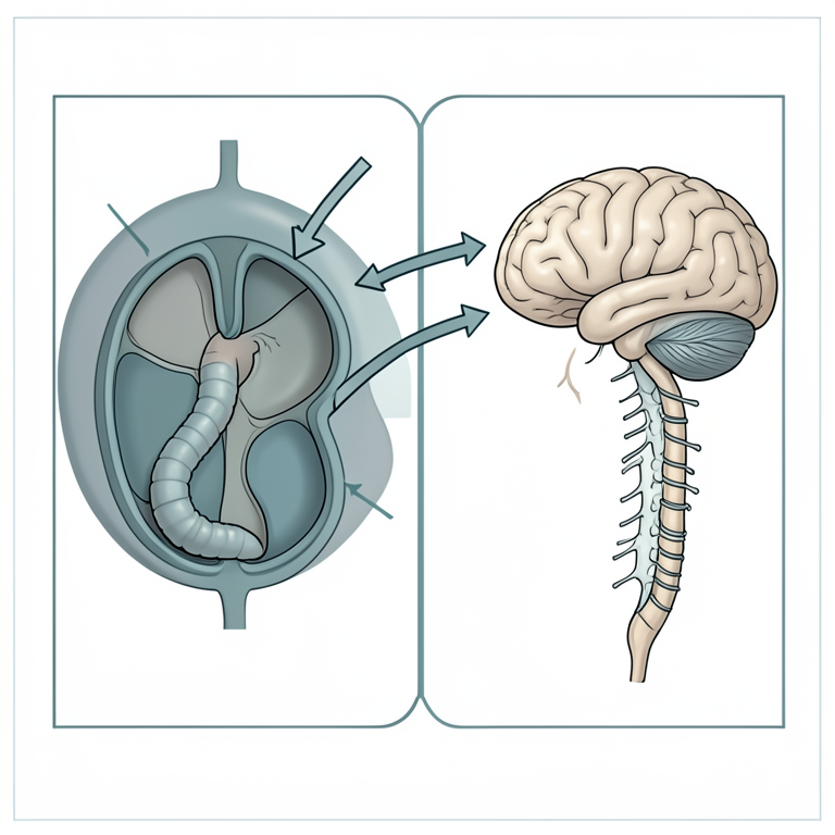
Aberrant Migration of Setaria digitata
AI-generated conceptual illustration (cattle (Bos taurus) and Setaria digitata nematodes). It explains the idea and does not show study data.
View source evidence ↗Figures & tables
From the original paper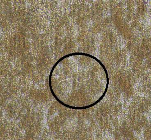 Figure 1
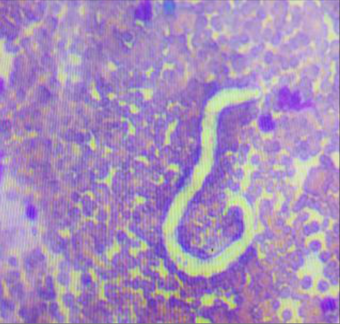 Figure 2
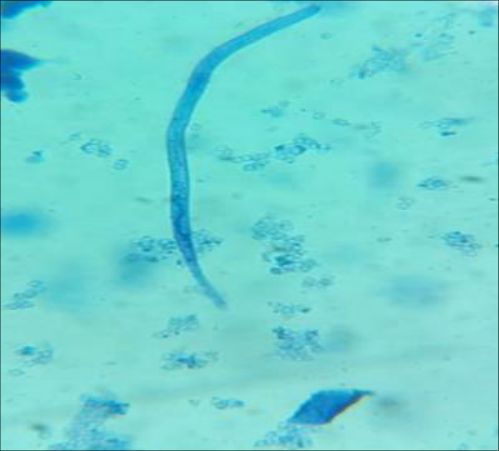 Figure 3
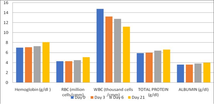 Figure 4
Introduction
Setaria digitata is a filarial nematode belonging to the family Onchocercidae that primarily parasitizes bovines, particularly cattle and buffaloes. In its natural hosts, the adult worms inhabit the peritoneal cavity, while the larval stages (microfilariae) circulate in the peripheral bloodstream. These adult worms are generally considered non-pathogenic in their usual anatomical location, although heavy infections may occasionally induce mild fibrinous peritonitis (Ronghang and Roy, 2015) [8] . The life cycle of S. digitata involves mosquito vectors, mainly species belonging to the genera Aedes, Culex and Anopheles, which act as intermediate hosts. Microfilariae ingested during a blood meal develop into infective larvae within the mosquito and are subsequently transmitted to susceptible hosts during subsequent feeding (Yadav and Tandon, 1991) [15] . Although bovines serve as the natural definitive hosts, the infective larvae may undergo aberrant migration in atypical hosts such as sheep, goats and horses (Soulsby, 1986) [12] . In these hosts, the larvae can invade the Central Nervous System (CNS), producing a neurological condition termed cerebrospinal nematodiasis, commonly referred to as "kumri" or lumbar paralysis. This disease is characterized by severe neurological manifestations including ataxia, hind limb paralysis, motor incoordination, drooping of eyelids or ears and varying degrees of paresis (Perumal et al.,2016) [6] . Cerebrospinal nematodiasis caused by S. digitata is of considerable veterinary importance in tropical and subtropical regions, where mosquito vectors are abundant. The disease has been reported in several Asian countries including India, Sri Lanka and Nepal, particularly affecting goats and horses. The pathogenesis primarily involves aberrant migration of developing larvae through neural tissues, leading to inflammation and damage within the spinal cord and brain, which ultimately results in neurological dysfunction (Senanayake et al., 2020) [9,10] . In cattle, infections with S. digitata are usually asymptomatic because the parasite remains confined to the peritoneal cavity. However, sporadic ectopic migrations and unusual clinical manifestations have occasionally been documented. Despite the widespread presence of this parasite in bovines, reports of cerebrospinal nematodiasis occurring in cattle are extremely rare, especially in southern India. Therefore, the present communication describes a rare occurrence of cerebrospinal nematodiasis in cattle from the Cauvery delta region of Tamil Nadu and its therapeutic management, thereby contributing to the existing knowledge on the aberrant pathogenic potential of S. digitata in its natural host.
Case history and clinical observations
A 4-year-old crossbred Jersey cow was presented to the Large Animal Medicine Referral Clinic, Veterinary Clinical Complex, VCRI, Orathanadu, TANUVAS with the history of circling, staggering gait, shivering of the hindquarters, excessive salivation, muscle twitching and hyperactivity for the past one week, without any apparent alteration in feed intake. A detailed clinical examination was performed, including musculoskeletal and neurological assessments. Cranial nerve evaluation revealed intact pupillary light reflexes bilaterally (both direct and consensual), normal menace response with intact palpebral reflexes. No abnormalities such as nystagmus or strabismus were observed. General physical examination was followed by laboratory investigations including faecal examination, wet blood film examination and hematobiochemical analysis of blood and serum samples. For wet blood examination, a drop of fresh blood was placed on a clean, grease-free glass slide, covered with a coverslip and examined under low magnification for motile microfilariae (Fig. 1). Additionally, thick blood smears were prepared, air-dried and stained with Giemsa stain (1:20 dilution) for 30 minutes (Fig. 2). Based on preliminary findings, the case was tentatively diagnosed as microfilariasis. Confirmation was achieved using the Modified Knott's concentration technique, which demonstrated the presence of microfilariae in the peripheral blood (Watanabe et al., 2004) [16] .
Treatment and Discussion
Based on the morphological characteristics, the microfilariae were identified as Setaria spp. (Fig. 3). The microfilariae were slender, elongated, unsheathed, and exhibited tapering anterior and posterior ends, with a smooth body outline and indistinct nuclei, which are typical features described for Setaria digitata in bovine hosts. The posterior end appeared pointed without terminal nuclei, aiding differentiation from other filarid microfilariae reported in domestic animals (Soulsby 1982; Bowman, 2021) [2,12] . Coprological examination did not reveal any parasitic ova. Hematological analysis indicated mild anemia along with lymphopenia. Serum biochemical evaluation revealed hypoproteinemia at the time of presentation which may be attributed to chronic parasitic stress and associated metabolic alterations. Based on clinical signs, detection of circulating microfilariae, and morphological identification, the case was diagnosed as microfilariasis due to Setaria spp. The animal was treated with Ivermectin @ 200 µg/kg body weight, subcutaneously as a single dose. Supportive therapy included Vitamin B-complex @12 mL, intramuscularly once daily. Fluid therapy was provided using intravenous Dextrose Normal Saline @ 1500 mL. Anti-inflammatory support was achieved with Meloxicam @ 0.2 mg/kg body weight intramuscularly once daily for 3-5 days. The case was followed up for 21 days, during which gradual clinical improvement was observed, with complete resolution of neurological signs and no recurrence. Hematobiochemical parameters also showed progressive normalization over the follow-up period (Fig. 4). Cattle, buffalo and deer act as natural definitive hosts for Setaria spp., particularly Setaria cervi and Setaria digitata, in which adult worms typically reside in the peritoneal cavity with minimal or no clinical significance (Soulsby, 1982; Lanková et al., 2021) [5,12] . These infections are generally considered non-pathogenic or only mildly associated with chronic peritonitis in natural hosts. However, the biological behavior of the parasite changes significantly when larval stages aberrantly migrate. Infective larvae (L3) introduced via vector transmission may occasionally deviate from their normal migratory pathway and invade the central nervous system, leading to cerebrospinal nematodiasis (Senanayake et al., 2020) [9,10] . This condition has been well documented to cause severe neurological disease, including posterior paralysis particularly in aberrant hosts such as sheep, goats and horses, where it is frequently fatal (Bazargani et al.,
. Although rare, similar migration into the cerebrospinal cavity has also been reported in cattle, where larvae of S. digitata and related species have been directly observed in association with paralysis. The pathogenesis primarily involves mechanical damage due to larval migration through neural tissues, combined with inflammatory responses (Tung et al., 2003) [13] . These observations are in agreement with the present study, where the affected animal exhibited characteristic neurological manifestations suggestive of aberrant larval migration and associated neural tissue damage, further supporting the role of Setaria spp. in the pathogenesis of cerebrospinal nematodiasis in cattle. At the molecular level, parasite-derived and host-mediated enzymatic mechanisms appear to play a crucial role in tissue damage. Filarial parasites such as S. cervi produce proteolytic enzymes, including collagenases and other metalloproteinases, which facilitate tissue invasion and immune evasion (Singh and Rathaur, 2003; Pokharel et al., 2006) [11] . In parallel, host inflammatory responses during parasitic invasion of the central nervous system are associated with upregulation of Matrix Metalloproteinases (MMPs). These enzymes degrade extracellular matrix components and disrupt structural integrity of neural tissues, including the myelin sheath, thereby contributing to demyelination and neurological dysfunction. The interaction between parasite-secreted proteases and host-derived MMPs likely exacerbates neural tissue injury, resulting in progressive paralysis (Bruschi and Pinto, 2013) [3] . Various pharmacological agents such as Levamisole, Diethylcarbamazine citrate and Anthimoline have been reported to be effective against microfilariasis in cattle and buffaloes, primarily by reducing circulating microfilarial load and limiting further transmission (Karki, 2008) [4] . These drugs act through different mechanisms, including neuromuscular paralysis of parasites and alteration of hostparasite interactions. However, Ivermectin has been widely documented as highly effective against circulating microfilariae, owing to its potent action on glutamate-gated chloride channels leading to parasite paralysis and death. The prolonged persistence in plasma further enhances its efficacy by maintaining therapeutic levels against microfilariae. Clinically, ivermectin administration is often associated with rapid reduction in parasitemia and marked improvement in clinical signs, frequently after a single dose, making it the drug of choice in field conditions (Urquhart et al., 1996; Bowman, 2021) [2,14] .
Acknowledgement
AcknowledgmentThe authors acknowledge The Dean, Veterinary College and Research Institute, Orathanadu, Tamilnadu and Tamilnadu Veterinary and Animal Sciences University, India to carry out the study successfully.
References
16 in the original paper- Bazargani T, et al. Cerebrospinal nematodiasis of cattle, sheep and goats in Iran. Iran J Parasitol. 2008;3(1):1620.
- Bowman DD. Georgis' parasitology for veterinarians. 11th ed. Elsevier; 2021.
- Bruschi F, Pinto B. The significance of matrix metalloproteinases in parasitic infections involving the central nervous system. Pathogens (Basel). 2013;2(1):105-129. DOI:10.3390/pathogens2010105.
- Karki K. A laboratory epidemiological outbreak investigation of Kumri (cerebrospinal nematodiasis) and use of diethylecarbamazin in treatment of goat in Banke district of Mid-Western Region of Nepal. Vet World. 2008;1(6):168-170.
- Lanková S, Vejl P, Melounová M, Čílová D, Vadlejch J, Miklisová D, et al. Setaria cervi (Filarioidea, Onchocercidae) undressing in ungulates: altered morphology of developmental stages, their molecular detection and complete sequence cox1 gene. Parasitology. 2021;148(5):598-611. DOI:10.1017/S0031182020002449.
- Perumal ANI, Gunawardene YINS, Dassanayake RS. Setaria digitata in advancing our knowledge of human lymphatic filariasis. J Helminthol. 2016;90(2):129-138. DOI:10.1017/S0022149X15000309.
- Pokharel DR, Rai R, Kumar P, Chaturvedi CM, Rathaur S. Tissue localization of collagenase and leucine aminopeptidase in the bovine filarial parasite Setaria cervi. Filaria J. 2006;5:7-7. DOI:10.1186/1475-2883-57.
- Ronghang B, Roy B. Filarial infections in semi-wild cattle of Northeast India: first record of Setaria digitata (von Linstow 1906) in Bos frontalis (Lambert 1804). J Parasit Dis. 2015;39(4):693-696. DOI:10.1007/s12639013-0404-4.
- Senanayake KS, Söderberg J, Põlajev A, Malmberg M, Karunanayake EH, Tennekoon KH, et al. The genome of Setaria digitata: a cattle nematode closely related to human filarial parasites. Genome Biol Evol. 2020;12(2):3971-3976. DOI:10.1093/gbe/evaa017.
- Senanayake KS, Söderberg J, Põlajev A, Malmberg M, Karunanayake EH, Tennekoon KH, et al. The genome of Setaria digitata: a cattle nematode closely related to human filarial parasites. Genome Biol Evol. 2020;12(2):3971-3976. DOI:10.1093/gbe/evaa017.
- Singh RN, Rathaur S. Setaria cervi: in vitro released collagenases and their inhibition by Wuchereria bancrofti infected sera. J Helminthol. 2003;77(1):77-81. DOI:10.1079/JOH2002164.
- Soulsby EJL. Helminths, arthropods and protozoa of domesticated animals. 7th ed. Baillière Tindall; 1982.
- Tung KC, Lai CH, Ooi HK, Yang CH, Wang JS. Cerebrospinal setariosis with Setaria marshalli and Setaria digitata infection in cattle. J Vet Med Sci. 2003;65(9):977-983. DOI:10.1292/jvms.65.977.
- Urquhart GM, Armour J, Duncan JL, Dunn AM, Jennings FW. Veterinary parasitology. 2nd ed. Blackwell Science; 1996.
- Yadav AK, Tandon V. Scanning electron microscopy of Setaria digitata (Nematoda: Filaroidea). J Vet Parasitol. 1991;5:76-80.
- Watanabe T, Kitani A, Murray PJ, Strober W. NOD2 is a negative regulator of Toll-like receptor 2-mediated T helper type 1 responses. Nature immunology. 2004 Aug 1;5(8):800-8.