
Research article · 2026 · Full text
International Journal of Biology SciencesPathological Investigation of Organ-Specific Lesions in Poultry Infected with Newcastle Disease
Volume 8(6) · Pages 40–45 · DOI: 10.33545/26649926.2026.v8.i6a.859 ↗
Research areasVirology and Viral DiseasesBird parasitology and diseasesAnimal Virus Infections Studies
Abstract
Author-writtenThis study was aimed to evaluate the pathological features of naturally occurring Newcastle disease in broiler chickens reared in commercial broiler farms of Srinagar and Ganderbal District of Kashmir during the year 2025. A total number of eight outbreaks were recorded across different poultry units and carcasses of dead birds were submitted to the Division of Veterinary Pathology for post-mortem examination. The diagnosis was confirmed based on characteristic pathomorphological changes observed in visceral organs, including proventriculitis, pneumonia, hemorrhagic enteritis, bursitis, nephritis, splenitis, encephalitis and lymphoid depletion in lymphoid tissues of affected birds. These findings emphasize that Newcastle disease remains a significant economic concern for the poultry industry, underscoring the need for continuous surveillance and preventive measures in poultry farms.
Summary
Source-checkedPlain-language reading of the full paper, prepared with AI and checked against the text. Each point links to the authors' own words.
Why it matters
- Newcastle disease is a highly contagious viral infection in birds that causes significant illness and death within the poultry industry. View evidence ↗
- In Jammu and Kashmir, the disease is a primary cause of economic losses for poultry farmers. View evidence ↗
- Depending on the strain's virulence and host susceptibility, the disease can potentially cause up to 100% morbidity and mortality. View evidence ↗
The question
- The study aimed to evaluate pathological changes in the organs of infected broiler chickens in Kashmir due to a lack of existing information. View evidence ↗
What they did
- Researchers performed detailed post-mortem examinations on 100 broiler bird carcasses. View evidence ↗
- Tissue samples from the liver, kidney, spleen, thymus, bursa of Fabricius, gizzard, and proventriculus were collected for microscopic analysis. View evidence ↗
- Samples were fixed in 10% formalin and stained with Hematoxylin and Eosin using routine histological techniques. View evidence ↗
What they found
- The proventriculus showed consistent congestion, hyperemia, and multiple petechial hemorrhages on the mucosal surface. View evidence ↗
- Gross examination of the brain revealed swelling, edema, congestion, and frequent petechial hemorrhages. View evidence ↗
- The liver was typically enlarged and congested, appearing mottled with red areas of hemorrhage. View evidence ↗
- Kidneys exhibited enlargement, marked congestion, and visible surface hemorrhages. View evidence ↗
- The bursa of Fabricius commonly showed congestion, edema, a mottled appearance, and frequent hemorrhages. View evidence ↗
- The spleen exhibited pronounced lymphoid depletion, specifically in the white pulp, which impairs immune function. View evidence ↗
- The thymus showed severe necrosis and lymphoid depletion, resulting in significant organ atrophy. View evidence ↗
- Microscopic analysis of the brain showed encephalitis characterized by widespread inflammatory cell infiltration in neural tissue and perivascular spaces. View evidence ↗
- The proventriculus showed an inflammatory response with dense infiltration of heterophils, plasma cells, and lymphocytes. View evidence ↗
What it means
- The authors conclude that strict biosecurity measures are essential to reduce the impact of Newcastle disease on the poultry industry. View evidence ↗
- The authors state that understanding the disease's pathological features is critical for accurate diagnosis and effective prevention. View evidence ↗
Figures & tables
From the original paper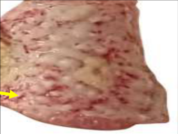 Figure 1A
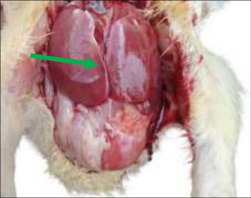 Figure 1B
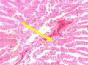 Fig2
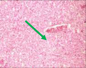 Figure 3
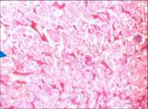 Figure 4
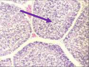 Figure 5
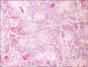 Figure 6
Introduction
Newcastle disease is a highly contagious and acute viral infection affecting a wide range of avian species, resulting in considerable morbidity and mortality in the poultry industry. In the Union Territory of Jammu and Kashmir, this disease is a major contributor to economic losses among poultry farmers, along with other bacterial, viral and fungal infections. It primarily affects both domestic and wild birds and is caused by the Newcastle disease virus (NDV), which belongs to the genus Avulavirus under the family Paramyxoviridae. The disease was first reported in Newcastle, England, in 1926, from which it derives its name. This disease affects multiple avian species including chickens, turkeys, pigeons, ducks, geese, as well as wild birds such as cormorants and sparrows. The transmission mainly occurs through direct contact with infected birds, although contaminated fences and respiratory secretions also serve as important sources of infection. Newcastle disease is considered one of the most devastating diseases of poultry, with the potential to cause up to 100% morbidity and mortality depending on the virulence of the strain and the susceptibility of the host species. The clinical severity ranges from mild respiratory signs to severe respiratory distress, neurological manifestations and high mortality. The common gross lesions include hemorrhages in the proventriculus, pneumonia, and thickening of air sacs, tracheal exudates, enteritis and splenitis. The disease has a worldwide distribution and is particularly prevalent in many Asian countries, including India. Due to limited available information on Newcastle disease in broiler chickens in Kashmir, this study was undertaken to evaluate the pathological alterations affecting the structure and function of various organs in infected broiler birds.
Materials and Methods
The study was carried out at the Division of Veterinary Pathology, SKUAST-Kashmir. During the study period, systematic visits were conducted to both organized and backyard poultry farms to monitor mortality patterns. A total of 100 carcasses of broiler birds were collected and submitted to the Division for detailed post-mortem examination. Diagnosis was confirmed based on the evaluation of gross and histopathological lesions observed in various organs. Necropsies were performed in accordance with the standard procedures described by Calnek (1997) [4] and all pathological findings were carefully recorded. The tissue samples from different organs exhibiting characteristic lesions including the proventriculus, gizzard, bursa of Fabricius, spleen, thymus, kidney and liver were collected for microscopic examination. The collected samples were fixed in 10% formalin, processed by using routine histological techniques and stained with Hematoxylin and Eosin following the method outlined by Lillie (1954) [9] .
Results
The pathological findings of this investigation are systematically described under clinical, gross and microscopic observations recorded in poultry infected with Newcastle disease. Gizzard: The gizzard shows varying degrees of epithelial damage, including erosions and ulcerations that appear as irregular disruptions of the koilin layer. Congestion is commonly present, imparting a reddish discoloration to the organ. The inflammatory processes often result in thickening of the muscular wall.
Hemorrhages may be visible as red patches within the mucosa or submucosa. In severe cases, fibrinous exudates may accumulate either on the surface or within the lumen. Additionally, the presence of lymphoid nodules, formed due to lymphocyte proliferation, indicates an active immune response to viral infection. Brain: Grossly, the brain is revealing congestion, edema and swelling. Petechial hemorrhages are frequently seen on the brain surface and within the parenchyma. Necrotic areas may develop due to neuronal damage. In advanced stages, the meninges often show thickening and fibrinous exudation, suggesting severe inflammation. Sometimes ventriculitis may be observed in the affected birds as characterized by fluid accumulation within the Brian ventricles, along with demyelination of nerve fibers, contributing to neurological dysfunction. Lungs: Pulmonary lesions include marked congestion and haemorrhage, often accompanied by edema that leads to swelling and impaired respiratory function. In severe infections, areas of consolidation may develop, indicating loss of normal lung architecture. The necrotic foci may also be present within the lung tissue. The air sacs frequently appear thickened and may contain caseous or fibrinous material, indicative of airsacculitis and chronic inflammatory processes. Heart|: The heart may exhibit cardiomegaly due to vascular congestion and accumulation of blood. Hemorrhages are commonly observed within the myocardium and focal areas of necrosis may develop as a result of tissue damage. In some cases, pericardial effusion is evident, characterized by the accumulation of serous or fibrinous fluid within the pericardial sac. These changes may impair cardiac function and contribute to systemic circulatory disturbances. Liver: The liver is often enlarged and congested, displaying a characteristic mottled appearance with areas of red discoloration due to haemorrhage. The multiple necrotic foci may be scattered throughout the parenchyma, giving the organ a friable consistency. In some cases, the liver appears swollen and rounded with granular surface changes. These alterations reflect hepatocellular injury, vascular disturbances and inflammatory responses associated with the infection. Kidneys: Renal lesions are characterized by enlargement and marked congestion, with visible hemorrhages on the surface. Necrotic areas may be present within the renal tissue, indicating cellular damage. In chronic cases, fibrosis may develop, resulting in a rough, irregular and scarred appearance of the kidneys. These structural changes can significantly impair renal function. Spleen: The spleen typically shows enlargement, congestion and a mottled appearance. Hemorrhages and necrotic lesions are frequently observed. Fibrin deposits may form nodular structures on the surface of the spleen. In severe infections, lymphoid depletion occurs, leading to splenic atrophy and reduced immunological function, making the birds more susceptible to secondary infections. Thymus: The thymus often undergoes atrophy, appearing pale and mottled in appearance. Hemorrhagic areas may be present, indicating vascular damage. Edema contributes to swelling of the organ, while necrosis may develop in advanced stages of the disease. These changes compromise the thymus's role in T-cell production, thereby weakening the immune response. Bursa of Fabricius: The bursa commonly exhibits edema, congestion and a mottled appearance. Hemorrhages are frequently present and in severe cases, mucosal striations and fibrin deposits may be observed. Gelatinous or serous exudate may accumulate within the lumen. In extreme cases, rupture of the bursa can occur, potentially leading to peritonitis due to leakage of infectious material into the abdominal cavity.
Discussion
Newcastle disease, commonly referred to as Ranikhet disease, is a highly infectious viral condition that affects both organized and backyard poultry systems. It is caused by virulent strains of avian avulavirus-1, also known as Newcastle disease virus (NDV) or avian paramyxovirus-1 (APMV-1). The disease is characterized by high rates of illness and death, particularly in unvaccinated flocks. Apart from domestic birds, numerous wild and captive avian species are also susceptible and can serve as reservoirs of infection. The transmission between infected chickens and pigeons, both domestic and wild, has also been documented (Alexander et al., 2005) [3] . The neurological signs observed in affected birds are mainly due to viral multiplication within the brain, resulting in neuronal damage and vascular injury. This triggers an inflammatory response marked by macrophage infiltration around blood vessels, which may extend to astrocytes and microglial cells. Similar observations have been reported in earlier studies, where encephalitis was consistently identified in infected birds. Clinically, affected birds show depression, lethargy, reduced feed intake, clustering behaviour, drooping wings, respiratory distress, lowered head carriage, ruffled feathers and weakness. In the early phase, yellowish diarrhoea is commonly seen, while surviving birds often display poor growth, reduced activity and a characteristic posture with closed eyes and fluffed feathers (Yadav et al. 2024 & Zachary et al., 2012) [16,21] . The proventriculus, which serves as the glandular stomach in poultry, is frequently involved in Newcastle disease, particularly in infections caused by highly virulent strains. Grossly, lesions include congestion and hemorrhages of the mucosa, with erosions and ulcerations in severe cases (Saidu et al., 2009 & Rafiq et al. (2023) [11,14] . Microscopically, there is significant infiltration of inflammatory cells, especially lymphocytes, along with necrosis of the glandular epithelium (King et al., 2009 & Naikoo et al. (2025) [8,17] . Although less commonly affected, the gizzard may exhibit hemorrhages, erosions and necrotic lesions, with mononuclear inflammatory cell infiltration evident around these areas. In the central nervous system, lesions such as perivascular cuffing, neuronal degeneration, gliosis and microglial proliferation are frequently observed (Susta et al., 2011) [18] , correlating with neurological signs like tremors, incoordination and paralysis. Pulmonary involvement typically includes congestion and haemorrhage, while histological examination reveals severe interstitial pneumonia with infiltration of lymphocytes, plasma cells and macrophages (Rafiq et al. (2024) [15] . Advanced cases may also demonstrate necrosis and haemorrhage of the respiratory epithelium (Miller et al., 2011) [18] . Cardiac involvement is characterized by myocarditis, with hemorrhages affecting both the epicardial and myocardial layers. Histologically, infiltration of mononuclear cells and multifocal necrosis of cardiac muscle fibers are commonly observed, which may compromise cardiac function and contribute to mortality (El-Tayeb et al., 2012) [6] . The liver often appears mottled due to congestion and haemorrhage and microscopically shows disrupted architecture, dilated sinusoids, hepatocellular degeneration and inflammatory infiltration (Wang et al., 2012) [19] .
Renal lesions typically include enlargement, congestion and occasional hemorrhages. Histopathological findings such as tubular degeneration, necrosis and interstitial lymphocytic infiltration may impair kidney function, clinically reflected by dehydration and increased uric acid levels (Kim et al., 2007 & Shafi et al. 2023). [7,12] Lymphoid organs including the caecal tonsils, spleen, thymus, and bursa of Fabricius are markedly affected, exhibiting lymphoid depletion that leads to immunosuppression ( Miller & Koch, 2013) [10] . Lesions in the crop are characterized by erosions and ulcerations of varying severity, sometimes forming raw surfaces, while the gizzard may show superficial erosions (Alexander et al., 2016) [1] . The intestines frequently exhibit severe hemorrhages in both mucosal and submucosal layers. [5,12] . These observations are in agreement with previous studies and reinforce the multisystemic nature of Newcastle disease, emphasizing its profound impact on poultry health and production.
Conclusion
Newcastle disease remains a significant viral infection of poultry, producing diverse pathological changes that vary with disease severity and the organs involved, ultimately leading to substantial economic losses, particularly for small-scale farmers. A clear understanding of its pathological features is essential for accurate diagnosis, effective control and prevention in avian populations. Therefore, strict implementation of appropriate biosecurity measures is essential to reduce the overall impact of this disease on the poultry industry.
Figures and tables from the paper
References
21 in the original paper- Alexander DJ. Newcastle disease and other avian paramyxoviruses. Rev Sci Tech. 2016;19(2):443-462.
- Akamatsu M, Sekizuka T, Horie M, Noda T, Kawaoka Y. Pathogenesis of Newcastle disease virus in chickens. J Comp Pathol. 2008;139(4):203-215.
- Alexander DJ. Newcastle disease. In: Saif YM, editor. Diseases of Poultry. 11th ed. Ames (IA): Blackwell Publishing; 2005. p. 63-87.
- Calnek BW. Diseases of Poultry. 10th ed. Ames (IA): Iowa State University Press; 1997.
- Dimitrov KM, Ramey A, Afonso CL. Updated classification and phylogenetic analysis of Newcastle disease virus. Infect Genet Evol. 2016;39:216-230.
- El-Tayeb B, Abd El-Hamid HS, Abd El-Aziz NK. Pathological and immunohistochemical studies on Newcastle disease. J Vet Diagn Invest. 2012;24(4):785793.
- Kim SH, et al. Pathobiology of Newcastle disease virus infection. Avian Pathol. 2007;36(2):131-139.
- King DJ, Seal BS. Biological and molecular characterization of Newcastle disease virus. Avian Dis. 2009;53(1):1-9.
- Lillie RD. Histopathologic Technic and Practical Histochemistry. New York (NY): McGraw-Hill; 1954.
- Miller PJ, Koch G. Newcastle disease. In: Diseases of Poultry. 13th ed. Ames (IA): Wiley-Blackwell; 2013.
- Saidu L, Abdu PA, Umoh JU, Abdullahi SU. Pathological findings in experimental Newcastle disease in chickens. Int J Poult Sci. 2009;8(3):263-267.
- Shafi M, Shah S, Bashir S, Kamil S, Mir M, Shah F, Mudasir S. Nephro-pathological hallmarks of metformin induced toxicity in Wistar rats. Pharma Innov J. 2023;12(6):4248-4255.
- Shafi M, Jan S, Basher A, Shah S, Kamil S, Mir M, Shah F, Mudasir S. Clinico-pathological assessment of Newcastle disease in broiler chicken. Pharma Innov J. 2023;12(8):1436-1449.
- Rafiq M, Kamil SA, Yaqub M, Shah SA, Kawoosa MS, Beigh AB. Newcastle disease in chicken of Kashmir Valley. Int J Vet Sci Anim Husb. 2023;9(3):1071-1073.
- Rafiq M, Goswami P, Yaqub M, Shafi M, Kamil SA, Shah SA. Nephropathy associated with bacterial diseases in broiler chicken. Indian J Vet Pathol. 2024;48(2):169-175.
- Yadav PK, Shah SA, Shafi M, et al. Etiological and histomorphological studies on early chick mortality. Open Vet J. 2024;14(11):3037-3046.
- Naikoo AH, Shafi M, Wani BM, et al. Molecular detection of inclusion body hepatitis in broilers. J Livest Sci. 2025;16(2):301-306.
- Susta L, Miller PJ, Afonso CL. Clinicopathological characterization of Newcastle disease virus strains. Vet Pathol. 2011;48(3):586-596.
- Wang Z, Ma B, Liu W. Pathogenesis of Newcastle disease virus in chickens. Vet Microbiol. 2012;158(12):1-9.
- World Organisation for Animal Health. Newcastle disease (infection with Newcastle disease virus). OIE Terrestrial Manual. Paris: World Organisation for Animal Health; 2021.
- Zachary JF. Pathologic Basis of Veterinary Disease. 5th ed. St. Louis (MO): Elsevier; 2012.