
Research article · 2026 · Full text
International Journal of Biology SciencesThe role of Visfatin and immunological biomarkers in patients with myocardial infarction in Ramadi city
Volume 8(6) · Pages 33–39 · DOI: 10.33545/26649926.2026.v8.i6a.858 ↗
Research areasCardiac Fibrosis and RemodelingAcute Myocardial Infarction ResearchAdipokines, Inflammation, and Metabolic Diseases
Abstract
Author-writtenObjectives: This study aimed to investigate the clinical significance of Visfatin and selected immunological biomarkers in conjunction with conventional cardiac biomarkers among patients diagnosed with myocardial infarction (MI) in Ramadi City, Iraq. Methods: A case-control study was conducted between November 2023 and January 2024, involving 80 participants aged 45-70 years. The study cohort included 50 patients with clinically confirmed myocardial infarction and 30 apparently healthy individuals who served as the control group. Serum levels of Creatine Kinase-MB (CK-MB), Troponin-T, N-terminal pro-brain natriuretic peptide (NT-proBNP), and Visfatin were measured. In addition, plasma concentrations of interleukin-32 (IL-32) and interleukin-37 (IL-37) were determined using standardized immunoassay techniques. Statistical analyses were performed to compare biomarker levels between patients and controls. Results: Patients with myocardial infarction demonstrated significantly elevated serum levels of CK-MB, Troponin-T, and NT-proBNP compared with healthy controls (P ≤ 0.0001). Furthermore, plasma concentrations of IL-32 and IL-37 were markedly increased in the MI group (P ≤ 0.0001), indicating enhanced inflammatory and immunological activity. In contrast, serum Visfatin levels were significantly decreased in patients with myocardial infarction relative to the control group (P ≤ 0.0001). Conclusions: The present findings indicate that IL-32, IL-37, and NT-proBNP are strongly associated with myocardial infarction and may serve as valuable diagnostic and prognostic biomarkers. The observed reduction in Visfatin levels among MI patients suggests a potential protective or regulatory role in cardiovascular pathology and highlights its possible utility as a novel biomarker for myocardial infarction in the Iraqi population.
Summary
Source-checkedPlain-language reading of the full paper, prepared with AI and checked against the text. Each point links to the authors' own words.
Why it matters
- Cardiovascular diseases are a significant global public health issue, serving as a primary cause of death and illness. View evidence ↗
- Myocardial infarction occurs when coronary artery obstruction leads to prolonged ischemia, causing permanent damage to heart muscle cells. View evidence ↗
- Inflammatory processes are central to how myocardial injury begins and advances. View evidence ↗
The question
- The study aimed to evaluate the clinical importance of Visfatin and specific immunological biomarkers alongside standard cardiac markers in patients with myocardial infarction in Ramadi City, Iraq. View evidence ↗
What they did
- This case-control study took place from November 2023 to January 2024, including 50 patients with confirmed myocardial infarction and 30 healthy controls, all aged between 45 and 70 years. View evidence ↗
- Researchers measured serum levels of CK-MB, Troponin-T, NT-proBNP, and Visfatin, as well as plasma levels of IL-32 and IL-37 using standardized immunoassays. View evidence ↗
- Data were analyzed using GraphPad Prism 7 and SPSS 24, with results expressed as mean ± SD and significance set at p ≤ 0.05. View evidence ↗
What they found
- Patients with myocardial infarction had significantly higher levels of Troponin-T (6.174 ± 0.8137 ng/mL vs 0.213 ± 0.0462 ng/mL) and CK-MB (196.87 ± 40.14 IU/mL vs 81.32 ± 32.18 IU/mL) than controls. View evidence ↗
- NT-proBNP levels were markedly higher in the myocardial infarction group (1512.5 ± 95.2 pg/mL) compared to the control group (33.8 ± 21.04 pg/mL). View evidence ↗
- Unlike other markers, serum Visfatin levels were significantly lower in patients with myocardial infarction (11.92 ± 4.641 ng/mL) than in healthy controls (19.61 ± 3.756 ng/mL). View evidence ↗
- Plasma levels of IL-32 (318.83 ± 4.591 pg/mL vs 88.98 ± 3.453 pg/mL) and IL-37 (245.36 ± 33.08 pg/mL vs 26.34 ± 43.84 pg/mL) were significantly increased in MI patients. View evidence ↗
- ROC analysis showed that Troponin-T (AUC 1.000), CK-MB (AUC 0.9859), IL-37 (AUC > 0.90), and Visfatin (AUC > 0.90) had high diagnostic accuracy, while NT-proBNP (AUC 0.8479) was good and IL-32 (AUC 0.6170) was moderate. View evidence ↗
What it means
- The authors conclude that the reduction of Visfatin in MI patients suggests it could be a novel biomarker for diagnosing and assessing risk of myocardial infarction in the Iraqi population. View evidence ↗
- The study emphasizes that both inflammatory and cardiac biomarkers play an important role in the pathophysiology of myocardial infarction. View evidence ↗
- The authors recommend further large-scale, multicenter studies to better understand the molecular mechanisms and therapeutic potential of Visfatin, IL-32, and IL-37. View evidence ↗
Visual summary
3 visualsIllustrations explain the paper; charts are replotted from the paper's own table values.
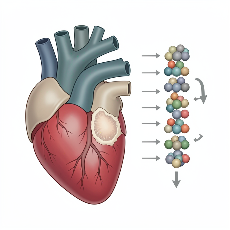
Biomarker Directionality in MI
AI-generated conceptual illustration (human heart tissue). It explains the idea and does not show study data.
View source evidence ↗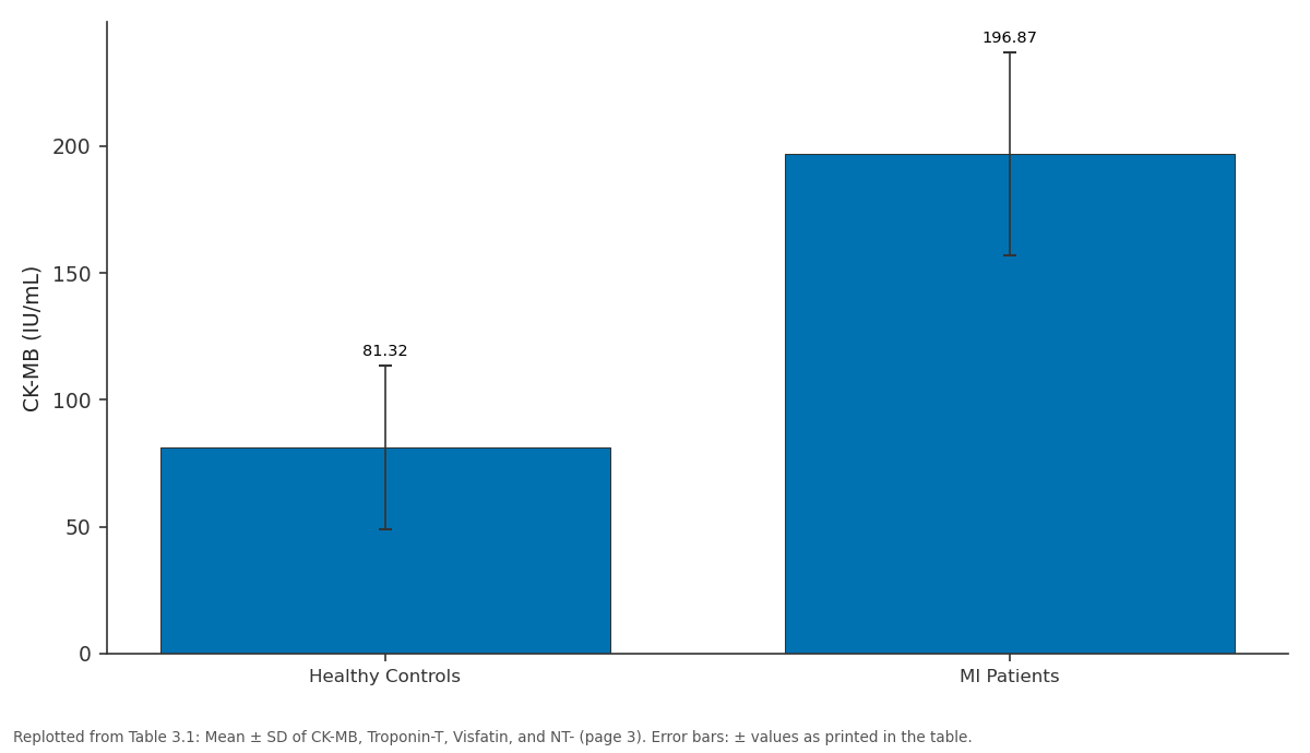
CK-MB (IU/mL): Healthy Controls vs MI Patients
Replotted from Table 3 (page 3) of the original paper. Every bar is the paper's own table value.
View source evidence ↗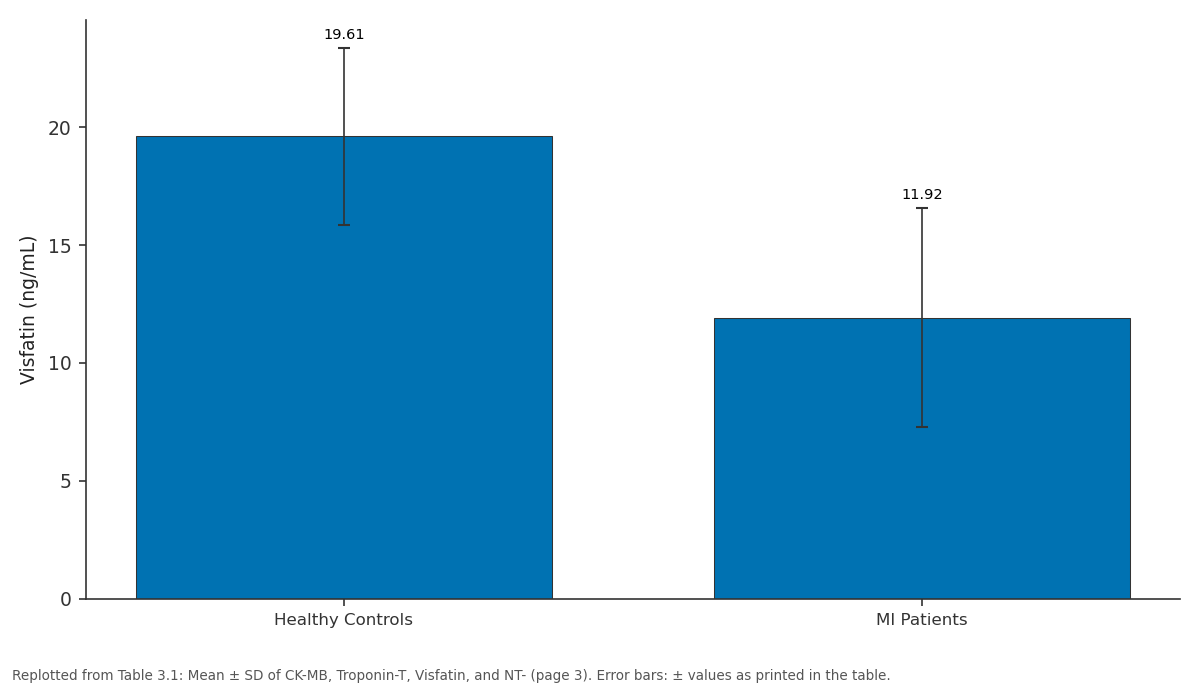
Visfatin (ng/mL): Healthy Controls vs MI Patients
Replotted from Table 3 (page 3) of the original paper. Every bar is the paper's own table value.
View source evidence ↗Figures & tables
From the original paper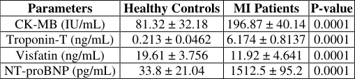 Table 3
 Table 3
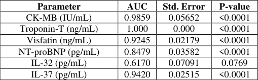 Table 3
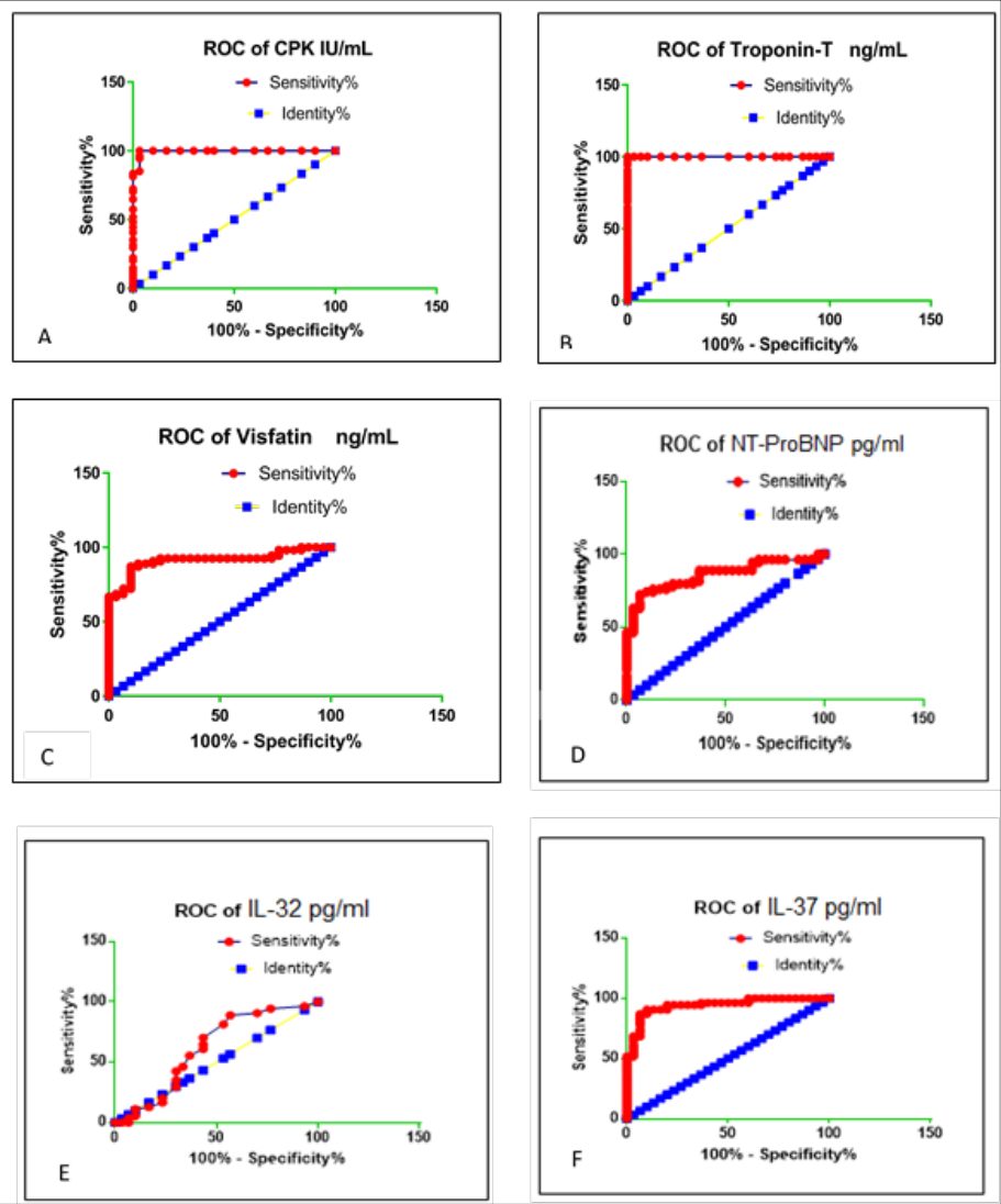 Figure 3
Introduction
Cardiovascular diseases (CVDs) remain one of the leading causes of morbidity and mortality worldwide and represent a major public health challenge. Although the prevalence of CVD has declined in many developed countries due to advances in preventive and therapeutic strategies, risk factors such as obesity, diabetes mellitus, sedentary lifestyle, and metabolic disorders continue to contribute significantly to disease burden and mortality rates [1,2] . Myocardial infarction (MI) is characterized by irreversible myocardial cell injury resulting from prolonged ischemia caused by coronary artery obstruction. Early ultrastructural alterations following ischemia include depletion of cellular glycogen, myofibrillar relaxation, and disruption of the sarcolemma within minutes after the ischemic event [3] . Acute myocardial infarction (AMI) may lead to severe complications, including cardiogenic shock, which is associated with impaired cardiac output, tissue hypoperfusion, and systemic hypoxia [4] . Inflammation plays a fundamental role in the initiation and progression of myocardial injury. Acute ischemic damage promotes the recruitment of inflammatory cells and stimulates the release of numerous cytokines involved in immune and inflammatory responses [5] . Excessive production of inflammatory mediators contributes to endothelial dysfunction, cardiomyocyte apoptosis, and deterioration of cardiac function [6,7] . Consequently, inflammatory biomarkers have attracted increasing attention as potential indicators of cardiovascular injury and prognosis.
Interleukin-37 (IL-37), a member of the IL-1 cytokine family, is recognized as an anti-inflammatory mediator with immunosuppressive properties. It regulates innate and adaptive immune responses and has demonstrated protective effects against inflammatory disorders in experimental models [8][9][10] . IL-37 is produced by several cell types, including macrophages, endothelial cells, smooth muscle cells, and dendritic cells [11] . Previous studies have suggested a potential association between elevated IL-37 levels and cardiovascular diseases, particularly acute myocardial infarction [12,13] . Interleukin-32 (IL-32) is another multifunctional cytokine involved in inflammatory signaling, apoptosis, and immune regulation. IL-32 has been implicated in atherosclerotic plaque instability through stimulation of matrix metalloproteinases and activation of inflammatory pathways such as nuclear factor kappa-B (NF-κB) and p38 mitogenactivated protein kinase (MAPK) (14-16). Furthermore, IL-32 may contribute to myocardial remodeling and fibrosis by enhancing the expression of procollagen and transforming growth factor-beta (TGF-β), thereby increasing the risk of adverse cardiac events following MI [17] . Cardiac biomarkers remain essential tools in the diagnosis and assessment of myocardial infarction. Cardiac troponin-T (cTn-T) is considered the gold standard biomarker for myocardial injury due to its high sensitivity and specificity, particularly with the development of high-sensitivity assays [18] . Creatine kinase-MB (CK-MB) also plays a valuable role in evaluating myocardial necrosis, although troponins demonstrate superior diagnostic performance [19] . In addition, N-terminal pro-brain natriuretic peptide (NT-proBNP) is widely used to assess cardiac dysfunction and ventricular stress, especially in patients with heart failure and acute coronary syndromes [20,21] . Visfatin, also known as nicotinamide phosphoribosyltransferase (NAMPT), is an adipocytokine involved in inflammatory and metabolic pathways. It has been associated with endothelial dysfunction, vascular inflammation, and atherosclerosis [22,23] . Visfatin may contribute to cardiovascular pathogenesis through its proinflammatory activity and its effects on vascular smooth muscle cells and endothelial integrity. However, studies investigating visfatin levels in myocardial infarction have reported inconsistent findings, suggesting the need for further investigation, particularly in different populations [24,25] . Therefore, the present study aimed to evaluate serum Visfatin levels and selected immunological biomarkers, including IL-32 and IL-37, in patients with myocardial infarction in Ramadi City, Iraq, and to investigate their association with conventional cardiac biomarkers and disease status.
Materials and Methods
Materials and Methods
Study Population Healthy Control Group
The healthy control group (Group B) consisted of 30 apparently healthy Iraqi males and females matched to the patient group in terms of age and gender. Individuals included in the control group had no history of myocardial infarction, ischemic heart disease, hypertension, diabetes mellitus, smoking, or alcohol consumption. In addition, none of the participants had acute or chronic inflammatory diseases, severe systemic disorders, or active infections at the time of sample collection. All healthy controls were free from medication use and had no clinically diagnosed medical conditions.
Myocardial Infarction Group
The patient group (Group A) included 50 individuals diagnosed with myocardial infarction (MI), with ages ranging from 45 to 70 years. Patients were recruited from the Cardiac Care Units (CCU) of the General Teaching Hospitals in Ramadi and Fallujah between November 2023 and January 2024. Diagnosis of MI was confirmed by specialist physicians based on clinical examination, electrocardiographic findings, and cardiac biomarker evaluation. Blood samples were collected within 48 hours after the onset of myocardial infarction following informed consent from all participants.
Blood Sample Collection
Venous blood samples were collected from both patients and healthy controls in the early morning between 8:00 and 10:30 a.m. after an overnight fasting period of approximately 12 hours. A total of 8 mL of venous blood was obtained from each participant under aseptic conditions. Blood samples were transferred into plain gel tubes without anticoagulant and allowed to clot at room temperature (23-27°C) for approximately one hour. Serum was separated by centrifugation at 3000 ×g for 15 minutes and subsequently stored at -20°C until biochemical and immunological analyses were performed.
Biochemical and Immunological Analysis Determination of Creatine Kinase-MB (CK-MB)
Serum CK-MB levels were measured using the FUJI DRI-CHEM ANALYZER according to the manufacturer's instructions. Quality control procedures were performed before sample analysis using the appropriate quality control card. Serum samples were loaded onto the analyzer, and the assay was performed automatically following entry of sample identification and sequence numbers.
Determination of Cardiac Troponin-T (cTn-T)
Serum cardiac Troponin-T concentrations were determined using the ichroma™ immunofluorescence assay system according to the manufacturer's protocol. Briefly, 75 μL of serum sample was mixed with 75 μL of detection buffer and vortexed thoroughly. Subsequently, 75 μL of the prepared mixture was dispensed into the cartridge well and incubated at 23-27°C for 12 minutes. The cartridge was then inserted into the ichroma™ Reader for automated fluorescence measurement and result interpretation.
Determination of Serum Visfatin Levels
Serum Visfatin concentrations were measured using a commercially available enzyme-linked immunosorbent assay (ELISA) kit (Catalog No: E0025Hu) according to the manufacturer's instructions. All reagents and samples were equilibrated to room temperature (18-25°C) prior to analysis. Briefly, standards and serum samples were added to ELISA plate wells followed by anti-Visfatin antibody and streptavidin-HRP conjugate. After incubation at 37°C for 60 minutes, the plate was washed five times using wash buffer. Substrate solutions A and B were added sequentially, and the plate was incubated in the dark for 10 minutes. The enzymatic reaction was terminated by adding stop solution, and absorbance was measured at 450 nm using a microplate reader.
Measurement of N-terminal Pro-Brain Natriuretic Peptide (NT-proBNP)
Serum NT-proBNP levels were measured using the Cobas e 411 analyzer (Roche Diagnostics, Germany) based on the electrochemiluminescence sandwich immunoassay technique. Device calibration was performed using CAL1 and CAL2 calibration solutions prior to sample analysis. Serum samples were loaded into the sample wheel, and all analyses were conducted automatically according to the manufacturer's instructions.
Measurement of Plasma IL-32 and IL-37 Levels
Plasma concentrations of interleukin-32 (IL-32) and interleukin-37 (IL-37) were quantified using commercially available enzyme-linked immunosorbent assay (ELISA) kits according to the manufacturers' protocols.
Statistical Analysis
Statistical analysis was performed using GraphPad Prism version 7 and the Statistical Package for Social Sciences (SPSS) version 24. Data were expressed as mean ± standard deviation (SD). Comparisons between groups were conducted using appropriate statistical tests, and a p-value ≤ 0.05 was considered statistically significant.
Results and Discussion
Serum Levels of CK-MB, Troponin-T, Visfatin, and NT-proBNP Table 3.1 presents the serum levels of CK-MB, Troponin-T, Visfatin, and NT-proBNP in myocardial infarction (MI) patients and healthy controls. Cardiac troponins are highly specific markers of myocardial injury due to their exclusive localization within cardiac muscle tissue. Following myocardial ischemia, disruption of cardiomyocyte membrane integrity results in the release of intracellular proteins into the circulation, including CK-MB and cardiac troponins [29,30] . Troponin-T typically rises within 6-8 hours after myocardial injury, reaches peak concentrations within 12-24 hours, and remains elevated for several days, making it a highly sensitive and specific biomarker for acute myocardial infarction [31,32] .
| Parameter | AUC | Std. Error | P-value |
|---|---|---|---|
| CK-MB (IU/mL) | 0.9859 | 0.05652 | <0.0001 |
| Troponin-T (ng/mL) | 1.000 | 0.000 | <0.0001 |
| Visfatin (ng/mL) | 0.9245 | 0.02179 | <0.0001 |
| NT-proBNP (pg/mL) | 0.8479 | 0.03582 | <0.0001 |
| IL-32 (pg/mL) | 0.6170 | 0.07091 | 0.0769 |
| IL-37 (pg/mL) | 0.9420 | 0.02515 | <0.0001 |
Table text extracted from the original PDF; superscript letters are shown inline. The original table image is below.
View original table image
| Parameters | Healthy Controls | MI Patients | P-value |
|---|---|---|---|
| IL-32 (pg/mL) | 88.98 ± 3.453 | 318.83 ± 4.591 | P < 0.001 |
| IL-37 (pg/mL) | 26.34 ± 43.84 | 245.36 ± 33.08 | P < 0.001 |
Table text extracted from the original PDF; superscript letters are shown inline. The original table image is below.
View original table image
| Parameters | Healthy Controls | MI Patients | P-value |
|---|---|---|---|
| CK-MB (IU/mL) | 81.32 ± 32.18 | 196.87 ± 40.14 | 0.0001 |
| Troponin-T (ng/mL) | 0.213 ± 0.0462 | 6.174 ± 0.8137 | 0.0001 |
| Visfatin (ng/mL) | 19.61 ± 3.756 | 11.92 ± 4.641 | 0.0001 |
| NT-proBNP (pg/mL) | 33.8 ± 21.04 | 1512.5 ± 95.2 | 0.0001 |
Table text extracted from the original PDF; superscript letters are shown inline. The original table image is below.
View original table image
Similarly, CK-MB remains an important indicator of myocardial necrosis, particularly during the early phase of infarction and for detection of reinfarction because of its shorter half-life and return to baseline within 36-48 hours [33] . The combined use of CK-MB and Troponin-T improves diagnostic sensitivity during the acute phase of myocardial infarction.
In contrast, serum Visfatin levels were significantly decreased in MI patients (11.92 ± 4.641 ng/mL) compared with healthy controls (19.61 ± 3.756 ng/mL) (P < 0.0001). These findings are consistent with previous studies reporting reduced Visfatin concentrations in patients with myocardial infarction [34] . However, contradictory findings have also been reported in the literature, where elevated Visfatin levels were associated with cardiovascular disease and atherosclerosis [35] .
Visfatin is a multifunctional adipocytokine involved in inflammatory regulation, endothelial dysfunction, and vascular remodeling. It is expressed in macrophages and foam cells within unstable atherosclerotic plaques and may contribute to plaque destabilization and inflammatory activation [36,37] . The reduction observed in Visfatin levels in the present study may be related to excessive inflammatory cell recruitment and altered metabolic responses during acute myocardial infarction. Serum NT-proBNP levels showed a marked elevation in MI patients compared with controls (1512.5 ± 95.2 pg/mL vs. 33.8 ± 21.04 pg/mL; P < 0.0001). NT-proBNP is secreted primarily in response to ventricular wall stress, myocardial ischemia, and impaired ventricular function. Elevated NT-proBNP concentrations reflect myocardial dysfunction and ventricular overload associated with acute myocardial infarction and heart failure [38] . The significant increase observed in the current study supports previous reports demonstrating that NT-proBNP is a valuable biomarker for assessing cardiac dysfunction, ventricular remodeling, and prognosis in MI patients [39] . Elevated NT-proBNP levels are strongly associated with increased mortality risk and reduced left ventricular function following myocardial infarction [40] .
Plasma Levels of IL-32 and IL-37 The present findings demonstrated a highly significant increase in plasma IL-32 and IL-37 levels in myocardial infarction patients compared with healthy controls (P < 0.001). Plasma IL-32 concentrations increased from 88.98 ± 3.453 pg/mL in controls to 318.83 ± 4.591 pg/mL in MI patients, while IL-37 levels increased from 26.34 ± 43.84 pg/mL to 245.36 ± 33.08 pg/mL. IL-32 is a pleiotropic cytokine involved in inflammatory signaling pathways and has been implicated in the progression of atherosclerosis and plaque instability. Previous studies demonstrated that IL-32 stimulates inflammatory mediators through activation of NF-κB and MAPK signaling pathways, thereby contributing to vascular inflammation and myocardial injury [41,42] . Elevated IL-32 levels may therefore reflect increased inflammatory activity during acute myocardial infarction.
The current results are consistent with previous investigations demonstrating a positive association between IL-32 levels and the severity of coronary artery disease [43] .
Increased IL-32 expression may contribute to macrophage activation, plaque destabilization, and adverse cardiovascular outcomes. Similarly, IL-37 levels were markedly elevated in MI patients. IL-37 is recognized as an anti-inflammatory cytokine with immunomodulatory properties. Increased IL-37 expression during myocardial infarction may represent a compensatory protective response aimed at limiting excessive inflammation and reducing myocardial injury [44] . Experimental studies have shown that IL-37 suppresses inflammatory cytokine production, reduces myocardial fibrosis, and protects cardiomyocytes against ischemic injury [45] . Despite its anti-inflammatory role, elevated IL-37 levels have also been associated with severe coronary artery disease and acute coronary syndrome, suggesting that increased circulating IL-37 may reflect heightened inflammatory burden and disease severity [46] . Therefore, IL-37 may serve as both a regulatory cytokine and a potential biomarker for cardiovascular disease progression.
Receiver Operating Characteristic (ROC) Curve Analysis
Receiver operating characteristic (ROC) curve analysis was performed to evaluate the diagnostic performance of the investigated biomarkers in differentiating myocardial infarction patients from healthy controls.
Conclusion
The present study demonstrated that serum CK-MB and Troponin-T levels were significantly elevated in patients with myocardial infarction, confirming their importance as sensitive and reliable biomarkers for the early diagnosis of acute myocardial injury. NT-proBNP levels also showed a marked increase in MI patients, highlighting its clinical value in assessing myocardial dysfunction and cardiac stress associated with infarction. A significant reduction in serum Visfatin levels was observed among MI patients compared with healthy controls, suggesting a potential role for Visfatin as a novel biomarker in the diagnosis and risk assessment of myocardial infarction in the Iraqi population. These findings indicate that alterations in Visfatin levels may be associated with inflammatory and metabolic disturbances accompanying cardiovascular disease. Furthermore, plasma IL-32 and IL-37 concentrations were significantly increased in MI patients, supporting their involvement in inflammatory and immune responses during myocardial infarction. Elevated IL-32 levels may contribute to atherosclerotic progression, plaque instability, and macrophage-mediated inflammation, whereas increased IL-37 levels may represent a compensatory anti-inflammatory response aimed at reducing myocardial injury and limiting inflammatory damage. Receiver operating characteristic (ROC) analysis demonstrated excellent diagnostic performance for Troponin-T, CK-MB, Visfatin, and IL-37, indicating their potential utility as diagnostic biomarkers for myocardial infarction. Overall, the findings of the present study emphasize the important role of inflammatory and cardiac biomarkers in the pathophysiology of myocardial infarction. Further largescale and multicenter studies are recommended to clarify the molecular mechanisms and therapeutic potential of Visfatin, IL-32, and IL-37 in cardiovascular diseases.
Informed Consent Statement
Informed consent was obtained from all participants included in the study prior to blood sample collection and laboratory analysis.
Figures and tables from the paper
References
64 in the original paper- Kopechek JA, Carson AR, McTiernan CF, Chen X, Klein EC, Villanueva FS. Cardiac gene expression knockdown using small inhibitory RNA-loaded microbubbles and ultrasound. PLoS One. 2016;11(7):e0159751.
- Patel BM, Mehta AA. Choice of antihypertensive agents in diabetic subjects. Diabetes Vasc Dis Res. 2013;10(5):385-396.
- Thygesen K, Alpert JS, Jaffe AS, Chaitman BR, Bax JJ, Morrow DA, et al. Fourth universal definition of myocardial infarction. J Am Coll Cardiol. 2018;72(18):2231-2264.
- Vahdatpour C, Collins D, Goldberg S. Cardiogenic shock. J Am Heart Assoc. 2019;8(8):e011991.
- Kain V, Prabhu SD, Halade GV. Inflammation revisited: inflammation versus resolution of inflammation following myocardial infarction. Basic Res Cardiol. 2014;109(6):444.
- Liu J, Wang H, Li J. Inflammation and inflammatory cells in myocardial infarction and reperfusion injury: a double-edged sword. Clin Med Insights Cardiol. 2016;10:79-84.
- Hori M, Nishida K. Oxidative stress and left ventricular remodelling after myocardial infarction. Cardiovasc Res. 2009;81(3):457-464.
- Garlanda C, Dinarello CA, Mantovani A. The interleukin-1 family: back to the future. Immunity. 2013;39(6):1003-1018.
- Nold MF, Nold-Petry CA, Zepp JA, Palmer BE, Bufler P, Dinarello CA. IL-37 is a fundamental inhibitor of International Journal of Biology Sciences https://www.biologyjournal.net innate immunity. Nat Immunol. 2010;11(11):10141022.
- McNamee EN, Masterson JC, Jedlicka P, McManus M, Grenz A, Collins CB, et al. Interleukin-37 expression protects mice from colitis. Proc Natl Acad Sci U S A. 2011;108(40):16711-16716.
- Teng X, Hu Z, Wei X, Wang Z, Guan T, Liu N, et al. IL-37 ameliorates the inflammatory process in psoriasis by suppressing proinflammatory cytokine production. J Immunol. 2014;192(4):1815-1823.
- Boraschi D, Lucchesi D, Hainzl S, Leitner M, Maier E, Mangelberger D, et al. IL-37: a new anti-inflammatory cytokine of the IL-1 family. Eur Cytokine Netw. 2011;22(3):127-147.
- Dinarello CA, Nold-Petry C, Nold M, Fujita M, Li S, Kim S, Bufler P. Suppression of innate inflammation and immunity by interleukin-37. Eur J Immunol. 2016;46(5):1067-1081.
- Zhuang X, Wu B, Li J, Shi H, Jin B, Luo X. The emerging role of interleukin-37 in cardiovascular diseases. Immun Inflamm Dis. 2017;5(3):373-379.
- Ji Q, Zeng Q, Huang Y, Shi Y, Lin Y, Lu Z, et al. Elevated plasma IL-37, IL-18, and IL-18BP concentrations in patients with acute coronary syndrome. Mediators Inflamm. 2014;2014:165742.
- Khawar B, Abbasi MH, Sheikh N. A panoramic spectrum of complex interplay between the immune system and IL-32 during pathogenesis of various systemic infections and inflammation. Eur J Med Res. 2015;20:7.
- Heinhuis B, Popa CD, van Tits BLJH, Kim SH, Zeeuwen PLJM, van den Berg WB, et al. Towards a role of interleukin-32 in atherosclerosis. Cytokine. 2013;64(1):433-440.
- Tsai CL, Chiu YM, Lee YJ, Hsu YJ, Chang HH, Huang PH, et al. Interleukin-32 plays an essential role in human calcified aortic valve cells. Eur Cytokine Netw. 2018;29(1):36-47.
- Xuan W, Huang W, Wang R, Liao Y, Wang Y, Zhang Y, et al. Elevated circulating IL-32 presents a poor prognostic outcome in patients with heart failure after myocardial infarction. Int J Cardiol. 2017;243:367-373.
- Aydin S, Ugur K, Aydin S, Sahin İ, Yardim M, Erman F, et al. Biomarkers in acute myocardial infarction: current perspectives. Vasc Health Risk Manag. 2019;15:1-10.
- Samal B, Sun Y, Stearns G, Xie C, Suggs S, McNiece I. Cloning and characterization of the cDNA encoding a novel human pre-B-cell colony-enhancing factor. Mol Cell Biol. 1994;14(2):1431-1437.
- Fukuhara A, Matsuda M, Nishizawa M, Segawa K, Tanaka M, Kishimoto K, et al. Visfatin: a protein secreted by visceral fat that mimics the effects of insulin. Science. 2005;307(5708):426-430.
- Ucgun T, Başar C, Memişoğulları R, Yılmaz H, Koca H, Korkmaz L. Serum visfatin and omentin levels in slow coronary flow. Rev Port Cardiol. 2014;33(12):789-794.
- Hayıroğlu Mİ, Çınar T, Bıçakçı B, Keskin M, Uzun AO, Kozan Ö. Predictors of femoral hematoma in patients undergoing elective coronary procedure: a trigonometric evaluation. Int J Cardiovasc Imaging. 2018;34(8):1177-1184.
- Sarhat ER. Acute myocardial infarction: melatonin, apelin, and visfatin as predictors of disease. Diyala J Med. 2017;13(2):11-17.
- Volpe M, Rubattu S, Burnett JC Jr. Natriuretic peptides in cardiovascular diseases: current use and perspectives. Eur Heart J. 2014;35(7):419-425.
- Bhatia V, Nayyar P, Dhindsa S. Brain natriuretic peptide in diagnosis and treatment of heart failure. J Postgrad Med. 2003;49(2):182-185.
- Al-Naffi KO. The role of NT-proBNP in the diagnostic workup of patients with chronic dyspnea of unexplained etiology. Med J Babylon. 2015;12(3):711-720.
- Basu S, Rani P, Srinivasan A. Association of creatine kinase-MB and troponin-I with electrocardiographic changes in acute myocardial infarction. Biomed Res. 2009;20(2):84-84.
- Weil BR, Young RF, Shen X, Suzuki G, Qu J, Malhotra S, et al. Brief myocardial ischemia produces cardiac troponin I release and focal myocyte apoptosis in the absence of pathological infarction in swine. JACC Basic Transl Sci. 2017;2(2):105-114.
- Mythili S, Malathi N. Diagnostic markers of acute myocardial infarction. Biomed Rep. 2015;3(6):743-748.
- Higgins JP, Higgins JA. Elevation of cardiac troponin I indicates more than myocardial ischemia. Clin Invest Med. 2003;26(3):133-147.
- Tucker JF, Collins RA, Anderson AJ, Hauser J, Kalas J, Apple FS, et al. Early diagnostic efficiency of cardiac troponin I and troponin T for acute myocardial infarction. Acad Emerg Med. 1997;4(1):13-21.
- Pochhi M, Muddeshwar MG. Serum enzyme markers in myocardial infarction: a study of rural area. Asian J Med Sci. 2017;8(2):34-37.
- Fukuhara A, Matsuda M, Nishizawa M, Segawa K, Tanaka M, Kishimoto K, et al. Visfatin: a protein secreted by visceral fat that mimics the effects of insulin. Science. 2005;307(5708):426-430.
- Yu F, Li J, Huang Q, et al. Increased peripheral blood visfatin concentrations may be a risk marker of coronary artery disease: a meta-analysis of observational studies. Angiology. 2018;69(9):825-834.
- Erten M, Çimenci İG, Kuloğlu T, et al. The relationship between visfatin and cardiac markers on induced myocardial infarction in rats. Cytokine. 2019;115:116120.
- Liu Z, Li X, Zhang M, et al. The role of mean platelet volume/platelet count ratio and neutrophil to lymphocyte ratio on the risk of febrile seizure. Sci Rep. 2018;8(1):1-10.
- Mazaherioun M, Hosseinzadeh AMJ, Janani L, et al. Elevated serum visfatin levels in patients with acute myocardial infarction. Arch Iran Med. 2012;15(11):688-692.
- Dalamaga M. Nicotinamide phosphoribosyltransferase/visfatin: a missing link between overweight/obesity and postmenopausal breast cancer? Med Hypotheses. 2012;79(5):617-621.
- Spiroglou SG, Kostopoulos CG, Varakis JN, et al. Adipokines in periaortic and epicardial adipose tissue: differential expression and relation to atherosclerosis. J Atheroscler Thromb. 2010;17(2):115-130.
- Macín SM, Reyes L, Lange J, et al. Prognostic value of NT-proBNP in patients with acute myocardial International Journal of Biology Sciences https://www.biologyjournal.net infarction with ST-segment elevation. COPD. 2016;68(10):50-58.
- Kunj M, Kumar B, Kumar A. N-terminal pro-brain natriuretic peptide as a predictor of complication and mortality in acute ST-segment elevation myocardial infarction. Int J Contemp Med Res. 2017;4(5):11001103.
- Khan SQ, Quinn P, Davies JE, Ng LL. N-terminal proB-type natriuretic peptide is better than TIMI risk score at predicting death after acute myocardial infarction. Heart. 2008;94(1):40-43.
- Damen MSMA, Popa CD, Netea MG, et al. Interleukin32 in chronic inflammatory conditions is associated with a higher risk of cardiovascular diseases. Atherosclerosis. 2017;264:83-91.
- Nold-Petry CA, Nold MF, Zepp JA, et al. IL-32dependent effects of IL-1β on endothelial cell functions. Proc Natl Acad Sci U S A. 2009;106(10):3883-3888.
- Yang Z, Shi L, Xue Y, et al. Interleukin-32 increases in coronary arteries and plasma from patients with coronary artery disease. Clin Chim Acta. 2019;497:104109.
- Yu X, Zhou B, Zhang Z, et al. Significant association between IL32 gene polymorphisms and susceptibility to endometrial cancer in Chinese Han women. Tumour Biol. 2015;36(7):5265-5272.
- Netea MG, Lewis EC, Azam T, et al. Interleukin-32 induces the differentiation of monocytes into macrophage-like cells. Proc Natl Acad Sci U S A. 2008;105(9):3515-3520.
- Ramji DP, Davies TS. Cytokines in atherosclerosis: key players in all stages of disease and promising therapeutic targets. Cytokine Growth Factor Rev. 2015;26(6):673-685.
- Ji Q, Zeng Q, Huang Y, et al. Elevated plasma IL-37, IL-18, and IL-18BP concentrations in patients with acute coronary syndrome. Mediators Inflamm. 2014;2014:165742.
- Kain V, Prabhu SD, Halade GV. Inflammation revisited: inflammation versus resolution of inflammation following myocardial infarction. Basic Res Cardiol. 2014;109:444.
- Liu J, Wang H, Li J. Inflammation and inflammatory cells in myocardial infarction and reperfusion injury: a double-edged sword. Clin Med Insights Cardiol. 2016;10:79-84.
- Zhu R, Sun H, Yu K, et al. Interleukin-37 and dendritic cells treated with interleukin-37 plus troponin I ameliorate cardiac remodeling after myocardial infarction. J Am Heart Assoc. 2016;5(12):e004406.
- Zhou P, Li Q, Su S, et al. Interleukin-37 suppresses M1 macrophage polarization through inhibition of the Notch1 and nuclear factor-kappa B pathways. Front Cell Dev Biol. 2020;8:56.
- Yang T, Fang F, Chen Y, et al. Elevated plasma interleukin-37 plays an important role in acute coronary syndrome through suppression of ROCK activation. Oncotarget. 2017;8(6):9686-9695.
- Shaoyuan C, Ming D, Yulang H, Hongcheng F. Increased IL-37 in atherosclerotic disease could be suppressed by atorvastatin therapy. Scand J Immunol. 2015;82(4):328-336.
- Sharma S, Kulk N, Nold MF, et al. The IL-1 family member 7b translocates to the nucleus and downregulates proinflammatory cytokines. J Immunol. 2008;180(8):5477-5482.
- Cavalli G, Koenders M, Kalabokis V, et al. Treating experimental arthritis with the innate immune inhibitor interleukin-37 reduces joint and systemic inflammation. Rheumatology (Oxford). 2016;55(12):2220-2229.
- Rudloff I, Ung HK, Dowling JK, et al. Parsing the IL37-mediated suppression of inflammasome function. Cells. 2020;9(1):178.
- Yang Z, Kang L, Wang Y, et al. Role of IL-37 in cardiovascular disease inflammation. Can J Cardiol. 2019;35(7):923-930.
- Chai M, Zhang HT, Zhou YJ, et al. Elevated IL-37 levels in the plasma of patients with severe coronary artery calcification. J Geriatr Cardiol. 2017;14(5):285291.
- Zafar IA, Jillella DV, Kempuraj D, et al. Measurement of elevated IL-37 levels in acute ischemic brain injury: a cross-sectional pilot study. Cureus. 2017;9:e1767.
- Li LY, Song Z, Cheng X, et al. Correlation between early plasma interleukin-37 responses with low inflammatory cytokine levels and benign clinical outcomes in severe acute respiratory syndrome coronavirus 2 infection. J Infect Dis. 2021;223(4):568580.