
Research article · 2026 · Full text
International Journal of Advanced Chemistry ResearchTernary Co3Cr2NiO8 nanocomposite synthesis by Olea europaea leaves extract and study the bacterial activity in dental decay
Volume 8(5) · Pages 35–40 · DOI: 10.33545/26646781.2026.v8.i5a.673 ↗
Research areasNanoparticles: synthesis and applicationsPolymer Nanocomposite Synthesis and IrradiationEssential Oils and Antimicrobial Activity
Abstract
Author-writtenNanomaterials have unique characteristics as compared to bulk materials. Nanomaterials have unique qualities due to their tiny size, such as increased reactivity, higher surface area to volume ratio, and enhanced structural properties. This research attempted to synthesize Cobalt-chrome-Nickel nanocomposite by using Olea europaea leaves extract. The synthesized Cobalt-chrome-Nickel nanocomposite from Olea europaea leaves was synthesized using a green method. The detailed characterization of Cobalt-chrome-Nickel nanocomposite was investigated using UV-visible spectroscopy, field emission Scanning Electron Microscopy (FE-SEM), X Ray Diffraction (XRD), FTIR and EDX. These measurements show that these Cobalt-chrome-Nickel nanocomposites were spherical in shape with a diameter of 58.15-71.92 nm and were stable for at least two months. Finally, the antibacterial activity of Cobalt-chrome-Nickel nanocomposite was determined by spread plate method and found that the synthesized Cobalt-chrome-Nickel nanocomposite has significant antibacterial activity against three bacterial species: Streptococcus mutans, Streptococcus mitis and Staphylococcus lentus in dental infections.
Summary
Source-checkedPlain-language reading of the full paper, prepared with AI and checked against the text. Each point links to the authors' own words.
Why it matters
- Dental caries, or tooth decay, is a common chronic global disease. View evidence ↗
- Tooth decay is driven by bacterial adhesion, biofilm formation, and local demineralization caused by acidic by-products from carbohydrate fermentation. View evidence ↗
- Using plant leaves for the biosynthesis of nanomaterials is a cost-effective, rapid, and environmentally friendly method where the leaves serve as reducing and stabilizing agents. View evidence ↗
The question
- The study aimed to synthesize a ternary nanocomposite using an eco-friendly method, characterize it, and test its antibacterial activity against Streptococcus mutans, Streptococcus mitis, and Staphylococcus lentus. View evidence ↗
What they did
- The ternary Co3Cr2NiO8 nanocomposite was prepared using a 1:1:1 stoichiometric ratio of 0.01 mole each of Co(NO3)2, Cr(NO33), and NiCl2 in 50 ml, mixed at 25 °C. View evidence ↗
- Bacterial samples were collected via oral swabs from patients with dental decay, with cavity depth standardized to 2 mm using a Williams periodontal probe. View evidence ↗
- Antibacterial activity was measured by placing 5 mm diameter paper disks soaked in either the nanocomposite or plant extract (control) on plates, which were then incubated for 24 hours at 37 °C. View evidence ↗
What they found
- XRD analysis using the Scherrer formula determined the average crystallite size of the nanocomposite to be approximately 2.7 nm. View evidence ↗
- FE-SEM imaging showed that the synthesized nanoparticles were spherical with an average size ranging from 58.15 to 71.92 nm. View evidence ↗
- EDX analysis revealed the nanocomposite composition to be 54.1% cobalt, 17% nickel, 20% copper, and 9.5% oxygen. View evidence ↗
- Streptococcus mutans showed sensitivity to the nanocomposite at concentrations of 50% and above, with inhibition zones of 12 mm (50%), 16 mm (75%), and 20 mm (100%). View evidence ↗
- Streptococcus mitis exhibited inhibition zones of 10 mm (50%), 12 mm (75%), and 15 mm (100%), with no inhibition observed at 25%. View evidence ↗
- Staphylococcus lentus was more resistant than the Streptococcus species, showing inhibition zones of 10 mm (75%) and 14 mm (100%), but no inhibition at 50% or 25%. View evidence ↗
- ANOVA confirmed a statistically significant dose-dependent correlation between the nanocomposite concentration and its antibacterial activity (F = 113.79, p < 0.001). View evidence ↗
What it means
- The authors conclude that the nanocomposite is more effective against oral streptococci than S. lentus and recommend its potential use as a concentration-sensitive antimicrobial agent for biomedical or dental applications. View evidence ↗
- The authors state that the green synthesis method used to prepare the nanocomposite is safe, cheap, easy to use, and fast. View evidence ↗
Visual summary
2 visualsIllustrations explain the paper; charts are replotted from the paper's own table values.
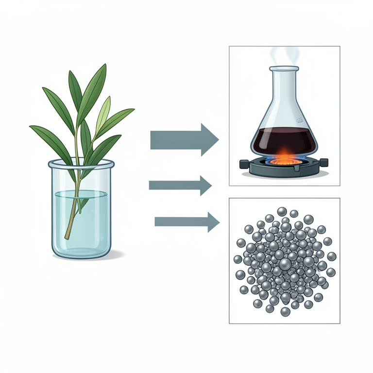
Green Synthesis Process
AI-generated conceptual illustration (Olea europaea leaves and Co3Cr2NiO8 nanocomposites). It explains the idea and does not show study data.
View source evidence ↗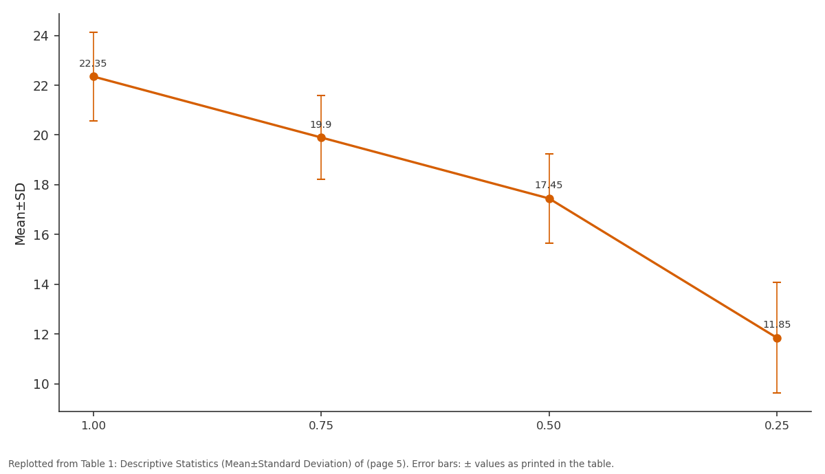
Mean±SD: 1.00 vs 0.75 vs 0.50 vs 0.25
Replotted from Table 1 (page 5) of the original paper. Every bar is the paper's own table value.
View source evidence ↗Figures & tables
From the original paper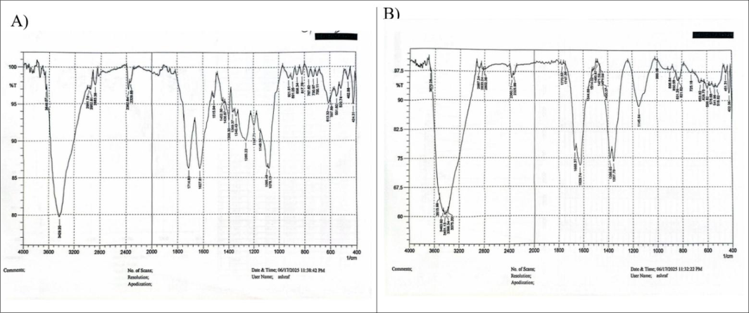 Figure 1
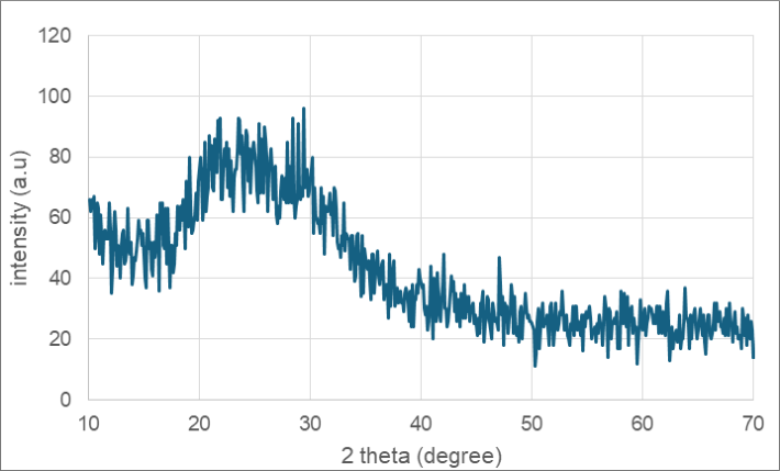 Figure 2
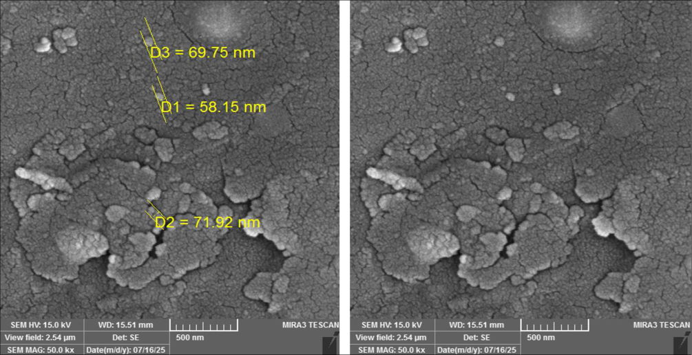 Figure 3
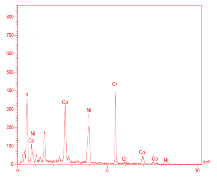 Figure 4
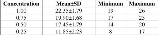 Table 1
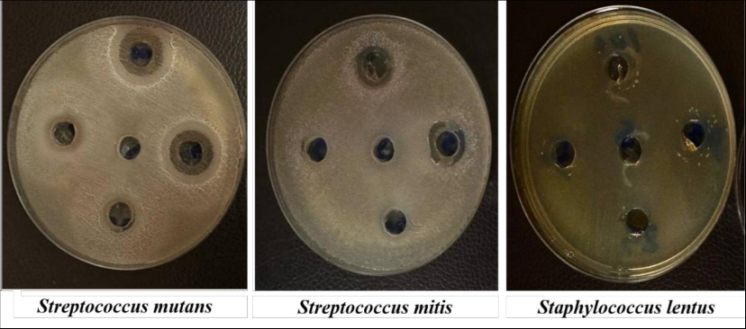 Figure 5
Introduction
Dental caries, sometimes called tooth decay, are one of the most widespread chronic diseases of people in the world. It is active, reversible, preventable and complicated biofilm-mediated multifactorial disease. Caries can affect all age groups throughout the life cycle [1] . Many factors lead to the development of dental caries, such as the adhesion of bacteria with the tooth surface, biofilm formation on the teeth (dental plaque), and the local demineralization by the acidic by-products from bacterial fermentation of carbohydrates initiated on the tooth surface [2] . Caries management requires dentists to show the degree of severity factors in the treatment. Many new technologies have been applied to the treatment of dental diseases. Caries Management by Risk Assessment (CAMBRA) system developed in 2002, Prevention is always better than cure, But the activity of caries may need to be treated [3] . In general, two approaches are used to carry removal from teeth with susceptible and asymptomatic pulps: nonselective caries removal (traditional approach) and selective caries removal [4] . Biosynthesis by plant leaves is an environmentally friendly synthesizing of nanomaterials, low cost and rapid synthesis. Plant leaves act as reducing and stabilizing agents to keep nanomaterials in nanoscales for a long period of time [5] . Metal salts are mixed with plant leaves mixed under control conditions such as temperature and pH. Biomolecules reduce the metal ion to nanoscale. Olea europaea leaves are rich in bioactive compounds such as polyphenols, flavonoids and oleuropein. These compounds act as reducing agents [6,7] . Joshua A. at el. in (2021) studied provides a comprehensive evaluation of current nanomaterial uses in dental fillings and procedures. The study compared standard and nanomaterial-based dental filling materials. Material comparisons consider biocompatibility, toxicity, reactivity, cost, and antimicrobial/antibacterial characteristics. Not one material has proven to have all the beneficial qualities/characteristics desired for the filling.
After analyzing three commonly used dental filling nanomaterials (Carbon-Based Nanotubes, Silica Nanoparticles, and Silver-Coated Nanoparticles), it was found that Silica Nanoparticles had the largest material benefit and should be suggested for further usage. Issues with future dental filling applications [8] . Zahra Vaseghi, Omid Tavakoli, Ali Nematollahzadeh in (2018) have evaluation of the antibacterial activity of the synthesized ternary nanoparticles and study antibacterial activity is tested against important oral strains, such as Streptococcus mutans associated with dental caries and Porphyromonas gingivalis associated with gingivitis. Evaluation is performed using laboratory tests such as the well diffusion method and the minimum inhibitory concentration (MIC). When comparing the three most currently used dental filling nanomaterials -Carbon-Based Nanotubes, Silica Nanoparticles and Silver-Coated Nanoparticles -it was observed that Silica Nanoparticles demonstrated material benefit performance [9] . The aim of this work is to synthesis eco-friendly, low cost, and rapid synthesis of ternary nanocomposite characterization nanocomposite synthesized by multiple techniques such as FTIR, XRD, FE-SEM and EDX. Apply ternary nanocomposite as antibacterial against three species of bacteria: against Streptococcus mutans, Streptococcus mitis and Staphylococcus lentus in dental infections
Experimental
Experimental
Nanocomposite synthesis
Nanoparticles: Individually the source of metals (cobalt, copper and Nickel) were used as a stoichiometric ratio (1:1:1) to prepare ternary oxides of Co 3 Cr 2 NiO 8 Nanocomposite. Accordingly, 50 ml, 0.01 mole of each Co(NO3)2, Cr(NO33) and NiCl2 were mixed at 25 °C. To synthesize the Cobalt-chrome-Nickel nanocomposite two milliliters of Olea europaea leaf extract were added drop by drop to five milliliters of the stock solution of Cobalt-Copper-Nickel. The volume was filled to 50 ml with distilled water, and the sample was heated for 15 minutes at 70℃.
The synthesis of Cobalt-chrome-Nickel nanocomposite is indicated by a shift in solution color from yellow to brownish yellow to deep brown. The sample was then dried, yielding the Cobalt-Copper-Nickel nanocomposite for characterization. Fourier transform infrared (FTIR) spectra were obtained at room temperature on a Shimadzu FTIR 84005 spectrometer for the plant extract containing Cobalt-chrome-Nickel nanocomposite (0.01 g) dried at 60 ºC for 4 hours using KBr [10] . An X-ray Diffraction (XRD) pattern was produced using a Shimadzu XRD-6000 diffractometer with Cu Kα (λ=1.54056 A°) to validate the biogenesis of nanocomposite [11] . The morphology and contact surface of the nanocomposite were examined using a SEM S-4160 [12,13] .
Application of nanocomposite for Antibacterial activity in dental decay 1. Sample collection
Oral swab were collected from patient with dental decay who visited Ibn Sina University of medical and pharmaceutical sciences with similar oral hygiene level were selected for data collected, each carious tooth examined using a Williams periodontal probe to ensure a cavity depth of 2 mm for Standardization ;all patients examined and sampled by the same dentist [14] .
Antibacterial activity
Studying the antibacterial activity for nanocomposite after collection and identification through the VITEK 2(bioMérieux, Inc.) system certain isolates were chosen for subsequent studies to examine the effect of the nanomaterial against Streptococcus mutans, Streptococcus mitis and Staphylococcus lentus. They were evaluated using method and inoculated by spread plate method. Bacteria were subculture on nutrient agar and incubated for 24 hours at 37 ℃. Mueller-Hinton Agar plates infected with pathogenic microorganisms were used. Each plate included sterile paper disks 5mm in diameter that had been soaked with plant extract (control) and nanocomposite. The plates were then incubated for 24 hours at 37 °C. We measured and documented the inhibitory zones [15] .
Results and discussion: Synthesis
The FT-IR analysis was utilized to identify the different functional groups present in Olea europaea leaves extract [16] . Fig1A shows a typical FT-IR spectrum for Olea europaea leaves extract along with a comparison with Fig1B which shows Co3Cr2NiO8 nanocomposite synthesized using this extract. Comparison of these two spectra indicated that the peak of synthesis nanocomposite and Olea europaea leaves extract have showed similar peaks with reduced intensity of OH and NH groups in the region between 4000-3000 cm⁻¹ with peak in the nanocomposite at (3515, 3463, 3431, 3398 cm⁻¹). In the region 3000-2800 cm⁻¹ (C-H stretching) peaks at 2887, 2853, 2602 cm⁻¹ were observed. The reduced intensity may indicate partial interaction or coverage of these groups on the nanocomposite surface. In the region 1800-1500 cm⁻¹ (C=O and aromatic C=C stretching) A strong carbonyl peak was observed at 1714 cm⁻¹ and aromatic C=C peaks at 1627 cm⁻¹. In the Olea europaea leaves extract, peaks in 1770, 1747, 1668 and 629 cm⁻¹ were observed in the nanocomposite. The shifts to higher wavenumbers (blue shift) may result from coordination between carbonyl oxygen and metal ions. Strong peaks were observed at 653, 628, 578, 541, 451 cm⁻¹ due to the M-O (metal-oxygen) vibrations. The references demonstrate the patterns of the nanocomposite [12,17] . The XRD pattern displays a considerable number of broadening lines, which are typical of nanoscale materials. The crystallite size may be estimated using the Debye-Scherrer equation [18] . The XRD pattern of Cobalt-chrome-Nickel nanocomposite reveals four distinct peaks at 2θ=18.27°, 21.13°, 30.06°, 35.40°, and 37.03°. These peaks correspond to the (111), ( 200), ( 220), ( 311) and ( 222) crystal planes of the hexagonal nanocomposite [19] . The data show that peak broadening indicates smaller size, indicating favorable circumstances for crystal formation.
The Scherrer formula was used to determine the average crystal size of the nanocomposite, which was around 2.7 nm. As seen in Fig. (3), the shape and size distribution of nanocomposites were examined using Field Emission Scanning Electron Microscopy (FE-SEM). Nanocomposites have a smooth, well-crystalline surface. According to the measurement pictures, the samples had a high porosity, indicating amorphous ratios. In the samples, the spherical form predominates [20] . The synthetic nanoparticles' average size ranges from 58.15 to 71.92 nm. Very small percentages of the samples have some clusters; however this happened when the sample was being prepared for testing. The FE-SEM study indicates that the samples created spherical aggregates with a roughly homogeneous distribution. Furthermore, the crystal character of produced nanocomposites of the same size is shown. Measurement photos include a high percentage of tiny sizes and uniform diameters. 4) Typical Co,Ni,Cr and oxygen peaks are present in the spectrum. The results confirm a high purity of the synthesized nanostructures [21,22] . Furthermore, the EDX measurement which showed cobalt (54.1%), Nickel (17%), copper (20%) and for oxygen was (9.5%). The EDX of Cobalt-Copper-Nickel nanocomposite synthesized shown in fig. (4). In the present study, the antibacterial effect of nanoparticles was tested against Streptococcus mutans, Streptococcus mitis and Staphylococcus lentus [23] . Diagnoses bacteria by The Vitek analyzer were found to be valuable for the speed and accuracy with which results were available for the common bacterial pathogens [24] . The nanomaterial tested in this study exhibited varying levels of antibacterial activity against the three isolated bacterial species, depending on the concentration used. The results on Streptococcus mutans showed clear sensitivity to the nanomaterial at concentrations of 50% and above. At 100%, a 20 mm inhibition zone was observed. At 75% a 16 mm inhibition zone was observed. At 50%, the inhibition zone was 12 mm, showing moderate effectiveness. No inhibition was observed at 25%, suggesting this concentration is insufficient to affect the growth of S. mutans. These findings indicate that this nanomaterial starts to be efficient at 50% and more potently at higher concentrations. In the case of Streptococcus mitis, the same trend was followed by S. mutans, but the sensitivity was a bit weaker in the intermediate concentrations. The inhibition zone was 15 mm at 100% which indicates good antibacterial activity. A 12 mm zone was seen at 75% and 10 mm zone at 50% with moderate inhibition. There was no inhibition at 25 percent meaning that there was low activity at this concentration. The progressive growth in the inhibition zone is a confirmation that the effect of the material improves with concentration [23] . In the case of Staphylococcus lentus, it was more resistant to the nanomaterial than Streptococcus species. A 14 mm inhibition zone was obtained at 100% and this shows good sensitivity with maximum concentration. A 10 mm zone was observed at 75%, but not at 50% or 25%. The findings indicate that the nanomaterial needs to be used in higher concentrations to inhibit S. lentus, perhaps because of the differences between the cell wall structure or resistance strategies. One-way analysis of variance (ANOVA) was used to analyse the experimental data to determine the effect of various concentrations on the measured response. Descriptive statistics revealed a gradual decrease in mean values with decreasing concentration, where the highest concentration (1) exhibited a mean of 22.35±1.79, followed by 0.75 (19.90±1.68), 0.5 (17.45±1.79), and 0.25 (11.85±2.23). The results of the ANOVA showed that there was a very significant difference between the groups that were tested (F = 113.79, p<0.001) and that the difference in concentrations did have a statistically significant effect on the measured response. The result of this finding supports the existence of a robust dosedependent correlation between concentration and activity. The high intensity of the decrease in response with low concentrations points to the fact that the performance of the tested system is directly dependent on the concentration levels. Additional post hoc test should be conducted to establish the particular differences between individual groups.
Conclusion
A nanocomposite was successfully prepared by green synthesis, and the method is considered cheap, safe, easy to use, and fast to produce. The nanocomposites were characterized by several techniques, the most important of which are FTIR, XRD, FE-SEM and EDX. Comparison of these two spectra indicated that the peak of synthesized nanocomposite and Olea europaea leaves extract are similar peaks but with reduced intensity in the coordination bands of OH and NH groups. The reduced intensity may indicate partial interaction or coverage of these groups on the nanocomposite surface. The crystallite size of nanocomposite was 2.7 nm calculated from XRD analysis, the nanoparticles size was found to be 58.15 to 71.92 nm based on FE-SEM. The difference between these numbers is the larger particle size compared to the crystallites; size indicates that the nanoparticles are composed of several crystallites, forming polycrystalline aggregates rather than single crystals. The antimicrobial evaluation of the Co 3 CrNi 2 O 8 Nanocomposite against Streptococcus mutans, Streptococcus mitis, and Staphylococcus lentus revealed a concentration-dependent inhibitory effect. S. mutans showed the highest sensitivity to the nanocomposite, with a noticeable inhibition starting at 50%, and a pronounced effect at 75% and 100%, indicating strong susceptibility. Overall, the results indicate that the synthesized nanocomposite is more effective against oral streptococci than S. lentus, and its antibacterial efficacy clearly enhanced with increasing concentration. This highlights its potential as a concentration-sensitive antimicrobial agent for dental or biomedical applications.
Figures and tables from the paper
| Concentration | Mean±SD | Minimum | Maximum |
|---|---|---|---|
| 1.00 | 22.35±1.79 | 19 | 26 |
| 0.75 | 19.90±1.68 | 17 | 23 |
| 0.50 | 17.45±1.79 | 14 | 20 |
| 0.25 | 11.85±2.23 | 8 | 17 |
Table text extracted from the original PDF; superscript letters are shown inline. The original table image is below.
View original table image
References
24 in the original paper- Yadav K, Prakash S. Dental caries: a microbiological approach. Journal of Clinical Infectious Diseases and Practice. 2017;2(1):1-15.
- Siddiqui S, Saba I. Demineralization and remineralization of teeth. EduBubs Publishing House; 2020.
- Swarna S, Nivedhitha M. Establishment of clinical protocol for early caries detection and minimal intervention using principles of CAMBRA. International Journal of Pharmaceutical Research. 2020;12(2).
- Barros MMAF, et al. Selective, stepwise, or nonselective removal of carious tissue: which technique offers lower risk for the treatment of dental caries in permanent teeth? A systematic review and metaanalysis. Clinical Oral Investigations. 2020;24(2):521532.
- Saratale RG, et al. New insights on the green synthesis of metallic nanoparticles using plant and waste biomaterials: current knowledge, their agricultural and environmental applications. Environmental Science and Pollution Research. 2018;25(11):10164-10183.
- Shafey AE. Green synthesis of metal and metal oxide nanoparticles from plant leaf extracts and their applications: a review. Green Processing and Synthesis. 2020;9(1):304-339.
- Khudiar SS, et al. Characterization of NO2 gas sensing for ZnO nanostructure grown hydrothermally on porous silicon. Optik. 2022;249:168300.
- Eisenstat J, et al. A comparative review of material properties for current and future dental filling nanomaterials. International Journal of Engineering Materials and Manufacture. 2021;6(4):225-241.
- Vaseghi Z, Tavakoli O, Nematollahzadeh A. Rapid biosynthesis of novel Cu/Cr/Ni trimetallic oxide nanoparticles with antimicrobial activity. Journal of Environmental Chemical Engineering. 2018;6(2):18981911.
- Khalaf RL, et al. Synthesis of silver nanoparticles using Rosa flowers extracts: thermodynamic and kinetic studies on the inhibitory effects of nanoparticles on creatine kinase activity. Iraqi Journal of Science. 2021:2486-2500.
- Khalaf RL, Ahmed EM. Biosynthesis of silver nanoparticles using plant flowers of Nerium oleander extract and its antibacterial activity. Biochemical and Cellular Archives. 2019;19(1).
- Hasanin MS, et al. Advanced functional Cu-Cr-CoNPs@gelatin (GLN)-hydroxyethyl cellulose (HEC) nanocomposites with antimicrobial and anticancer activities. RSC Advances. 2024;14(50):37633-37643.
- Khalaf RL, Almousawi IM, Rheima AM. Dye pollution removal in aqueous solution using novel photochemically synthesized CoZnFe2O5 nanocomposite. Desalination and Water Treatment. 2021;243:287-293.
- Hamad AM, Atiyea QM. In vitro study of the effect of zinc oxide nanoparticles on Streptococcus mutans isolated from human dental caries. Journal of Physics: Conference Series. 2021.
- Lavaee F, Motamedifar M, Rafiee G. The effect of photodynamic therapy by gold nanoparticles on Streptococcus mutans and biofilm formation: an in vitro study. Lasers in Medical Science. 2022;37(3):17171725.
- Pasieczna-Patkowska S, Cichy M, Flieger J. Application of Fourier transform infrared (FTIR) spectroscopy in characterization of green synthesized nanoparticles. Molecules. 2025;30(3):684.
- Issa HK, et al. On the structure-properties relationship of amorphous and crystalline silica nanoparticles reinforced magnesium matrix nanocomposites. Journal of Alloys and Compounds. 2022;924:166605.
- Fatimah S, et al. How to calculate crystallite size from X-ray diffraction (XRD) using Scherrer method. ASEAN Journal of Science and Engineering. 2022;2(1):65-76.
- Selvanayaki R, et al. Structural, optical and electrical conductivity studies of pure and Fe doped zinc oxide (ZnO) nanoparticles. Materials Today: Proceedings. 2022;49:2628-2631.
- Liu A, et al. Characterizing mechanical heterogeneity of coal at nano-to-micro scale using combined nanoindentation and FESEM-EDS. International Journal of Coal Geology. 2022;261:104081.
- Sudha D, et al. Structural, optical, morphological and electrochemical properties of ZnO and graphene oxide blended ZnO nanocomposites. Ceramics International. 2023;49(5):7284-7288.
- Moro D, Ulian G, Valdre G. SEM-EDS nanoanalysis of mineral composite materials: a Monte Carlo approach. Composite Structures. 2021;259:113227.
- Mendes CR, et al. Antibacterial action and target mechanisms of zinc oxide nanoparticles against bacterial pathogens. Scientific Reports. 2022;12(1):2658.
- Shetty N, Hill G, Ridgway G. The Vitek analyser for routine bacterial identification and susceptibility testing: protocols, problems, and pitfalls. Journal of Clinical Pathology. 1998;51(4):316-323.