
Research article · 2019 · Full text
International Journal of Biology SciencesSynthesis and characterization of copper nanoparticles by chemical method and evaluation of their antibacterial and antibiofilm activities using the agar diffusion method
Volume 1(2) · Pages 137–140 · DOI: 10.33545/26649926.2019.v1.i2a.843 ↗
Research areasNanoparticles: synthesis and applicationsCopper-based nanomaterials and applicationsAntimicrobial agents and applications
Abstract
Author-writtenNanotechnology is a really new scientific field with real potential to solve many problems in many areas of science. Nanotechnology, when combined with several other scientific fields like chemistry, physics and biology has characterized metals in the form of nanoparticles. Copper nanoparticles are particularly concerning, as they exhibit more significant bioactivity among these. Their high surface-area-to-volume ratio renders them into excellent candidates to be used as an antibacterial agent in diverse biomedical applications. On the other hand, their universal use, especially in excess amounts may induce serious environmental threats; and therefore, they should be treated with attention to their possible influence on the environment.
Summary
Source-checkedPlain-language reading of the full paper, prepared with AI and checked against the text. Each point links to the authors' own words.
Why it matters
- Copper nanoparticles are highly valued for biomedical use as antibacterial agents because their high surface-area-to-volume ratio enhances their bioactivity. View evidence ↗
- The ongoing prevalence of bacterial diseases drives the research into copper nanoparticles as potential antimicrobial tools. View evidence ↗
- While useful, the widespread or excessive use of these nanoparticles may pose serious threats to the environment. View evidence ↗
The question
- The study aims to synthesize and characterize copper nanoparticles and evaluate their effectiveness against bacteria and biofilms using an agar diffusion assay. View evidence ↗
What they did
- The researchers used a green one-pot chemical reduction method, employing copper (II) sulfate pentahydrate as the starting precursor. View evidence ↗
- Sodium borohydride was used as the reducing agent, with the mixture stirred at room temperature for two hours. View evidence ↗
- Nanoparticles were isolated via centrifugation at 10,000 rpm for 15 minutes, washed four times, and dried at 50 °C for 24 hours. View evidence ↗
- Antibacterial activity was tested by loading 50 µL of CuNP solutions at concentrations of 0.1, 0.5, and 1.0 mg/mL into 6-mm diameter wells and incubating them at 37 °C for 24 hours. View evidence ↗
- Antibiofilm activity was evaluated by treating biofilms with 0.1, 0.5, and 1.0 mg/mL of Cu-NPs for 24 hours, followed by staining with 0.1% crystal violet for 15 minutes. View evidence ↗
What they found
- UV-Vis spectroscopy confirmed the formation of copper nanoparticles, showing a surface plasmon resonance peak at approximately 570 nm. View evidence ↗
- SEM imaging revealed that the synthesized nanoparticles were spherical with an average size between 30 and 50 nm. View evidence ↗
- XRD analysis confirmed the nanoparticles have a crystalline, face-centered cubic structure, indicated by peaks at the (111), (200), and (220) cubic planes. View evidence ↗
- The antibacterial effect was dose-dependent for both tested strains, with the largest inhibition zone occurring at the highest concentration of 1.0 mg/mL. View evidence ↗
- The nanoparticles demonstrated more significant antibacterial activity against Staphylococcus aureus than against Escherichia coli. View evidence ↗
- Cu-NPs reduced biofilm formation in both bacterial strains, with the maximum inhibition achieved at a concentration of 1.0 mg/mL. View evidence ↗
What it means
- The authors conclude that the synthesized copper nanoparticles are effective against both Gram-positive and Gram-negative bacteria and biofilms, making them suitable for medical and environmental applications. View evidence ↗
Visual summary
2 visualsIllustrations explain the paper; they do not show study data.
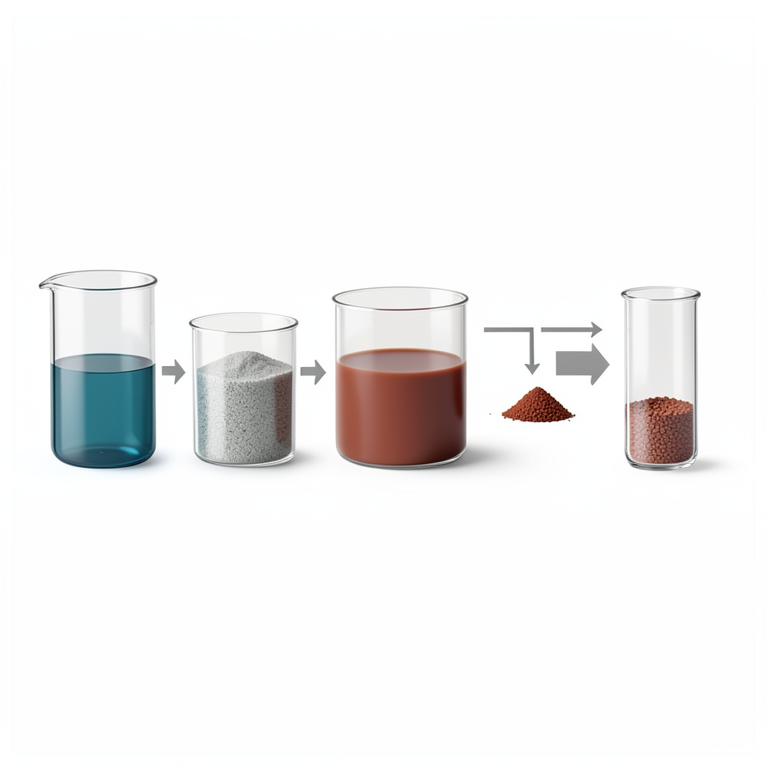
Synthesis Workflow
AI-generated conceptual illustration (copper nanoparticles). It explains the idea and does not show study data.
View source evidence ↗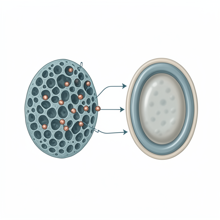
Differential Bacterial Susceptibility
AI-generated conceptual illustration (Staphylococcus aureus and Escherichia coli bacteria with copper nanoparticles). It explains the idea and does not show study data.
View source evidence ↗Figures & tables
From the original paper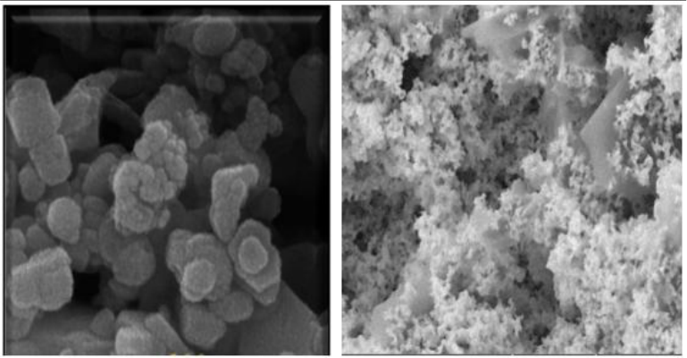 Figure 1
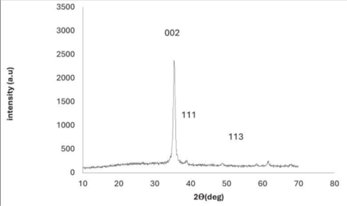 Figure 2
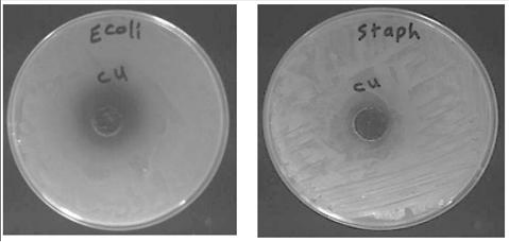 Figure 3
Introduction
Nanotechnology is also a field of science in rapid change, which holds immense potential for solving numerous challenges and has relevance of cross-cutting nature to many scientific as well technological sectors. Nanotechnology is integrated with natural sciences such as chemistry biology and physics to make nanoparticles from matching metals. Copper nanoparticles (Cu-NPs) have received great attention due to their remarkable bioactivity and diverse biological applications. Bacterial diseases occur even today, which has led to the great interest in preparing copper nanoparticles (Cu-NPs) as a potential antimicrobial agent due to their high surface area-to-volume ratio. (Chen and others, 2015) [1] .
While metallic nanoparticles have many favorable features, it is important to understand that extensive usage of these nanostructures could be detrimental to humans, animals and environment (Jalilian et al., 2020) [3] . Amongst the many copper-based nanomaterials (i.e., prefixes), one fascinating class are copper oxide nm′-materials, whose antibacterial activity is orders of magnitude higher than for typical nano powders and thus a particularly interesting alternative to traditional coarse copper oxide powders. Due to the great potential in many application fields, such as dye removal, gas sensors, semiconductors, organic catalysis and solar energy conversion giving exceptionally different chemical properties as compared with bulk counterparts (Ravindra et al., 2018) [4] . It can be used as CuO-NPs in heat transfer applications in the future. Many experimental researches conducted, reveal that CuO based nanofluids have been managed to increase thermal conductivity by as much as 12.4 wt% compared with deionized water (Chandrasekar et al., 2019) [2] .
Beyond industrial and catalytic applications, the CuO-NPs has been established to be a potential tool in preventing the establishment of bacterial biofilm with their considerable biomedical applications. Their capacity to form biofilms further allows their persistence in medical and environmental situations where this trait would make them desirable for use. CuO-NPs are prepared from numerous procedures: microwave irradiation, electrochemical methods, hydrothermal method, wet chemical method and precipitation pyrolysis. (Yuan et al., 2020) [6] . The principal aim of this work is the preparation and characterization of copper oxide nanoparticles (CuO-NPs), as well as to demonstrate their antibacterial and antibiofilm effects by means of an agar diffusion assay. This study aims to establish a simple and effective method of synthesizing CuO-NPs and evaluate their efficacy in preventing bacterial infections and biofilm formulation.
Material and Methods
Experimental Validation of Materials and Laboratory Equipment The experimental validation was conducted to confirm the sterilization of materials and laboratory equipment by means of UV irradiation and heat methods before using them in experiments. Culture media, buffers and heat-stable materials were autoclaved (121 °C for 15-20 min at 103 kPa) (Suman et al., 2020) [5] . All experiments performed in a laminar flow hood with both used 70% ethanol as sterilizable.
Setting Up Cultural Media
The media used to grow the bacterial cultures were nutrient agar (NA) and nutrient broth (NB), routinely applied for nonselective cultivation of bacteria.
Synthesis of Nanomaterial (Cu Nanoparticles)
Copper nanoparticles (Cu-NPs) were synthesized in this study via a facile, green one-pot chemical reduction method. We selected copper (II) sulfate pentahydrate [Cu-SO₄•5H₂O] as a precursor for the synthesis of nanoparticles. Copper sulfate: A 0.1 M copper sulfate solution was prepared by dissolving an accurately weighed quantity of copper sulfate in distilled water. An aqueous NaOH solution (1 M) was gradually added to the copper sulfate solution under continuous stirring until the pH value reached around 12. It resulted in production of copper hydroxide precipitate (Cu (OH)₂), which underwent reduction Sodium borohydride (NaBH₄) solution was used as the reducing agent to reduce producing copper nanoparticles from copper hydroxide. The reaction mixture was stirred at room temperature for two hours. Immediately After the Reaction, Copper Nanoparticles were Isolated by Centrifugation at 10,000 rpm for 15 Minutes; The supernatant was discarded, and the nanoparticles were washed four times with distilled water to remove residual chemicals. The copper nanoparticles obtained were dried at 50 °C for 24 hr. Characterization of Synthesized Copper Nanosized Particles. A UV-Vis spectrophotometer was used to analyze the surface plasmon resonance and confirm that copper nanoparticles had been generated. Spectrophotometry measurements were carried out at the range of 200-800 nm (Suman et al., 2020) [5] . The morphology and particle size of the nanoparticles were characterized by scanning electron microscopy (SEM). SEM (Scanning Electron Microscope): SEM Images were taken at an appropriate voltage. The crystalline structure of the nanoparticles was investigated by X-ray diffraction (XRD). XRD pattern was recorded using a CuKα radiation source within the 2θ range of 10° to 80°, at a scan rate of 2° per minute (Ravindra et al., 2018) [4] . Dynamic light scattering (DLS) was used to analyze the samples for average particle size and polydispersity index, a measure of uniformity in particle size distribution (Yuan et al., 2020) [6] .
The agar diffusion technique was used to assess the antibacterial activity of the copper nanoparticles. Culturing of Escherichia coli (E. coli) and Staphylococcus aureus (S. aureus) in nutrient broth at 37 °C for 24 hours on the day of the experiment, bacterial suspensions were adjusted to a concentration of 10⁸ CFU/mL. The bacterial suspension (100 of the passenger-fusion strain with Lpt6-lacZ353 remained stable for 78 and 56 days, respectively) or sterile saline as a control was evenly spread on sterile agar plates with obtained from Escherichia coli suspension were initially diluted in L media. Wells of 6-millimeter diameter were made on the agar plates and 50 µL of CuNP solution (0.1, 0.5 and 1.0 mg/mL, prepared in dimethyl sulfoxide DMSO; Merck, Germany) was loaded onto each well. They incubated the plates at 37 °C for 24h. Antibacterial activity was measured based on the zone of inhibition (mm) surrounding each well. The experiments were conduct, in triplicate, and the inhibition zone is reported as the average of three (Chen et al., 2015) [1] . The antibiofilm effect of copper nanoparticles was determined by crystal violet staining. Method: A total of 9 strains were used to generate the reference MIC from which only one bacteria culture was incubated into a microtiter plate (96-well) and left for 24 h at 37 °C. The planktonic cells were then discarded, and the wells were washed with PBS to remove non-attached cells. Then specific concentrations of Cu-NPs (0.1, 0.5 and 1•0 mg/mL) were added to the wells and incubated for 24 h. Crystal violet staining of the biofilm was then stained with crystal violet solution (0.1%) at room temperature for 15 min. Excess dye was subsequently washed off and the biofilm was dissolved using ethanol. Biofilm formation was quantified by measuring the absorbance at 595 nm with a microplate reader (Chandrasekar et al., 2019) [2] .
All data were expressed as mean ± SD. Statistical significance was evaluated by one-way analysis of variance (ANOVA) and post hoc Tukey's test at p<0.05. The experiments were performed in triplicates to ensure reproducibility and accuracy of results.
Results and Discussion
Synthesis and characterization UV-Vis spectroscopy, SEM, XRD and DLS confirmed the successful synthesis of copper nanoparticles (Cu-NPs) by means of the chemical reduction method. The UV-Vis spectra of the synthesis Cu-NPs showed a surface plasmon resonance (SPR) peak with about 570nm, which indicates the formation of copper nanoparticles (Suman et al., 2020) [5] . The SPR peak also hinted that the nanoparticles had a size-distribution relatively close to uniform. The morphology of the Cu-NPs is shown in Fig 1, which represented spherical appearance with the average size of about 30-50 nm by scanning electron microscopy (SEM) images. This is in accordance with the characterization by dynamic light scattering (DLS) analysis revealing a correspondingly sized particle range with low PDI confirming relatively monodispersed. From X-ray diffraction (XRD) pattern analysis, fig 2 the obtained copper nanoparticles showed distinct peaks that could be correlated to the cubic planes of (111), ( 200) and (220); these cubic planes confirm their crystalline nature and they possess a face-centered cubic structure (Ravindra et al.2018) [4] .
Antibacterial Activity
The antibacterial activity of the synthesized Cu-NPs was measured against Escherichia coli (E. coli) and Staphylococcus aureus (S. aureus) by the means of agar diffusion method. This result indicated the antibacterial effect of Cu-NPs are dosedependent against both bacterial strains as shown in the results section, a greater concentration of Cu-NPs resulted in an increase zone of inhibition where the widest iron spreader (1.0 mg/mL) The significant antibacterial activity of Cu-NPs was determined on S. aureus while it was less effective on E. coli Fig 3 . These finding implies that Cu-NPs exhibits an antibacterial activity against various bacterial strain and the difference in reaction may be linked to the variations of cell wall composition and ability of bacteria to interact with nanoparticles (Chen et al., 2015) [1] . These findings were in agreement with previous studies, which had shown that copper nanoparticles were quite potent for killing several species of bacteria due to the copper ions which can penetrate through cell membranes and the consequent production of reactive oxygen species (ROS) causing cellular death (Jalilian et al., 2020) [3] . Also, could be due to uncle size of Cu-NPs which might performed more surface area for more interaction with bacterial cell and antimicrobial activity because size (Yuan et al., 2020) [6] .
Antibiofilm Activity
Evaluation of antibiofilm activity the crystal violet staining method used to assess the efficacy of Cu-NPs. Both bacterial strains were affected by Cu-NPs in reducing biofilm formation, the results shown that. However, biofilm inhibition increased with Cu-NPs concentrations and reached the maximum at 1.0 mg/mL concentration level. The inhibition of biofilm formation accompanied the antibacterial results, with more significant reductions observed for S. aureus than E. coli. These results indicate that Cu-NPs are able to induce the inhibition of not only planktonic growth, but also the prevention of bio-film formation, which is a mechanism associated with chronic infections and bacterial resistance to antibiotics. Copper nanoparticles have also been shown in previous studies to prevent biofilm formation by disrupting the adhesion of bacterial cells on surfaces (Ravindra et al., 2018) [4] . The role of biofilm inhibition brought by Cu-NPs are likely associated with damaging structural features of the biofilm matrix, as well as the interaction between the bacteria and cell membranes.
Discussion
The outcome of this study shows that copper nanoparticles synthesized from the simple chemical reduction method have significant antibacterial and antibiofilm activity. The small size and spherical shape of Cu-NPs thus directly determine their large surface area, providing the high activity that characterizes these materials in antimicrobial action. The antibacterial activity was observed to be dose dependent against both E. coli and S. aureus, indicating that the inhibition zone increased with increasing concentrations of Cu-NPs, which also establish the preventative potential of Cu-NPs as an effective positive antibacterial agent. Besides, the capacity of Cu-NPs for biofilm inhibition makes them promising candidates in medicine particularly to overcome biofilm-associated infections. The better antibacterial activity of Cu-NPs in comparison with Gram-negative bacteria (E. coli) might be due to the structural properties of the bacterial cell wall. E. coli has an outermembrane (OM) which may effectively provide resistance to Cu-NPs penetration, whereas S. aureus possesses a relatively thick peptidoglycan layer making it possibly more vulnerable towards action of the Cu-NPs via diffusion or through pores in the bacterial cell envelope. These results were in accordance with the previous reports, which demonstrated higher effectiveness of copper nanomaterials against Gram-positive bacteria (Jalilian et al., 2020) [3] .
In summary, synthesized copper nanoparticles were effective against Gram-positive and Gram-negative bacteria as well as biofilm formation; therefore, they can be considered for use in medicine and the environment to treat bacterial infections and preserving materials from being fouled by biofilms. Whereas more investigations are needed to determine in vivo toxicity and long-term chemical stability of the Cu-NPs for proper appraisal of its application in clinics.
References
7 in the original paper- Chen CH, Hsieh SH, Wu J. Antibacterial activity of copper nanoparticles: Synthesis and mechanisms of action. Journal of Nanotechnology. 2015;2015:1-7.
- Chandrasekar M, Karthik R, Rajendran R. Thermal conductivity enhancement of CuO nanofluid: A review. International Journal of Heat and Mass Transfer. 2019;135:700-715.
- Jalilian M, Kian MA, Asgarian S. Environmental and health risks of nanomaterials: A comprehensive review. Environmental Toxicology and Chemistry. 2020;39(8):1891-1900.
- Ravindra R, Kumar MR, Meena S. The role of copper nanoparticles in wastewater treatment and environmental protection. Journal of Environmental Management. 2018;210:72-81.
- Suman S, Yadav S, Singh R. Copper nanoparticles: Synthesis, properties, and biomedical applications. Materials Science and Engineering. 2020;87:156-167.
- Yuan X, Li H, Zhang L. Synthesis of copper oxide nanoparticles and their applications in catalysis and antibacterial activity. Journal of Nanomaterials. 2020;2020:1-12.
- Zhu Y, Wang L, Zhang L. Synthesis of CuO-CeO2 nanocomposites as efficient catalysts for environmental applications. Journal of Environmental Science. 2020;52:146-154