
Research article · 2026 · Full text
International Journal of Biology SciencesCharacterization and antimicrobial analysis of biogenically synthesized zinc oxide nanoparticles from Datura stramonium leaf extract
Volume 8(5) · Pages 98–103 · DOI: 10.33545/26649926.2026.v8.i5b.835 ↗
Research areasNanoparticles: synthesis and applicationsEssential Oils and Antimicrobial ActivityMedicinal Plant Research
Abstract
Author-writtenGreen synthesis of metal oxide nanoparticles has emerged as an eco-friendly and sustainable alternative to conventional chemical and physical synthesis methods. In the present study, zinc oxide nanoparticles (ZnONPs) were synthesized using aqueous leaf extract of Datura stramonium as a reducing and stabilizing agent. The synthesized nanoparticles were characterized using UV-Visible spectroscopy, Fourier Transform Infrared Spectroscopy (FTIR), Scanning Electron Microscopy (SEM), Transmission Electron Microscopy (TEM), and X-Ray Diffraction (XRD). The antimicrobial activity of the synthesized ZnONPs was evaluated against selected bacterial pathogens using the agar well diffusion method. The UV-Visible spectrum confirmed nanoparticle formation with characteristic absorption in the UV region. FTIR analysis indicated the presence of phytochemicals such as alkaloids, flavonoids, phenolics, and terpenoids involved in reduction and stabilization. SEM and TEM studies revealed predominantly spherical nanoparticles with nanoscale dimensions. The synthesized ZnONPs exhibited significant antimicrobial activity against both Gram-positive and Gramnegative bacterial strains. The findings suggest that Datura stramonium-mediated ZnONPs possess promising antimicrobial potential and can be utilized in biomedical and pharmaceutical applications.
Summary
Source-checkedPlain-language reading of the full paper, prepared with AI and checked against the text. Each point links to the authors' own words.
Why it matters
- Nanotechnology is a rapidly growing field with wide-ranging applications in areas such as medicine, agriculture, and environmental science. View evidence ↗
- Zinc oxide nanoparticles (ZnONPs) are of particular interest due to their biocompatibility and strong antimicrobial and photocatalytic properties. View evidence ↗
- Green synthesis using plant extracts is a sustainable alternative to conventional chemical and physical methods, which often require high energy and use hazardous chemicals. View evidence ↗
The question
- The study aims to synthesize, characterize, and evaluate the antimicrobial activity of zinc oxide nanoparticles produced using Datura stramonium leaf extract. View evidence ↗
What they did
- Purification of the synthesized nanoparticles was achieved through centrifugation at 17,000 rpm for 20 minutes. View evidence ↗
- The agar well diffusion method was used for antimicrobial assessment, with plates incubated at 37°C for 24 hours. View evidence ↗
What they found
- UV-Visible spectroscopy confirmed the formation of ZnONPs, showing a characteristic absorption peak between 350 and 380 nm. View evidence ↗
- Transmission Electron Microscopy (TEM) showed that the nanoparticles were predominantly spherical and ranged in size from 20 to 80 nm. View evidence ↗
- X-Ray Diffraction (XRD) analysis confirmed that the synthesized nanoparticles have a hexagonal wurtzite crystalline structure. View evidence ↗
- The nanoparticles showed antimicrobial activity across all tested strains, with Escherichia coli exhibiting the largest zone of inhibition at 20 ± 0.6 mm. View evidence ↗
- Gram-negative bacteria were found to be more sensitive to the nanoparticles than Gram-positive bacteria, likely due to differences in their cell wall structures. View evidence ↗
What it means
- The authors conclude that Datura stramonium is an effective and eco-friendly biological source for synthesizing zinc oxide nanoparticles. View evidence ↗
- The synthesized nanoparticles are recommended for potential use in pharmaceutical industries, biomedical devices, and environmental remediation. View evidence ↗
- The authors state that further research is needed regarding biocompatibility, cytotoxicity, and the feasibility of large-scale production. View evidence ↗
Visual summary
1 visualsIllustrations explain the paper; charts are replotted from the paper's own table values.
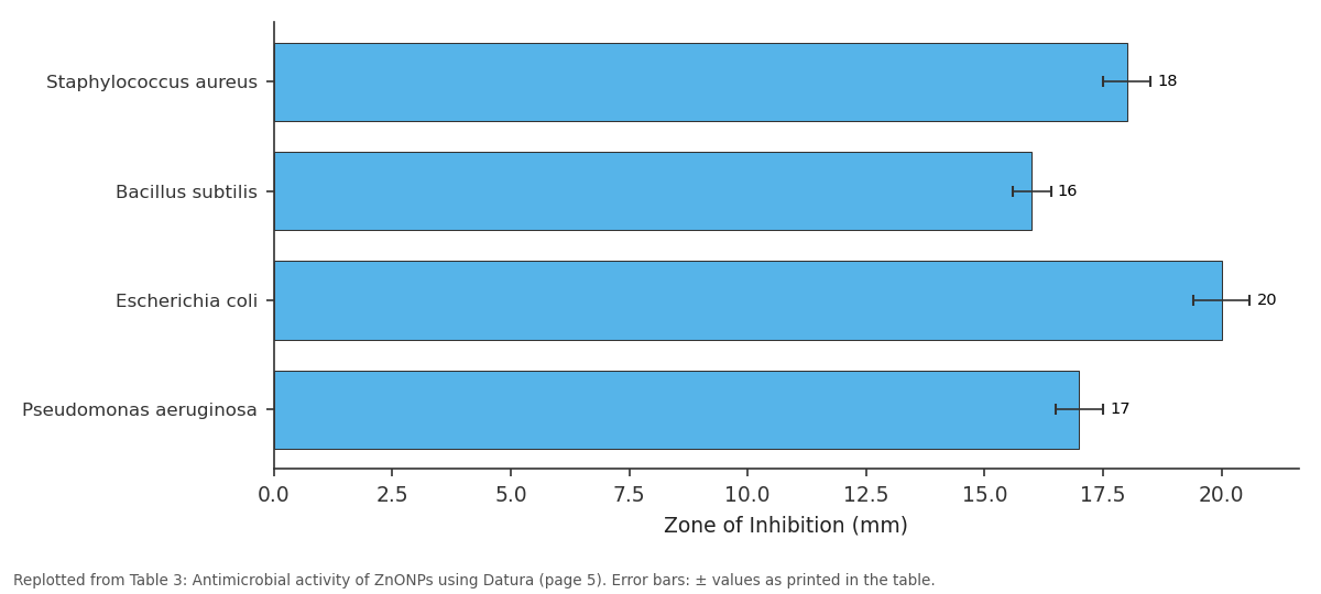
Zone of Inhibition (mm): Staphylococcus aureus vs Bacillus subtilis vs Escherichia coli vs Pseudomonas aeruginosa
Replotted from Table 3 (page 5) of the original paper. Every bar is the paper's own table value.
View source evidence ↗Figures & tables
From the original paper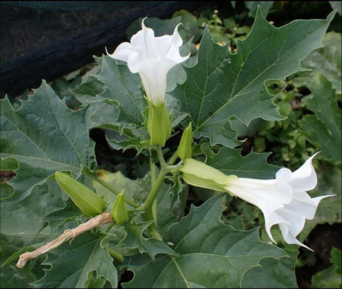 Figure 1
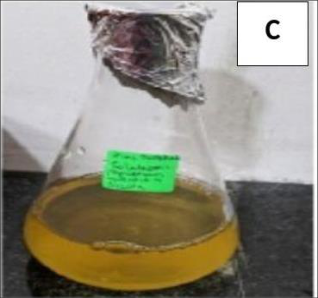 Figure 2
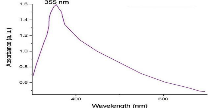 Figure 3
 Table 1
 Table 2
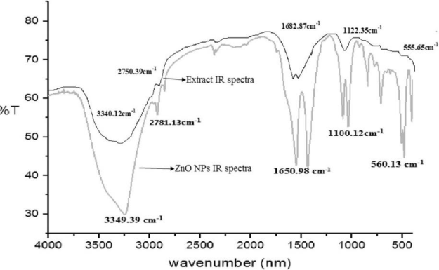 Figure 4
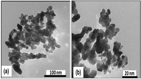 Figure 5
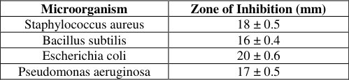 Table 3
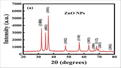 Figure 6
Introduction
Nanotechnology is one of the rapidly expanding fields due to its extensive applications in medicine, catalysis, agriculture, biotechnology, and environmental sciences. Nanoparticles are particles having dimensions below 100 nm and exhibit unique physical, chemical, and biological properties due to their high surface area to volume ratio (Rai et al., 2008) [14] . Metal oxide nanoparticles, particularly zinc oxide nanoparticles (ZnONPs), have attracted significant attention because of their remarkable antimicrobial, photocatalytic, and biocompatible properties. Zinc oxide nanoparticles are considered promising inorganic nanomaterials due to their high chemical stability, broad radiation absorption spectrum, electrochemical coupling coefficient, and photostability (Agnieszka and Jesionowski, 2014) [1] . ZnONPs have been widely employed in cosmetics, food additives, pharmaceuticals, ceramics, textiles, and biomedical applications (Sirelkhatim et al., 2015; Eixenberger et al., 2017) [18,6] . Their antimicrobial properties arise from the generation of reactive oxygen species (ROS), release of Zn2+ ions, and interaction with microbial cell membranes. Conventional physical and chemical methods for nanoparticle synthesis often involve hazardous chemicals, high energy requirements, and toxic by-products. Green synthesis using plant extracts offers a sustainable and eco-friendly alternative by utilizing naturally occurring phytochemicals as reducing and stabilizing agents (Raveendran et al., 2003) [16] . Plant-mediated synthesis is advantageous because plant extracts are readily available, safe to handle, economical, and rich in bioactive compounds. Datura stramonium, commonly known as Jimson weed or Thorn apple, belongs to the family Solanaceae and contains diverse phytochemicals including alkaloids, flavonoids, tannins, terpenoids, phenolics, glycosides, and steroids (Muller and Arfmann, 1987; Evan and Lampard, 1972) [8,13] . These bioactive compounds play a crucial role in the reduction and stabilization of nanoparticles. The present study focuses on the green synthesis, characterization, and antimicrobial evaluation of ZnO nanoparticles synthesized using Datura stramonium leaf extract. The work highlights the role of plant phytochemicals in nanoparticle synthesis and explores the biomedical significance of the synthesized nanoparticles.
Materials and Methods
Materials and Methods
Collection of Plant Material
Fresh leaves of Datura stramonium were collected from healthy plants and thoroughly washed with distilled water to remove dust and impurities. The leaves were shade dried and powdered using a sterile grinder.
Preparation of Plant Extract
Approximately 20 g of powdered leaf material was mixed with 100 mL distilled water and heated at 60 -70°C for 30 minutes. The extract was filtered using Whatman No.1 filter paper and stored at 4°C for further use.
Green Synthesis of ZnO Nanoparticles
DZn ONPs (Zn ONPs synthesized using Datura stramonium leaf extract) were synthesized by addition of 10 ml aqueous DLE (Datura stramonium leaf extract) to 40 ml aqueous 1 m M zinc nitrate solution by constant stirring at 75mathrm °C for 40 minutes at neutral p H (Figure 2C). The synthesized DZn ONPs were purified by centrifugation at 17000 rpm for 20 minutes.
Characterization of ZnO Nanoparticles 3.1 UV-Visible Spectroscopy
UV-Visible spectroscopy was performed to confirm nanoparticle formation. The absorption spectrum of ZnO nanoparticles showed a characteristic absorption peak in the range of 350-380 nm, indicating successful synthesis of ZnO nanoparticles.
Fourier Transform Infrared Spectroscopy (FTIR)
FTIR analysis was conducted to identify the functional groups involved in nanoparticle synthesis and stabilization. The spectrum revealed peaks corresponding to hydroxyl groups, amines, phenolic compounds, and alkaloids present in the plant extract. The Zn-O stretching vibration confirmed the formation of ZnO nanoparticles.
Transmission Electron Microscopy (TEM)
TEM analysis confirmed the nanoscale size distribution of the synthesized ZnO nanoparticles.
X-Ray Diffraction (XRD)
XRD analysis confirmed the crystalline nature of the synthesized ZnO nanoparticles.
Antimicrobial Activity 4.1 Test Microorganisms
The antimicrobial activity of synthesized ZnO nanoparticles was evaluated against selected Gram-positive and Gramnegative bacterial strains including:
Agar Well Diffusion Method
The antimicrobial activity was assessed using the agar well diffusion method. Nutrient agar plates were inoculated with bacterial cultures. Wells were prepared and loaded with different concentrations of ZnO nanoparticle suspension. Plates were incubated at 37°C for 24 hours.
Results and Discussion
Results and Discussion
Preliminary Confirmation of Nanoparticle Formation
The formation of zinc oxide nanoparticles was initially confirmed by visual observation of color change during the synthesis process. Upon addition of Datura stramonium leaf extract to the zinc nitrate solution, the reaction mixture gradually changed from pale yellow to whitish cream color, indicating the reduction of zinc ions and formation of ZnO nanoparticles. The color change occurs due to excitation of surface electrons and formation of nanosized zinc oxide particles. The phytochemicals present in the plant extract such as flavonoids, phenolics, alkaloids, and terpenoids acted as reducing and stabilizing agents during nanoparticle synthesis. Similar observations were reported by Mohanpuria et al. (2008) [12] and Dhillon et al. (2012) [5] , who described visible color change as a preliminary indication of successful biosynthesis of metal nanoparticles using plant extracts. The visual change therefore served as an initial confirmation for ZnO nanoparticle formation prior to spectroscopic and microscopic characterization.
UV-Visible Spectroscopic Analysis
The synthesis of zinc oxide nanoparticles (Zn ONPs) was initially confirmed by UV-Visible spectroscopic analysis.
The absorption spectrum exhibited a characteristic peak in the range of 350-380 nm (Figure 3), which is typical for Zn O nanoparticles and confirms the successful formation of Zn ONPs from zinc ions. Similar absorption peaks have been reported by Song and Kim (2009) [19] and Sabir et al. (2014) [17] . Likewise, Medina et al. (2018) [11] and Kumar et al. (2021) observed blue-shifted absorption bands attributed to quantum confinement effects in nanosized Zn O particles. The observed absorption band is associated with the surface plasmon resonance (SPR) of ZnO nanoparticles. The sharp and intense peak indicated the formation of stable and uniformly dispersed nanoparticles. Minor shifts in the absorption peak may result from variations in particle size, morphology, agglomeration state, and dielectric properties of the surrounding medium. The reduction and stabilization of ZnO nanoparticles were facilitated by phytochemicals present in the Datura stramonium leaf extract, including alkaloids, flavonoids, phenolics, tannins, and terpenoids, which acted as reducing and capping agents. The UV-Visible analysis further suggested the nanoscale nature of the synthesized particles, as bulk ZnO generally exhibits absorption at longer wavelengths. The observed blue shift in the absorption region may be attributed to quantum confinement effects. Overall, the results confirmed the successful green synthesis of ZnO nanoparticles using Datura stramonium leaf extract.
FTIR Analysis
Fourier Transform Infrared Spectroscopy (FTIR) analysis was performed to identify the functional groups present in Datura stramonium leaf extract responsible for the reduction, capping, and stabilization of zinc oxide nanoparticles (ZnONPs). The FTIR spectrum exhibited several characteristic peaks corresponding to various phytochemical constituents (Table 1 &Table 2). A broad absorption peak around 3200-3400 cm ¹ was attributed to O-H stretching vibrations ⁻ of alcohols and phenolic compounds, indicating the involvement of hydroxyl groups in the reduction of zinc ions. Peaks observed near 2900 cm ¹ corresponded to C-H stretching ⁻ vibrations of alkanes and aromatic compounds. The peaks in the region of 1600-1650 cm ¹ were assigned to C=C stretching of aromatic⁻ rings and N-H bending vibrations of amine groups, suggesting the participation of proteins, alkaloids, and flavonoids in nanoparticle stabilization. Peaks between 1000-1400 cm ¹⁻ represented C-O stretching vibrations of alcohols, ethers, and phenolic compounds. A characteristic absorption peak below 600 cm ¹ confirmed Zn-O stretching vibrations, ⁻ indicating the successful formation of ZnO nanoparticles (Figure 4). The presence of phytochemical functional groups on the nanoparticle surface demonstrated that bioactive compounds from Datura stramonium acted as natural capping and stabilizing agents. The FTIR findings are consistent with reports by Kulkarni and Muddapur (2014) [10] , who demonstrated the role of flavonoids, phenolics, alkaloids, and terpenoids in nanoparticle synthesis and stabilization. Jha and Prasad (2009) [9] also reported the involvement of phenolic compounds in nanoparticle reduction, while the Zn-O stretching peak below 600mathrmcm⁻¹ agrees with the findings of Agnieszka and Jesionowski (2014) [1] . Overall, the FTIR analysis confirmed that biomolecules present in the plant extract not only facilitated the biosynthesis of ZnO nanoparticles but also contributed to their stabilization and prevention of agglomeration, thereby enhancing their biocompatibility and biological activity.
| S.No. | Peak values | Bond | Functional group | Group frequency |
|---|---|---|---|---|
| 1. | 3340.12 | O-H stretching | Hydroxyl (-OH) | 3200-3600 |
| 2. | 2781.13 | C-H stretching | Aldehyde (-CHO) | 2700-2850 |
| 3 | 1650.98 | C=C / C=O stretching | Alkenes / Carbonyl (Amide I) | 1600-1680 |
| 4. | 1100.12 | C-O stretching | Alcohols / Ethers | 1000-1300 |
| 5. | 560.13 | Zn-O stretching | Metal-Oxygen (Zn-O) | 400-600 |
Table text extracted from the original PDF; superscript letters are shown inline. The original table image is below.
View original table image
| S.No. | Peak values | Bond | Functional group | Group frequency |
|---|---|---|---|---|
| 1. | 3340.12 | O-H stretching | Hydroxyl (-OH) | 3200-3600 |
| 2. | 2750.39 | C-H stretching | Aldehyde (-CHO) | 2700-2850 |
| 3. | 1682.87 | C=O stretching | Carbonyl (ketones, amides) | 1650-1750 |
| 4. | 1122.35 | C-O stretching | Alcohols / Ethers / Esters | 1000-1300 |
| 5. | 555.65 | Aromatic C-H bending | Aromatics | 400-600 |
Table text extracted from the original PDF; superscript letters are shown inline. The original table image is below.
View original table image
TEM Analysis
Transmission Electron Microscopy (TEM) analysis was performed to determine the size, shape, and distribution of the synthesized ZnO nanoparticles. TEM images revealed predominantly spherical nanoparticles with sizes ranging from 20-80 nm (Figure 5). The nanoparticles were well dispersed with minimal agglomeration, indicating effective capping and stabilization by phytochemicals present in the Datura stramonium extract.
The nanoscale particle size is significant as smaller nanoparticles possess a higher surface area-to-volume ratio, enhancing interaction with microbial cell membranes and promoting the generation of reactive oxygen species (ROS), thereby improving antimicrobial activity. TEM analysis also confirmed the crystalline nature of the nanoparticles, with clearly defined particle boundaries. The controlled particle size distribution demonstrates the effectiveness of the green synthesis approach in producing stable ZnO nanoparticles without the use of toxic chemical stabilizers. The present findings are in agreement with studies by Rasmussen et al. [15] , Espitia et al. [7] , and Medina et al. [11] , who reported spherical ZnO nanoparticles within the 10-100 nm range synthesized using plant extracts. Similar stabilization effects of plant phytochemicals were also reported by Kulkarni and Muddapur [10] . The XRD results confirm the successful synthesis of highly crystalline Zn O nanoparticles using Datura stramonium extract. The present findings are consistent with reports by Chen et al. [4] , Dutta et al., and Agnieszka and Jesionowski [1] , who described the characteristic hexagonal wurtzite crystalline structure of Zn O nanoparticles. Similar nanoscale crystalline Zn O synthesized through biological methods was also reported by Rasmussen et al. [15] .
Fig 6:
XRD pattern of ZnONPs using Datura stramonium leaf extract.
Antimicrobial Activity Analysis
The antimicrobial activity of the synthesized ZnO nanoparticles was evaluated against selected Gram-positive and Gram-negative bacterial pathogens using the agar well diffusion method. The nanoparticles exhibited significant antibacterial activity against all tested organisms. Among the bacterial strains, Escherichia coli showed the highest zone of inhibition, followed by Staphylococcus aureus, Pseudomonas aeruginosa, and Bacillus subtilis (Table 3). The present findings are consistent with earlier reports by Bedi and Kaur [2] , who demonstrated strong antibacterial activity of Zn O nanoparticles against E. coli, S. aureus, and P. aeruginosa. Similar antimicrobial mechanisms involving ROS generation and mathrm Zn²⁺ i on release were reported by Sirelkhatim et al. [18] and Sabir et al. [17] . Furthermore, Espitia et al. [7] observed greater susceptibility of Gramnegative bacteria toward Zn O nanoparticles, supporting the present results. The nanoscale size and increased surface area of ZnO nanoparticles enhance their interaction with bacterial cells and improve antibacterial efficiency. Gram-negative bacteria showed comparatively higher sensitivity due to differences in cell wall structure.
| Microorganism | Zone of Inhibition (mm) |
|---|---|
| Staphylococcus aureus | 18 ± 0.5 |
| Bacillus subtilis | 16 ± 0.4 |
| Escherichia coli | 20 ± 0.6 |
| Pseudomonas aeruginosa | 17 ± 0.5 |
Table text extracted from the original PDF; superscript letters are shown inline. The original table image is below.
View original table image
The phytochemicals adsorbed on nanoparticle surfaces may also contribute synergistically to antimicrobial activity. Naturally capped ZnO nanoparticles synthesized using Datura stramonium extract therefore possess improved biological effectiveness.
Conclusion
The present study successfully demonstrated the green synthesis of zinc oxide nanoparticles using Datura stramonium leaf extract. The synthesized nanoparticles were effectively characterized using UV-Visible spectroscopy, FTIR, TEM, and XRD techniques. The nanoparticles exhibited significant antimicrobial activity against selected bacterial pathogens.
The study confirms that Datura stramonium is a promising biological source for eco-friendly synthesis of ZnO nanoparticles. The synthesized nanoparticles possess potential applications in antimicrobial formulations, biomedical devices, pharmaceutical industries, and environmental remediation. Further investigations on cytotoxicity, biocompatibility, and large-scale production may facilitate the development of novel nanoparticle-based therapeutic agents.
Figures and tables from the paper
References
20 in the original paper- Agnieszka K, Jesionowski T. Zinc oxide from synthesis to application: A review. Materials. 2014;7:2833-2881.
- Bedi P, Kaur J. Antimicrobial activity of zinc oxide nanoparticles against pathogenic bacteria. International Journal of Nano Dimension. 2015;6(2):123-129.
- Cao G. Nanostructures and nanomaterials: Synthesis, properties and applications. Imperial College Press. 2004.
- Chen, Y. B., D. M., Koh, H. J., Park, K. T., Hiraga, K., Zhu, Z., & Yao, T. (1998). Plasma assisted International Journal of Biology Sciences https://www.biologyjournal.net molecular beam epitaxy of ZnO on c-plane sapphire: Growth and characterization. Journal of Applied Physics, 34, 3912-3918.
- Dhillon GS, et al. Green synthesis of metallic nanoparticles using plant extracts. Biotechnology Advances. 2012;30:50-62.
- Eixenberger J, et al. Applications of zinc oxide nanoparticles in biomedical systems. Nanomedicine. 2017;12:123-138.
- Espitia, P. J. P., Soares, N. D. F. F., Coimbra, J. S. D. R., de Andrade, N. J., Cruz, R. S., & Medeiros, E. A. A. (2012). Zinc oxide nanoparticles: Synthesis, antimicrobial activity and food packaging applications. Food and Bioprocess Technology, 5, 1447-1464.
- Evan, W. C., & Lampard, J. F. (1972). Alkaloids of Datura stramonium. Phytochemistry, 11, 3293-3298.
- Jha AKP, Prasad K. A green low-cost biosynthesis of Sb2O3 nanoparticles. Biochemical Engineering Journal. 2009;43:303-306.
- Kulkarni N, Muddapur U. Biosynthesis of metal nanoparticles: A review. Journal of Nanotechnology. 2014;2014:1-8.
- Medina C, et al. Applications of zinc oxide nanoparticles in agriculture and medicine. Materials Science Forum. 2018;912:23-35.
- Mohanpuria P, Rana NK, Yadav SK. Biosynthesis of nanoparticles: Technological concepts and future applications. Journal of Nanoparticle Research. 2008;10(3):507-517.
- Muller M, Arfmann HA. Phytochemical analysis of Datura stramonium. Journal of Ethnopharmacology. 1987;19:321-327.
- Rai M, et al. Silver nanoparticles as a new generation of antimicrobials. Biotechnology Advances. 2008;27:7683.
- Rasmussen JW, Martinez E, Louka P, Wingett DG. Zinc oxide nanoparticles for selective destruction of tumor cells and potential for drug delivery applications. Expert Opinion on Drug Delivery. 2010;7(9):10631077.
- Raveendran P, Fu J, Wallen S. Completely green synthesis and stabilization of metal nanoparticles. Journal of the American Chemical Society. 2003;125:13940-13941.
- Sabir S, et al. Zinc oxide nanoparticles for revolutionizing agriculture and medicine. Scientific World Journal. 2014;2014:1-8.
- Sirelkhatim A, et al. Review on zinc oxide nanoparticles: Antibacterial activity and toxicity mechanism. Nano-Micro Letters. 2015;7:219-242.
- Song JY, Kim HK, Kim BS. Biological synthesis of gold nanoparticles using Magnolia kobus and Diospyros kaki leaf extracts. Process Biochemistry. 2009;44:1133-1138.
- Xu ZP, et al. Inorganic nanoparticles as carriers for efficient cellular delivery. Advanced Materials. 2006;18:314-318.