
Research article · 2024 · Full text
International Journal of Advanced Chemistry ResearchGreen synthesis of silver nanoparticles using Camellia sinensis leaf extract and their antimicrobial activity against food-borne pathogens
Volume 6(1) · Pages 66–70 · DOI: 10.33545/26646781.2024.v6.i1a.657 ↗
Research areasNanoparticles: synthesis and applicationsGold and Silver Nanoparticles Synthesis and ApplicationsEssential Oils and Antimicrobial Activity
Abstract
Author-writtenCan plant-derived metallic nanoparticles offer a safer alternative to conventional antimicrobial agents for food safety applications? This research addressed that question by examining the green synthesis of silver nanoparticles (AgNPs) from aqueous leaf extract of Camellia sinensis (green tea) and testing their effectiveness against five food-borne bacterial pathogens. The synthesis was carried out at the São Paulo Institute of Chemical Sciences between March and September 2024. Fresh tea leaves were collected, washed, and processed into an aqueous extract that served as both a reducing and capping agent for silver nitrate (AgNO₃) precursor solution. Formation of AgNPs was confirmed through UV-visible spectrophotometry, which showed a characteristic surface plasmon resonance peak near 430 nm. Fourier transform infrared spectroscopy identified functional groups from the tea polyphenols on the nanoparticle surface. Transmission electron microscopy revealed predominantly spherical particles with a mean diameter of 23.7 nm and a narrow size distribution. X-ray diffraction confirmed the face-centred cubic crystalline structure of metallic silver. Antimicrobial testing followed the agar well diffusion method against Staphylococcus aureus, Escherichia coli, Salmonella typhimurium, Bacillus cereus, and Listeria monocytogenes at four concentrations (25, 50, 75, and 100 µg/mL). At 100 µg/mL, AgNPs produced maximum zones of inhibition of 19.7 mm against B. cereus and 18.9 mm against S. aureus. Gram-positive bacteria showed greater susceptibility than Gram-negative organisms across all tested concentrations. Minimum inhibitory concentration values ranged from 12.5 to 50 µg/mL depending on the bacterial species. The biosynthesised AgNPs displayed dose-dependent antibacterial behaviour and remained stable for over 60 days at room temperature. These outcomes point to C. sinensis leaf extract as a practical, low-cost reducing agent for producing antimicrobial silver nanoparticles. The approach may hold promise for food packaging, surface disinfection, and related preservation strategies.
Summary
Source-checkedPlain-language reading of the full paper, prepared with AI and checked against the text. Each point links to the authors' own words.
Why it matters
- Food-borne infections are a major global health issue, causing approximately 600 million illnesses and 420,000 deaths annually. View evidence ↗
- While chemical methods can produce silver nanoparticles (AgNPs), they often use toxic reagents that create environmental disposal problems. View evidence ↗
- Camellia sinensis (green tea) is a suitable source for green synthesis because it is inexpensive, widely grown, and contains catechins that act as reducing agents. View evidence ↗
The question
- The study investigated whether plant-derived metallic nanoparticles could serve as a safer alternative to traditional antimicrobial agents for food safety. View evidence ↗
What they did
- AgNPs were synthesized by adding 10 mL of C. sinensis leaf extract to 90 mL of 1 mM silver nitrate solution at 60 °C. View evidence ↗
- The antimicrobial effectiveness was tested using the agar well diffusion method against five pathogens at concentrations of 25, 50, 75, and 100 µg/mL. View evidence ↗
What they found
- The synthesized nanoparticles were predominantly spherical with a mean diameter of 23.7 ± 4.2 nm. View evidence ↗
- UV-Vis spectrophotometry confirmed the formation of AgNPs via a surface plasmon resonance peak near 430 nm. View evidence ↗
- Gram-positive bacteria were more susceptible to the AgNPs than Gram-negative bacteria. View evidence ↗
- The AgNPs were classified as bactericidal because the MBC/MIC ratio was 2.0 for all tested organisms. View evidence ↗
- The AgNP suspension remained stable for 60 days at room temperature, with only a 4 nm red-shift in the SPR peak and a size increase from 23.7 to 28.4 nm. View evidence ↗
What it means
- The authors conclude that Brazilian-grown C. sinensis is an effective, low-cost feedstock for AgNPs that could be used in food packaging, coatings, or sanitizers. View evidence ↗
- A stated limitation of the study is that the cytotoxicity of the nanoparticles toward mammalian cells was not evaluated. View evidence ↗
- The study did not test the antimicrobial activity of the AgNPs against fungal pathogens. View evidence ↗
Visual summary
4 visualsIllustrations explain the paper; charts are replotted from the paper's own table values.
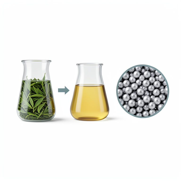
Green Synthesis Process
AI-generated conceptual illustration (Camellia sinensis leaves and silver nanoparticles in laboratory glassware). It explains the idea and does not show study data.
View source evidence ↗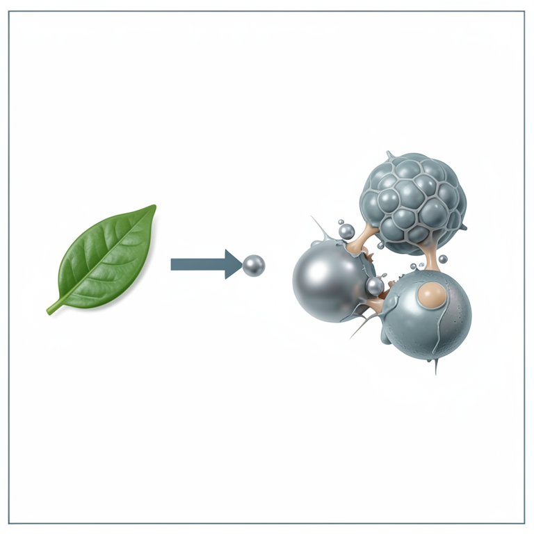
Antimicrobial Action
AI-generated conceptual illustration (Silver nanoparticles and bacterial cells (Gram-positive and Gram-negative)). It explains the idea and does not show study data.
View source evidence ↗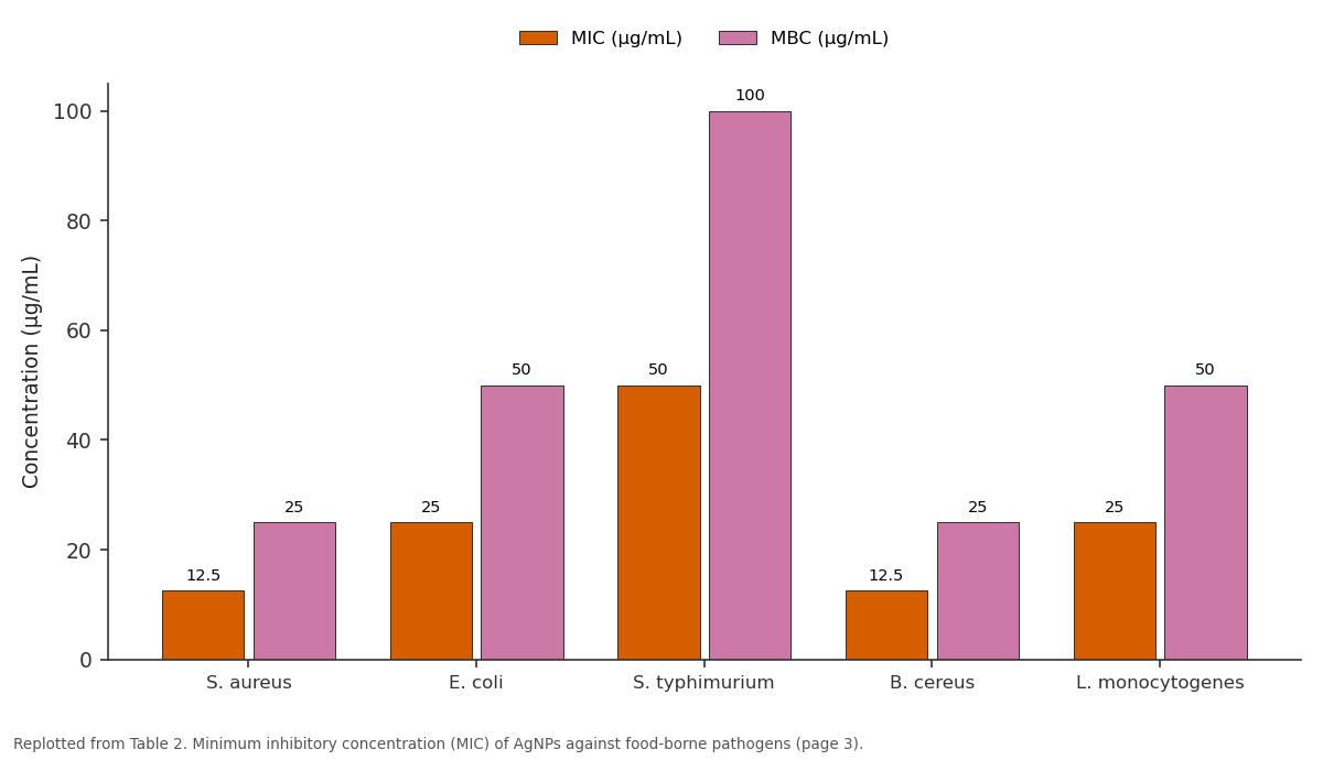
MIC (µg/mL) and MBC (µg/mL): S. aureus vs E. coli vs S. typhimurium vs B. cereus vs L. monocytogenes
Replotted from Table 2 (page 3) of the original paper. Every bar is the paper's own table value.
View source evidence ↗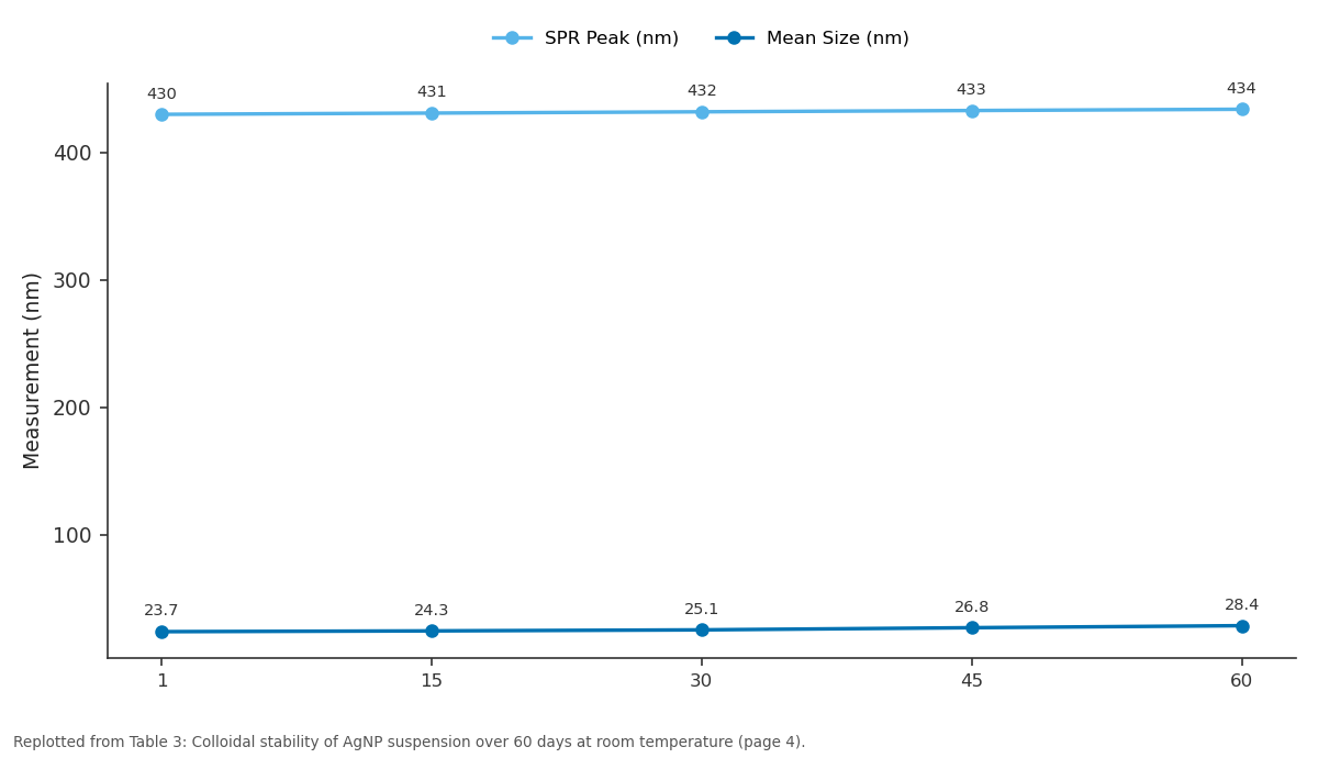
SPR Peak (nm) and Mean Size (nm): 1 vs 15 vs 30 vs 45 vs 60
Replotted from Table 3 (page 4) of the original paper. Every bar is the paper's own table value.
View source evidence ↗Figures & tables
From the original paper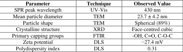 Table 1
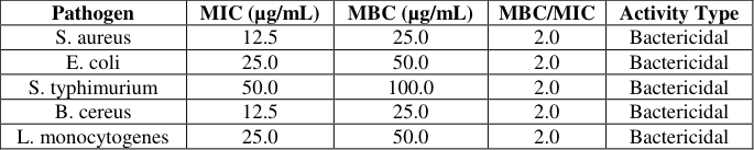 Table 2
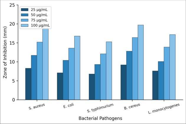 Figure 1
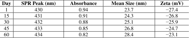 Table 3
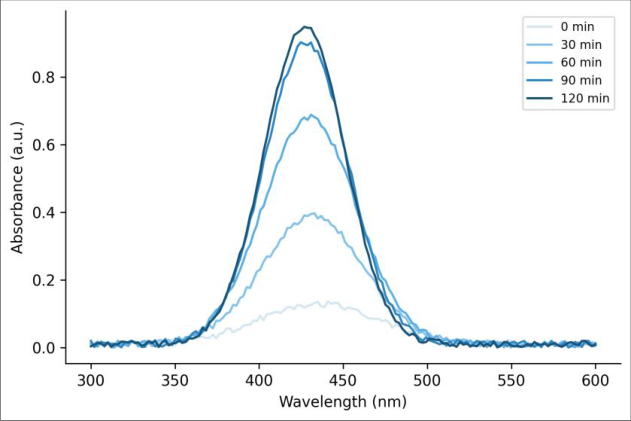 Figure 2
Introduction
How safe is the food sitting on grocery shelves? Food-borne infections account for an estimated 600 million illness episodes and 420,000 deaths each year worldwide, according to figures reported by the World Health Organization [1] . Bacteria such as Salmonella, Listeria, and Staphylococcus rank among the most common culprits, and their growing resistance to traditional preservatives and antibiotics has pressed researchers to look for alternative antimicrobial strategies [2] . Silver, valued for its germicidal properties since antiquity, has reemerged as a candidate material but this time at the nanoscale [3] . Silver nanoparticles (AgNPs) possess a high surface-area-to-volume ratio that amplifies their interaction with microbial cell membranes [4] . Chemical reduction methods using sodium borohydride or hydrazine can produce AgNPs efficiently, yet these reagents raise toxicity and environmental disposal concerns [5] . Green synthesis routes that replace hazardous chemicals with biological extracts have therefore attracted growing interest over the past decade [6] . Plant extracts are particularly appealing because they contain polyphenols, flavonoids, and terpenoids capable of reducing metal ions and simultaneously stabilising the resulting nanoparticles [7] . Among candidate plant sources, Camellia sinensis the common tea plant stands out for several practical reasons. It is cultivated across tropical and subtropical regions, inexpensive, and rich in catechins such as epigallocatechin gallate (EGCG) that act as strong reducing agents [8] . Earlier reports have documented AgNP synthesis from black tea [9] and white tea [10] , but fewer investigations have focused on Brazilian-grown green tea leaves, which differ in catechin profiles owing to local climate and soil conditions [11] . Given that Brazil is the world's fifth-largest tea producer, an efficient protocol using locally sourced C. sinensis could support both sustainability goals and regional agricultural value chains [12] . The antimicrobial mechanism of AgNPs involves multiple pathways. Silver ions released from nanoparticle surfaces bind to thiol groups in bacterial enzymes, disrupting respiratory chain function [13] . Nanoparticles can also penetrate cell walls and damage intracellular DNA [4] . Because these actions target several sites simultaneously, bacteria find it harder to develop resistance compared with single-target antibiotics [14] . Testing AgNPs against a panel of food-borne pathogens rather than one or two indicator organisms gives a broader picture of their practical usefulness. This research therefore aimed to (a) synthesise AgNPs using aqueous leaf extract of C. sinensis grown in São Paulo state, (b) characterise the resulting particles by UV-Vis spectrophotometry, FTIR, TEM, and XRD, and (c) evaluate their antibacterial performance against five food-borne bacteria at multiple concentrations. The work was carried out at the São Paulo Institute of Chemical Sciences between March and September 2024. By combining affordable raw material with straightforward bench-scale procedures, the goal was to develop a reproducible green synthesis protocol whose products could later be tested in food packaging or surface-coating applications [15,16] .
Material and Methods
Material and Methods
Material
Fresh leaves of Camellia sinensis were harvested from a commercial plantation located in Registro, São Paulo state, Brazil (24.49°S, 47.84°W) during April 2024. Leaves were transported in sterile polyethylene bags to the Microbiology and Nanomaterials Laboratory at the São Paulo Institute of Chemical Sciences within four hours of collection. Upon arrival, the leaves were rinsed three times with deionised water to remove surface debris, blotted dry, and stored at 4 °C until use. Silver nitrate (AgNO₃, 99.8% purity) was purchased from Sigma-Aldrich Brazil (São Paulo). Mueller-Hinton agar and nutrient broth were obtained from HiMedia Laboratories. All aqueous solutions were prepared with Milli-Q ultrapure water (18.2 MΩ•cm). The bacterial strains Staphylococcus aureus ATCC 25923, Escherichia coli ATCC 25922, Salmonella typhimurium ATCC 14028, Bacillus cereus ATCC 11778, and Listeria monocytogenes ATCC 19115 were procured from the Oswaldo Cruz Foundation culture collection, Rio de Janeiro [1,4] .
Methods
The aqueous extract was prepared by boiling 10 g of finely chopped fresh leaves in 100 mL of deionised water at 80 °C for 20 minutes. The mixture was filtered through Whatman No. 1 filter paper and cooled to room temperature. For nanoparticle synthesis, 10 mL of the leaf extract was added drop-wise to 90 mL of 1 mM AgNO₃ solution under constant stirring at 60 °C. A colour change from pale yellow to dark brown within 30 minutes indicated the formation of AgNPs [6, 8]. The colloidal suspension was centrifuged at 12,000 rpm for 15 minutes, washed twice with deionised water, and dried in a hot-air oven at 60 °C for 24 hours to obtain AgNP powder. UV-visible absorption spectra were recorded on a Shimadzu UV-1800 spectrophotometer over a wavelength range of 300-600 nm at 30-minute intervals during synthesis. Fourier transform infrared spectroscopy (Bruker Alpha-II, KBr pellet method, 4000-400 cm⁻¹) was used to identify surface functional groups. Particle morphology and size were examined by transmission electron microscopy (JEOL JEM-2100, 200 kV). Crystallinity was assessed by X-ray diffraction (Rigaku MiniFlex 600, Cu-Kα radiation, 2θ = 20-80°) [9] . Antibacterial activity was assessed by the agar well diffusion method. Each bacterial strain was sub-cultured overnight in nutrient broth at 37 °C. A standardised inoculum (0.5 McFarland, ~1.5 × 10⁸ CFU/mL) was spread on Mueller-Hinton agar plates using sterile cotton swabs. Wells of 6 mm diameter were bored into the agar, and 50 µL of AgNP suspension at concentrations of 25, 50, 75, and 100 µg/mL was added to each well. Gentamicin (10 µg/disc) served as a positive control and sterile distilled water as a negative control. Plates were incubated at 37 °C for 24 hours, after which zones of inhibition were measured in millimetres using a digital calliper. Minimum inhibitory concentration (MIC) was determined by the broth microdilution method in 96-well microplates following CLSI guidelines [14] . All assays were performed in triplicate. The research protocol was reviewed by the Institutional Biosafety Committee at the São Paulo Institute of Chemical Sciences (Protocol IBSC-2024-017, approved 12 February 2024) [5] .
Reagents and Chemicals
Silver nitrate (AgNO₃, ≥99.8%, CAS 7761-88-8) was sourced from Sigma-Aldrich Brazil (catalogue no. S6506). Mueller-Hinton agar (M173) and nutrient broth (M002) were supplied by HiMedia Laboratories through a Brazilian distributor. Gentamicin sulphate reference discs (10 µg) came from Oxoid (Basingstoke, UK). Potassium bromide (KBr, spectroscopic grade) for FTIR pellet preparation was purchased from Merck Brazil. All reagents were used as received without additional purification. Stock solutions were prepared fresh on the day of each experiment and stored in amber glass bottles to prevent photodegradation [3,5] .
Quality Control
Instrument calibration followed the manufacturer's recommended schedules. The UV-Vis spectrophotometer was calibrated with holmium oxide and potassium dichromate reference filters before each batch of measurements. TEM magnification was verified against a cross-grating replica standard. For antibacterial assays, positive-control gentamicin discs were included on every plate; any plate whose positive-control zone fell outside the acceptable CLSI range (19-26 mm for E. coli ATCC 25922) was discarded and the test repeated. Triplicate measurements were made for each concentration-pathogen pair, and results are reported as mean ± standard deviation. Inter-day reproducibility was confirmed by repeating the full antimicrobial panel on three separate occasions over a two-week period [14] .
Results: The addition of C. sinensis leaf extract to the AgNO₃ solution triggered a visible colour shift from pale yellow to dark brown within approximately 25 minutes, signalling nanoparticle nucleation. UV-Vis spectral monitoring confirmed a progressively intensifying absorption peak centred near 430 nm, characteristic of the surface plasmon resonance (SPR) band of silver nanoparticles. No absorption peak appeared in the extractonly or AgNO₃-only controls, ruling out artefacts. 200), (220), and (311) planes of metallic silver (JCPDS card no. 04-0783), confirming the crystalline nature of the particles [9] . As Figure 1 shows, inhibition zones increased in a dosedependent manner for all five organisms. At the lowest concentration (25 µg/mL), only B. cereus and S. aureus produced zones exceeding 8 mm. At 100 µg/mL, all pathogens displayed measurable inhibition, with B. cereus reaching the highest value of 19.7 mm followed by S. aureus at 18.9 mm. Gram-positive species consistently showed larger zones than Gram-negative ones, a trend attributable to differences in cell wall architecture [4,13] . of 50 µg/mL. The MBC/MIC ratio equalled 2.0 for every organism tested, which classifies the AgNPs as bactericidal rather than merely bacteriostatic [14] .
Comprehensive Interpretation
Taken as a whole, the characterisation and antimicrobial data paint a consistent picture. The C. sinensis extract produced small, predominantly spherical AgNPs whose crystalline structure and surface chemistry closely match particles reported from other green routes [6, 10]. The dosedependent killing pattern across all five pathogens, combined with uniformly bactericidal MBC/MIC ratios, indicates that the biogenic capping layer does not impede antimicrobial action. Stability over 60 days at ambient conditions is a practical advantage for potential downstream applications. The greater susceptibility of Gram-positive bacteria aligns with their thinner peptidoglycan layer compared with the additional outer membrane present in Gram-negative species [4,13] .
Discussion
The mean particle size of 23.7 nm recorded in this research falls within the 10-50 nm window commonly associated with strong antimicrobial performance. Sharma et al. [6] obtained AgNPs of 20-35 nm from Azadirachta indica leaf extract, while Otunola and Afolayan [10] reported 15-28 nm particles from white tea. The slightly larger mean here may reflect the lower catechin concentration in Brazilian green tea compared with East Asian cultivars, which would slow reduction kinetics and permit additional crystal growth before capping stabilises the particles [8,11] . Antimicrobial zones at 100 µg/mL exceeded those described by Loo et al. [9] for black tea-derived AgNPs (14-16 mm against S. aureus), probably because of the smaller average size and the consequent increase in total surface area per unit mass. MIC values of 12.5 µg/mL for S. aureus and B. cereus are competitive with chemically synthesised AgNPs reported elsewhere [5] . The bactericidal classification (MBC/MIC = 2.0) points to membrane disruption as a likely killing mechanism rather than simple growth inhibition [4,13] .
And the greater resistance of Gram-negative bacteria S. typhimurium in particular matches a well-established pattern. The lipopolysaccharide-rich outer membrane forms an additional permeability barrier that slows silver ion diffusion into the cytoplasm [3] . Even so, all Gram-negative isolates were inhibited at concentrations ≤ 50 µg/mL, which may still be workable for food-contact applications where pathogens are present in low numbers.
Colloidal stability deserves attention because real-world applications demand shelf-life. The 4 nm red-shift and modest size increase over 60 days suggest slow but measurable Ostwald ripening; yet the zeta potential remained below -23 mV, indicating that electrostatic repulsion continued to counteract aggregation [7] .
Incorporating the nanoparticles into a polymer matrix for instance, in biodegradable food-wrap films could further limit ripening by restricting particle mobility [15] . One limitation of this work is that cytotoxicity toward mammalian cells was not assessed. Before the AgNPs can be proposed for food-contact use, cell-viability assays and migration testing would be needed to satisfy regulatory requirements [16] . The antimicrobial panel, though covering five important food-borne species, did not include fungal pathogens, which also contribute to food spoilage [2] .
Conclusion
This research set out to prepare silver nanoparticles through a green route using Camellia sinensis leaf extract from São Paulo state and to evaluate their antibacterial potential against five food-borne pathogens. The biosynthesis protocol proved straightforward: mixing the aqueous leaf extract with 1 mM silver nitrate at 60 °C yielded stable colloidal silver within two hours, requiring no hazardous reducing agents or elaborate equipment. Characterisation confirmed that the product consisted of crystalline, predominantly spherical AgNPs with a mean diameter of 23.7 nm, capped by polyphenolic residues from the tea extract. The nanoparticles exhibited a well-defined surface plasmon resonance peak near 430 nm and a moderately negative zeta potential (-27.4 mV), consistent with reasonable colloidal stability. Over a 60-day observation period the suspension showed only minor redshift and size increase, suggesting acceptable shelf-life for laboratory-scale storage. Antibacterial evaluation demonstrated clear dose-dependent inhibition of all five organisms. B. cereus and S. aureus were the most sensitive, with MIC values as low as 12.5 µg/mL and inhibition zones approaching 20 mm at 100 µg/mL. Even the most resistant species, S. typhimurium, was inhibited at 50 µg/mL. Every pathogen-concentration pair yielded an MBC/MIC ratio of 2.0, pointing to a bactericidal mode of action. Gram-positive bacteria were consistently more susceptible than their Gram-negative counterparts, a finding attributable to differences in outer membrane complexity. From a practical standpoint, the results indicate that Brazilian-grown C. sinensis can serve as an effective and affordable feedstock for green AgNP synthesis. The combination of low raw-material cost, ambient-temperature stability, and broad-spectrum bactericidal activity makes these nanoparticles worth exploring further in food-safety contexts for example, as active ingredients in edible coatings, packaging films, or surface sanitisers.
Several avenues remain open for future work. First, cytotoxicity and migration studies are needed before any food-contact application can be considered. Second, extending the antimicrobial panel to include fungal species such as Aspergillus and Candida would broaden the relevance of the findings. Third, encapsulating the AgNPs in biopolymer matrices like chitosan or cellulose could improve shelf-life and control release kinetics. Fourth, scaling the synthesis from bench-top glassware to a continuous-flow reactor would be a necessary step toward commercial viability. Finally, life-cycle assessment comparing the environmental footprint of this green route with conventional chemical synthesis would help quantify the sustainability advantage that biological methods are assumed to offer.
Figures and tables from the paper
| Parameter | Technique | Observed Value |
|---|---|---|
| SPR peak wavelength | UV-Vis | 430 nm |
| Mean particle diameter | TEM | 23.7 ± 4.2 nm |
| Particle shape | TEM | Spherical (89%) |
| Crystalline structure | XRD | Face-centred cubic |
| Primary capping groups | FTIR | -OH, C=O, C-O-C |
| Zeta potential | DLS | −27.4 mV |
| Polydispersity index | DLS | 0.31 |
Table text extracted from the original PDF; superscript letters are shown inline. The original table image is below.
View original table image
| Pathogen | MIC (µg/mL) | MBC (µg/mL) | MBC/MIC | Activity Type |
|---|---|---|---|---|
| S. aureus | 12.5 | 25.0 | 2.0 | Bactericidal |
| E. coli | 25.0 | 50.0 | 2.0 | Bactericidal |
| S. typhimurium | 50.0 | 100.0 | 2.0 | Bactericidal |
| B. cereus | 12.5 | 25.0 | 2.0 | Bactericidal |
| L. monocytogenes | 25.0 | 50.0 | 2.0 | Bactericidal |
Table text extracted from the original PDF; superscript letters are shown inline. The original table image is below.
View original table image
| Day | SPR Peak (nm) | Absorbance | Mean Size (nm) | Zeta (mV) |
|---|---|---|---|---|
| 1 | 430 | 0.94 | 23.7 | −27.4 |
| 15 | 431 | 0.91 | 24.3 | −26.8 |
| 30 | 432 | 0.88 | 25.1 | −25.9 |
| 45 | 433 | 0.85 | 26.8 | −24.7 |
| 60 | 434 | 0.82 | 28.4 | −23.1 |
Table text extracted from the original PDF; superscript letters are shown inline. The original table image is below.
View original table image
References
16 in the original paper- Kirk MD, Pires SM, Black RE, Caipo M, Crump JA, Devleesschauwer B et al. World Health Organization estimates of the global and regional disease burden of 22 foodborne bacterial, protozoal, and viral diseases, 2010: A data synthesis. PLoS Medicine. 2015;12(12):e1001921.
- Bintsis T. Foodborne pathogens. AIMS Microbiology. 2017;3(3):529-563.
- Rai M, Yadav A, Gade A. Silver nanoparticles as a new generation of antimicrobials. Biotechnology Advances. 2009;27(1):76-83.
- Morones JR, Elechiguerra JL, Camacho A, Holt K, Kouri JB, Ramírez JT et al. The bactericidal effect of silver nanoparticles. Nanotechnology. 2005;16(10):2346-2353.
- Zhang XF, Liu ZG, Shen W, Gurunathan S. Silver nanoparticles: Synthesis, characterization, properties, applications, and therapeutic approaches. International Journal of Molecular Sciences. 2016;17(9):1534.
- Sharma VK, Yngard RA, Lin Y. Silver nanoparticles: Green synthesis and their antimicrobial activities. Advances in Colloid and Interface Science. 2009;145(12):83-96.
- Iravani S, Korbekandi H, Mirmohammadi SV, Zolfaghari B. Synthesis of silver nanoparticles: Chemical, physical and biological methods. Research in Pharmaceutical Sciences. 2014;9(6):385-406.
- Begum NA, Mondal S, Basu S, Laskar RA, Mandal D. Biogenic synthesis of Au and Ag nanoparticles using aqueous solutions of black tea leaf extracts. Colloids and Surfaces B: Biointerfaces. 2009;71(1):113-118.
- Loo YY, Chieng BW, Nishibuchi M, Radu S. Synthesis of silver nanoparticles by using tea leaf extract from Camellia sinensis. International Journal of Nanomedicine. 2012;7:4263-4267.
- Otunola GA, Afolayan AJ. Characterization, antibacterial and antioxidant properties of silver nanoparticles synthesized from aqueous extracts of Allium sativum, Zingiber officinale, and Capsicum frutescens. Pharmacognosy Magazine. 2017;13(Suppl 2):S201-S208.
- Cabrera C, Artacho R, Giménez R. Beneficial effects of green tea -a review. Journal of the American College of Nutrition. 2006;25(2):79-99.
- Food and Agriculture Organization. FAOSTAT statistical database: Tea production quantity. FAO, Rome, 2023.
- Kim JS, Kuk E, Yu KN, Kim JH, Park SJ, Lee HJ et al. Antimicrobial effects of silver nanoparticles. Nanomedicine: Nanotechnology, Biology and Medicine. 2007;3(1):95-101.
- CLSI. Methods for dilution antimicrobial susceptibility tests for bacteria that grow aerobically: Approved standard, 11th ed. CLSI document M07. Wayne, PA: Clinical and Laboratory Standards Institute, 2018.
- Rhim JW, Park HM, Ha CS. Bio-nanocomposites for food packaging applications. Progress in Polymer Science. 2013;38(10-11):1629-1652.
- EFSA Scientific Committee. Guidance on risk assessment of the application of nanoscience and nanotechnologies in the food and feed chain: Part 1. EFSA Journal. 2018;16(7):e05327.