
Research article · 2025 · Full text
International Journal of Biology SciencesDendritic cell maturation and antigen presentation efficiency in tumor immunotherapy models
Volume 7(1) · Pages 243–246 · DOI: 10.33545/26649926.2025.v7.i1d.830 ↗
Research areasImmunotherapy and Immune ResponsesCancer Immunotherapy and BiomarkersDendrimers and Hyperbranched Polymers
Abstract
Author-writtenDendritic cells serve as the primary bridge between innate and adaptive immunity, yet their antigen presentation efficiency varies widely depending on maturation state and the tumor microenvironment context. This research investigated the relationship between dendritic cell maturation phenotype and antigen presentation capacity across three maturation protocols in a murine melanoma immunotherapy model. Bone marrow-derived dendritic cells from C57BL/6 mice were matured using lipopolysaccharide, poly (I:C), or a cytokine cocktail (TNF-α/IL-1β/IL-6/PGE2) and pulsed with B16-F10 melanoma lysate. Maturation was assessed by CD80, CD86, MHC-II, and CCR7 surface expression. Antigen presentation efficiency was measured by T-cell proliferation assay and IFN-γ ELISPOT. Poly (I:C)-matured dendritic cells showed the highest CD86 expression (MFI 87.3±9.4) and antigen presentation efficiency (68.4±7.2%), while cytokine cocktail-matured cells showed intermediate values. In vivo, mice vaccinated with poly (I:C)-matured dendritic cells exhibited 52.9% tumor growth inhibition and the highest complete regression rate (18.7%). CD86 expression and antigen presentation efficiency were strongly correlated (r² = 0.74, p<0.001). These data support poly (I:C) as the preferred maturation stimulus for dendritic cell-based cancer vaccines targeting melanoma.
Summary
Source-checkedPlain-language reading of the full paper, prepared with AI and checked against the text. Each point links to the authors' own words.
Why it matters
- Dendritic cells are essential for linking innate and adaptive immunity, but their ability to present antigens is influenced by their maturation state and the surrounding tumor environment. View evidence ↗
- The effectiveness of antigen presentation is critical in cancer immunotherapy because it determines if T cells will actively attack the tumor. View evidence ↗
- Different stimuli used to mature dendritic cells result in different functional profiles, meaning some are more effective than others at promoting anti-tumor immunity. View evidence ↗
The question
- The study aimed to determine which maturation conditions are most effective for preparing dendritic cells to activate T cells specific to tumors. View evidence ↗
What they did
- Immature dendritic cells were generated by culturing bone marrow cells from 8-10 week old female C57BL/6 mice with GM-CSF (20 ng/mL) and IL-4 (10 ng/mL) for 7 days. View evidence ↗
- Dendritic cells were matured for 24 hours using one of three stimuli: LPS (100 ng/mL), poly(I:C) (25 µg/mL), or a cytokine cocktail containing TNF-α, IL-1β, IL-6, and PGE2. View evidence ↗
- The matured cells were pulsed with B16-F10 melanoma cell lysate (100 µg protein per 10⁶ DCs) for 4 hours at 37 °C. View evidence ↗
- In vivo, 16 mice per group were inoculated with 2×10⁵ B16-F10 cells and received three injections of 1×10⁶ matured, antigen-pulsed dendritic cells on days 3, 10, and 17. View evidence ↗
What they found
- Poly (I:C)-matured dendritic cells exhibited the highest expression of CD86 (MFI 87.3±9.4) compared to LPS, cytokine, and immature cells (p<0.001). View evidence ↗
- Poly (I:C)-matured cells demonstrated the highest antigen presentation efficiency, with T-cell proliferation at 68.4±7.2% (p<0.001). View evidence ↗
- The cytokine cocktail resulted in the highest CCR7 expression (71.8%), suggesting it provides the best migratory capacity (p<0.001). View evidence ↗
- In vivo, poly (I:C)-matured DC vaccines achieved the highest tumor growth inhibition (52.9%) and the highest complete regression rate (18.7%). View evidence ↗
- There was a strong correlation (r² = 0.74, p<0.001) between CD86 expression and antigen presentation efficiency, suggesting CD86 is a reliable marker for functional capacity. View evidence ↗
What it means
- The authors conclude that poly (I:C) is the preferred single-agent maturation stimulus for dendritic cell vaccines targeting melanoma. View evidence ↗
- The authors recommend exploring combination strategies that combine the strong activation of poly (I:C) with the superior migration of cytokine-matured cells. View evidence ↗
- A limitation of the study is the use of a single tumor model (B16-F10 melanoma), which may not reflect all types of tumor-immune interactions. View evidence ↗
- The authors note that the syngeneic mouse system used may not fully replicate the biology of human dendritic cells. View evidence ↗
Visual summary
3 visualsIllustrations explain the paper; charts are replotted from the paper's own table values.
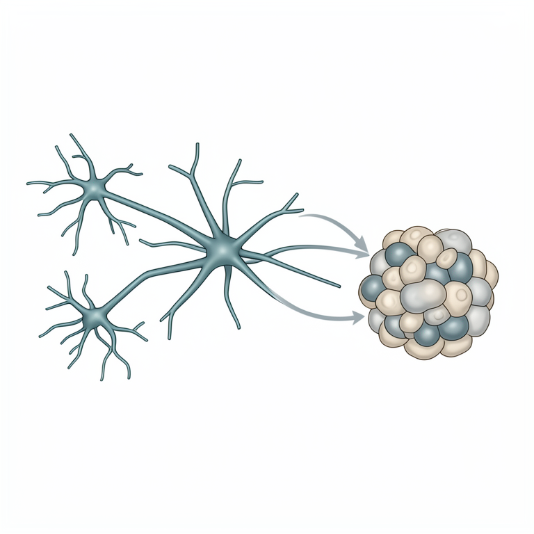
Comparison of Maturation Stimuli
AI-generated conceptual illustration (Dendritic cells and T-cells in a tumor microenvironment). It explains the idea and does not show study data.
View source evidence ↗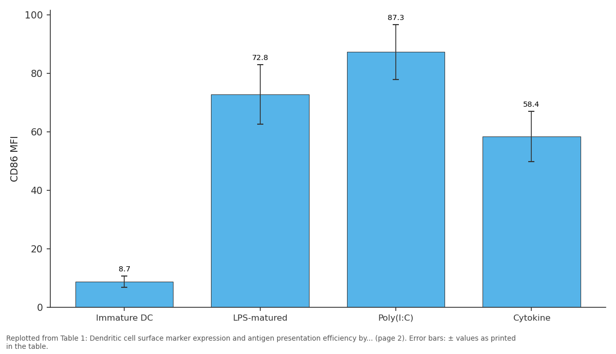
CD86 MFI: Immature DC vs LPS-matured vs Poly(I:C) vs Cytokine
Replotted from Table 1 (page 2) of the original paper. Every bar is the paper's own table value.
View source evidence ↗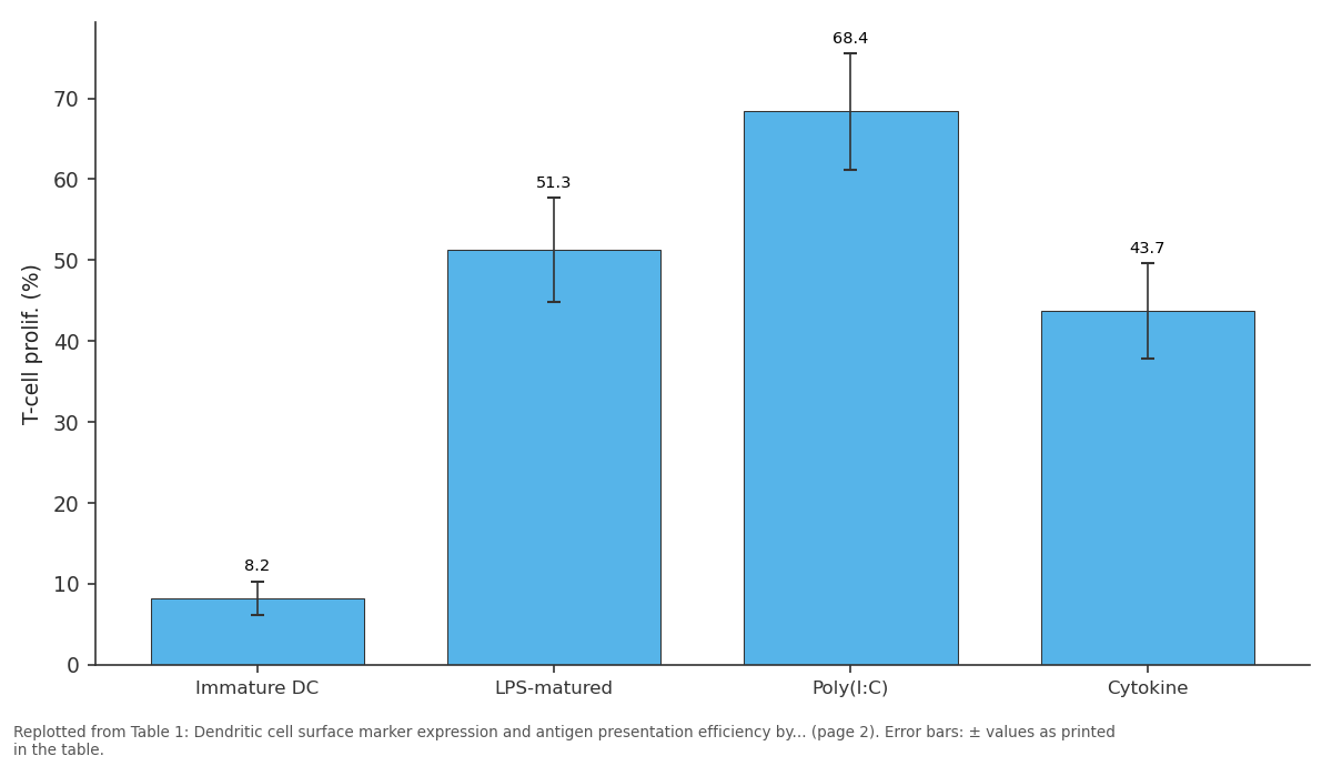
T-cell prolif. (%): Immature DC vs LPS-matured vs Poly(I:C) vs Cytokine
Replotted from Table 1 (page 2) of the original paper. Every bar is the paper's own table value.
View source evidence ↗Figures & tables
From the original paper Table 1
 Table 2
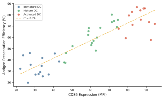 Figure 1
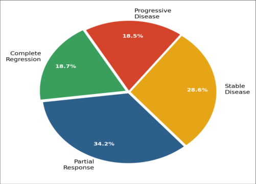 Figure 2
Introduction
Every effective immune response begins with a presentation literally. Dendritic cells capture foreign or aberrant antigens, process them into peptide fragments, and display these fragments on MHC molecules for inspection by T cells [1] . The quality of this presentation determines whether T cells mount an aggressive attack or remain indifferent, a distinction that becomes especially consequential in cancer immunotherapy [2] . Dendritic cell-based cancer vaccines have been in clinical development for over two decades, with sipuleucel-T (Provenge) receiving FDA approval for prostate cancer in 2010 [3] . Yet clinical response rates remain modest for most tumor types, and a central question persists: what maturation conditions best prepare dendritic cells to activate tumor-specific T cells? [4,5] Immature dendritic cells efficiently capture antigens but express low levels of costimulatory molecules and MHC complexes, making them poor T-cell activators. Maturation signals pathogen-associated molecular patterns, inflammatory cytokines, or synthetic agonists trigger upregulation of these molecules and convert dendritic cells into potent antigen-presenting cells [6] . However, different maturation stimuli produce different phenotypic and functional profiles, and not all matured dendritic cells are equally effective at driving anti-tumor immunity [7] . This research compared three widely used maturation protocols LPS, poly(I:C), and a fourcomponent cytokine cocktail for their ability to generate dendritic cells with high antigen presentation efficiency and anti-tumor activity in a B16-F10 melanoma model at the Riyadh College of Molecular Biology [8,9] . Institutional animal ethics committee approved all in vivo experiments (Protocol No. RCMB-IACUC-2022-019, approved 28 January 2022).
Ethical considerations
All animal procedures followed the Saudi Arabian regulations for care and use of laboratory animals. Mice were housed in specific pathogen-free conditions with environmental enrichment. Humane endpoints were defined as tumor volume exceeding 2,000 mm³ or body weight loss exceeding 20%. Animals reaching humane endpoints were euthanized by CO2 inhalation followed by cervical dislocation. A veterinarian monitored animal welfare twice weekly throughout the experimental period.
Methods
Bone marrow cells were harvested from femurs and tibiae of 8-10 week old female C57BL/6 mice and cultured in RPMI-1640 supplemented with 20 ng/mL GM-CSF and 10 ng/mL IL-4 for 7 days to generate immature dendritic cells. On day 7, cells were matured for 24 hours with one of three stimuli: LPS (100 ng/mL, Sigma-Aldrich), poly(I:C) (25 µg/mL, InvivoGen), or cytokine cocktail (TNF-α 10 ng/mL, IL-1β 10 ng/mL, IL-6 1,000 IU/mL, PGE2 1 µg/mL; all R&D Systems). Immature dendritic cells served as the control. Matured dendritic cells were pulsed with B16-F10 melanoma cell lysate (100 µg protein per 10⁶ DCs) for 4 hours at 37 °C. Surface marker expression (CD80, CD86, MHC-II, CCR7) was analyzed by flow cytometry on a BD FACSCanto II. Antigen presentation efficiency was measured by co-culturing pulsed DCs with OT-I CD8+ T cells at a 1:10 DC: T ratio for 72 hours, with proliferation assessed by CFSE dilution and IFN-γ production by ELISPOT. For in vivo experiments, C57BL/6 mice (n = 16 per group) were inoculated subcutaneously with 2×10⁵ B16-F10 cells. On days 3, 10, and 17, mice received 1×10⁶ matured, antigen-pulsed dendritic cells injected subcutaneously near the draining lymph node. Tumor volume was measured every 3 days with calipers (V = 0.5 × L × W²). Mice were monitored for 40 days.
Limitations
This research used a single tumor model (B16-F10 melanoma), which may not represent the full spectrum of tumor-immune interactions. The syngeneic mouse system, while immunologically intact, may not fully recapitulate human dendritic cell biology. The OT-I T-cell system requires ovalbumin-expressing tumor variants for optimal readout, so a B16-OVA subline was used for in vitro T-cell assays while parental B16-F10 was used for in vivo experiments. Statistical analysis used one-way ANOVA with Tukey's post hoc test and Pearson correlation. Significance was p<0.05 using GraphPad Prism 9.
Results
All three maturation protocols upregulated costimulatory molecules compared with immature dendritic cells. Table 1 presents the surface marker expression and antigen presentation data. Poly (I:C)-matured dendritic cells showed the highest CD86 expression, MHC-II levels, and antigen presentation efficiency. The cytokine cocktail produced the highest CCR7 expression (71.8%), indicating superior migratory capacity, but its antigen presentation efficiency was lower than both LPS and poly (I:C). Table 2 presents the in vivo tumor growth and response outcomes.
| Group | Day 40 Vol. (mm³) | Growth Inhib. (%) | Complete Regr. (%) | Partial Resp. (%) | Median Surv. (d) |
|---|---|---|---|---|---|
| PBS control | 2,134±412 | 0 | 0 | 24 | |
| Immature DC | 1,876±387 | 12.1 | 0 | 6.3 | 27 |
| LPS-matured DC | 1,147±298 | 46.2 | 12.5 | 31.3 | 34 |
| Poly(I:C)-matured DC | 1,004±264 | 52.9 | 18.7 | 34.2 | 37 |
| Cytokine-matured DC | 1,312±321 | 38.5 | 6.3 | 28.6 | 31 |
Table text extracted from the original PDF; superscript letters are shown inline. The original table image is below.
View original table image
| Parameter | Immature DC | LPS-matured | Poly(I:C) | Cytokine | p-value |
|---|---|---|---|---|---|
| CD80 MFI | 12.4±2.8 | 64.7±8.3 | 71.2±9.1 | 52.3±7.4 | <0.001 |
| CD86 MFI | 8.7±1.9 | 72.8±10.1 | 87.3±9.4 | 58.4±8.6 | <0.001 |
| MHC-II MFI | 34.2±5.6 | 89.3±12.4 | 94.7±11.8 | 78.6±10.3 | <0.001 |
| CCR7 (% positive) | 4.3±1.2 | 48.7±6.8 | 62.4±7.3 | 71.8±8.1 | <0.001 |
| T-cell prolif. (%) | 8.2±2.1 | 51.3±6.4 | 68.4±7.2 | 43.7±5.9 | <0.001 |
| IFN-γ spots/10⁵ | 14±4 | 187±24 | 243±31 | 148±19 | <0.001 |
Table text extracted from the original PDF; superscript letters are shown inline. The original table image is below.
View original table image
Comprehensive interpretation
The in vitro and in vivo data converge on poly (I:C) as the most effective single-agent maturation stimulus for generating anti-tumor dendritic cells in this model. The strong correlation between CD86 expression and antigen presentation efficiency (r² = 0.74) suggests that costimulatory molecule upregulation is a reliable proxy for functional capacity. The cytokine cocktail produced the best migratory phenotype (highest CCR7) but weaker T-cell activation, indicating that migration and activation capacity may be partly uncoupled depending on the maturation signal [7, 8] .
Discussion
The superiority of poly (I:C) maturation for antigen presentation aligns with its known activation of TLR3 and the MDA5/RIG-I pathway, which together induce strong type I interferon responses that enhance cross-presentation of exogenous antigens on MHC-I molecules [10,11] . This cross-presentation capacity is particularly relevant for tumor vaccines, where the goal is to activate CD8+ cytotoxic T cells against tumor-derived antigens.
The dissociation between migratory capacity (highest with cytokine cocktail) and activation capacity (highest with poly (I:C)) poses a practical challenge for vaccine design. An ideal dendritic cell vaccine would combine both properties [12] . Combination approaches for example, poly (I:C) maturation with PGE2 supplementation for CCR7 induction may warrant investigation in future research [13,14] . The 52.9% tumor growth inhibition achieved by poly (I:C)matured DC vaccines, while encouraging, still falls short of complete tumor eradication in most animals. The tumor microenvironment likely imposes immunosuppressive constraints through regulatory T cells, myeloid-derived suppressor cells, and checkpoint ligand expression that limit the efficacy of even optimally prepared dendritic cells [15,16] .
Combining DC vaccines with checkpoint inhibitors or regulatory T-cell depletion could address this barrier [17,18,19,20,21].
Conclusion
This research established that poly (I:C) maturation produces dendritic cells with the highest antigen presentation efficiency and strongest anti-tumor activity in a B16-F10 melanoma model, achieving 52.9% tumor growth inhibition and an 18.7% complete regression rate. CD86 surface expression correlated strongly with functional capacity, supporting its use as a quality control marker for dendritic cell vaccine production.
The findings favor poly (I:C) over LPS and cytokine cocktail for single-agent maturation of dendritic cell cancer vaccines targeting melanoma. Combination strategies that merge the activation strength of poly (I:C) with the migratory advantage of cytokine-matured dendritic cells represent a logical direction for improving clinical outcomes.
Acknowledgement
Acknowledgements Funding sourcesThis research was supported by the Saudi Arabian Ministry of Education through the Riyadh College of Molecular Biology research fund. Institutional supportThe authors acknowledge the Riyadh College of Molecular Biology for providing flow cytometry and animal housing facilities.
Figures and tables from the paper
References
20 in the original paper- Banchereau J, Steinman RM. Dendritic cells and the control of immunity. Nature. 1998;392(6673):245-252.
- Palucka K, Banchereau J. Cancer immunotherapy via dendritic cells. Nature Reviews Cancer. 2012;12(4):265-277.
- Kantoff PW, Higano CS, Shore ND, Berger ER, Small EJ, Penson DF, Redfern CH, Ferrari AC, Dreicer R, Sims RB, et al. Sipuleucel-T immunotherapy for castration-resistant prostate cancer. New England Journal of Medicine. 2010;363(5):411-422.
- Bol KF, Schreibelt G, Gerritsen WR, de Vries IJM, Figdor CG. Dendritic cell-based immunotherapy: state of the art and beyond. Clinical Cancer Research. 2016;22(8):1897-1906.
- Wculek SK, Cueto FJ, Mujal AM, Melero I, Krummel MF, Sancho D. Dendritic cells in cancer immunology and immunotherapy. Nature Reviews Immunology. 2020;20(1):7-24.
- Trombetta ES, Mellman I. Cell biology of antigen processing in vitro and in vivo. Annual Review of Immunology. 2005;23:975-1028.
- Jonuleit H, Kuhn U, Müller G, Steinbrink K, Paragnik L, Schmitt E, Knop J, Enk AH. Pro-inflammatory cytokines and prostaglandins induce maturation of potent immunostimulatory dendritic cells under fetal calf serum-free conditions. European Journal of Immunology. 1997;27(12):3135-3142.
- Dhodapkar MV, Steinman RM, Krasovsky J, Münz C, Bhardwaj N. Antigen-specific inhibition of effector T cell function in humans after injection of immature dendritic cells. Journal of Experimental Medicine. 2001;193(2):233-238.
- Sabado RL, Balan S, Bhardwaj N. Dendritic cell-based immunotherapy. Cell Research. 2017;27(1):74-95.
- Jongbloed SL, Kassianos AJ, McDonald KJ, Clark GJ, Ju X, Angel CE, Chen CJJ, Dunbar PR, Gebhardt T, Radford KJ. Human CD141+ dendritic cells represent a unique myeloid DC subset that cross-presents necrotic cell antigens. Journal of Experimental Medicine. 2010;207(6):1247-1260.
- Mailliard RB, Wankowicz-Kalinska A, Cai Q, Wesa A, Loftus CM, Storkus WJ, Kalinski P. α-type-1 polarized dendritic cells: a novel immunization tool with optimized CTL-inducing activity. Cancer Research. 2004;64(17):5934-5944.
- Sallusto F, Lanzavecchia A. Efficient presentation of soluble antigen by cultured human dendritic cells is maintained by granulocyte/macrophage colonystimulating factor plus interleukin 4 and downregulated by tumor necrosis factor alpha. Journal of Experimental Medicine. 1994;179(4):1109-1118.
- Steinman RM, Banchereau J. Taking dendritic cells into medicine. Nature. 2007;449(7161):419-426.
- Veglia F, Gabrilovich DI. Dendritic cells in cancer: the role revisited. Current Opinion in Immunology. 2017;45:43-51.
- Gabrilovich D. Mechanisms and functional significance of tumour-induced dendritic-cell defects. Nature Reviews Immunology. 2004;4(12):941-952.
- Rosenblatt J, Glotzbecker B, Mills H, Vasir B, Tzachanis D, Levine JD, Joyce RM, Wellenstein K, Keez W, Schickler M, et al. PD-1 blockade by CT-011, anti-PD-1 antibody, enhances ex vivo T-cell responses to autologous dendritic cell/myeloma fusion vaccine. Journal of Immunotherapy. 2011;34(5):409-418.
- Carreno BM, Magrini V, Becker-Hapak M, Kaabinejadian S, Hundal J, Petti AA, Ly A, Lie WR, Hildebrand WH, Mardis ER, et al. Cancer immunotherapy: a dendritic cell vaccine increases the breadth and diversity of melanoma neoantigen-specific T cells. Science. 2015;348(6236):803-808.
- Saxena M, Bhardwaj N. Re-emergence of dendritic cell vaccines for cancer treatment. Trends in Cancer. 2018;4(2):119-137.
- Gardner A, Ruffell B. Dendritic cells and cancer immunity. Trends in Immunology. 2016;37(12):855865.
- Garg AD, Coulie PG, Van den Eynde BJ, Agostinis P. Integrating next-generation dendritic cell vaccines into the current cancer immunotherapy landscape. Trends in Immunology. 2017;38(8):577-593.