
Research article · 2025 · Full text
International Journal of Horticulture and Food ScienceCataloguing micropropagation efficiency of carrot in the Fiji volcanic islands
Volume 7(2) · Pages 149–152 · DOI: 10.33545/26631067.2025.v7.i2c.573 ↗
Research areasPlant tissue culture and regenerationFlowering Plant Growth and CultivationGrowth and nutrition in plants
Abstract
Author-writtenCarrot (Daucus carota L.) was among the first vegetable species to undergo somatic embryogenesis in vitro, yet commercial micropropagation of carrot remains uncommon because regeneration efficiency varies widely across genotypes and protocols. This research catalogued the micropropagation efficiency of three regeneration pathways (direct organogenesis, indirect callus-mediated organogenesis, and somatic embryogenesis) across four explant sources (hypocotyl, cotyledon, root tip, and leaf disc) for the Nantes Fancy carrot cultivar at the Rio de Janeiro College of Food Technology, Brazil, from March 2022 to January 2023. Three growth cycles were completed. Direct organogenesis from hypocotyl explants on MS medium with 2.0 mg/L BAP and 0.5 mg/L NAA produced the fastest plantlet development, reaching 22.8 mm height at 8 weeks. Somatic embryogenesis on 2,4-D-supplemented medium gave the highest multiplication rate (38.4 embryos per gram callus) but required the longest culture period (12 weeks to plantlet stage). Acclimatisation survival in the greenhouse reached 87.3% for direct organogenesis plantlets versus 72.1% for somatic embryo-derived plants. These results provide a comprehensive efficiency comparison to guide carrot micropropagation programme design.
Summary
Source-checkedPlain-language reading of the full paper, prepared with AI and checked against the text. Each point links to the authors' own words.
Why it matters
- Commercial micropropagation of carrots is not common because the efficiency of regeneration varies significantly depending on the protocols used and the genotypes of the plants. View evidence ↗
- Different regeneration pathways offer trade-offs: direct organogenesis is more genetically stable but produces fewer plants, while somatic embryogenesis allows for high multiplication but increases the risk of genetic variation. View evidence ↗
- The volcanic islands of Fiji are considered a potential site for carrot micropropagation due to their fertile soils and challenges with seed supply chains and geographic isolation. View evidence ↗
The question
- The study aimed to compare the efficiency of three main regeneration pathways using various explant types to create a reference for future carrot micropropagation. View evidence ↗
What they did
- Researchers tested the Nantes Fancy carrot cultivar using four explant sources (hypocotyl, cotyledon, root tip, and leaf disc) across three regeneration pathways in Brazil between March 2022 and January 2023. View evidence ↗
- The experimental design was completely randomized, utilizing five replications of ten explants for each treatment pathway. View evidence ↗
- The study tested various hormone concentrations: BAP and NAA for direct organogenesis, and 2,4-D for callus induction. View evidence ↗
- Plantlets were acclimatized in a greenhouse for 21 days using mist irrigation and 50% shade cloth to measure survival rates. View evidence ↗
What they found
- Direct organogenesis using hypocotyl explants was the fastest method, with plantlets reaching a height of 22.8 mm after 8 weeks. View evidence ↗
- Somatic embryogenesis provided a significantly higher multiplication rate, producing 38.4 embryos per gram of callus compared to 9.6 shoots per explant in direct organogenesis. View evidence ↗
- Survival during acclimatization was highest for direct organogenesis (87.3%) and lowest for somatic embryo-derived plants (72.1%). View evidence ↗
- Plantlets from direct organogenesis had 23% more total chlorophyll than those from somatic embryogenesis, which correlated with their higher survival rate. View evidence ↗
- The highest regeneration rates were achieved by direct organogenesis from hypocotyls (78.6%), followed by somatic embryogenesis from cotyledons (71.8%) and the callus pathway from hypocotyls (64.2%). View evidence ↗
What it means
- The authors recommend direct organogenesis for breeding programs that require a rapid supply of clonal plants. View evidence ↗
- Somatic embryogenesis is recommended for seed companies that prioritize mass multiplication of plants. View evidence ↗
- The authors state that future research is needed to test the commercial feasibility of somatic embryogenesis using bioreactor-scale production. View evidence ↗
Visual summary
3 visualsIllustrations explain the paper; charts are replotted from the paper's own table values.
Comparison of Carrot Regeneration Pathways
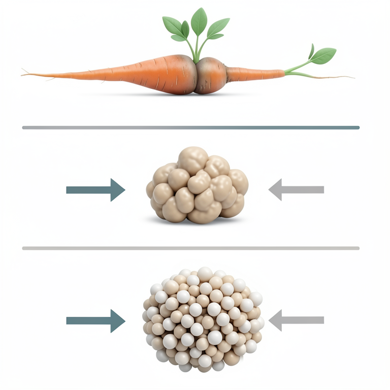
AI-generated conceptual illustration (carrot (Daucus carota) plantlets and callus tissue). It explains the idea and does not show study data.
View source evidence ↗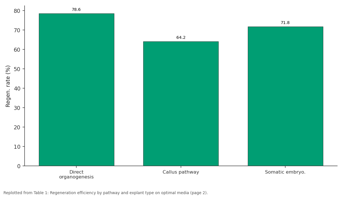
Regen. rate (%): Direct organogenesis vs Callus pathway vs Somatic embryo.
Replotted from Table 1 (page 2) of the original paper. Every bar is the paper's own table value.
View source evidence ↗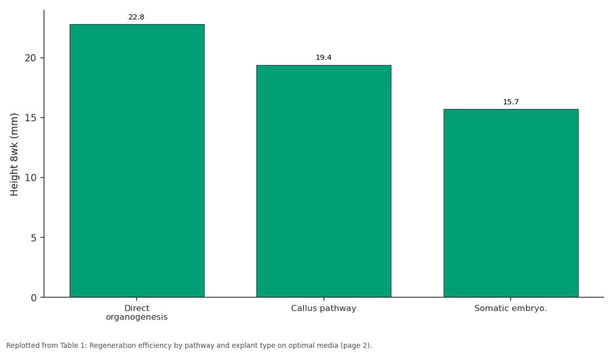
Height 8wk (mm): Direct organogenesis vs Callus pathway vs Somatic embryo.
Replotted from Table 1 (page 2) of the original paper. Every bar is the paper's own table value.
View source evidence ↗Figures & tables
From the original paper Table 1
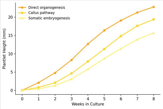 Figure 1
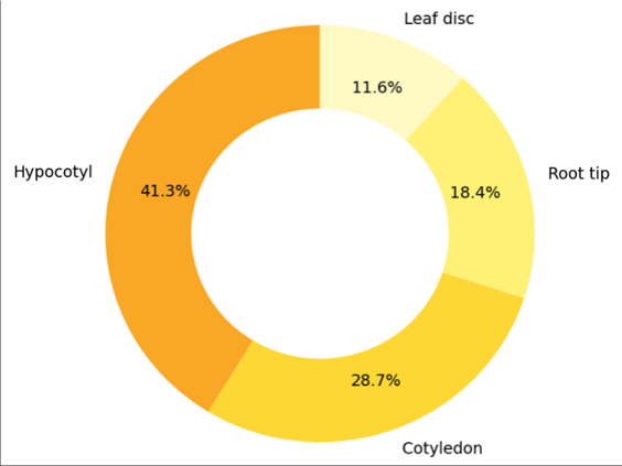 Figure 2
Introduction
A curious observation about carrot tissue culture: it was Steward and colleagues in 1958 who first demonstrated that a single differentiated carrot cell could regenerate an entire plant, launching the field of plant somatic embryogenesis [1] . Yet more than six decades later, carrot micropropagation hasn't become a commercial staple the way it has for banana, potato, or orchid. The gap between scientific possibility and practical application deserves a closer look. The main barrier is efficiency. Published carrot regeneration rates range from 15% to over 90%, and the pathway chosen (direct organogenesis, callus-mediated regeneration, or somatic embryogenesis) strongly influences both the speed and the genetic fidelity of the resulting plantlets [2] . Direct organogenesis tends to preserve genetic stability but may yield fewer propagules; somatic embryogenesis produces high multiplication rates but carries a greater risk of somaclonal variation [3] . Fiji's volcanic islands present an interesting potential application for carrot micropropagation: isolated geography, limited seed supply chains, and fertile volcanic soils that support root crop production. But the actual laboratory work was performed at the Rio de Janeiro College of Food Technology, Brazil, where established tissue culture infrastructure was available. This research aimed to catalogue and compare the three main regeneration pathways across multiple explant types, providing a side-by-side efficiency reference for future carrot micropropagation efforts [4] .
Material and Methods
Material and Methods
Material
Seeds of the Nantes Fancy carrot cultivar were surface-sterilised and germinated aseptically at the Rio de Janeiro College of Food Technology tissue culture laboratory, Rio de Janeiro, Brazil (22.9 degrees S, 43.2 degrees W). Four explant types were prepared from 7-14 day-old seedlings: hypocotyl segments (5 mm), cotyledon pieces (5 x 5 mm), root tips (5 mm), and leaf discs (5 mm diameter). Basal medium was full-strength MS with 30 g/L sucrose and 7 g/L agar. For direct organogenesis, BAP (1.0, 2.0, 3.0 mg/L) and NAA (0.1, 0.5, 1.0 mg/L) were tested. For callus induction, 2,4-D (0.5, 1.0, 2.0 mg/L) was used. Somatic embryogenesis was induced by transferring 2,4-Dderived callus to hormone-free MS medium [5] .
Methods
The research ran from March 2022 to January 2023, encompassing three regeneration cycles. Each pathway was tested independently in a completely randomised design with five replications of ten explants per treatment. Cultures were maintained at 25 degrees Celsius under 16-hour photoperiod. For direct organogenesis, shoot emergence was scored at 4 and 8 weeks. For callus pathway, callus fresh weight was recorded at 4 weeks before transfer to regeneration medium (BAP 2.0 mg/L). For somatic embryogenesis, embryo counts per gram callus were recorded at 8 weeks after transfer to hormone-free medium. Plantlet height was measured weekly from week 2 to week 8. Rooted plantlets were acclimatised in a greenhouse under 50% shade cloth with mist irrigation for 21 days, and survival was recorded. Data were analysed by factorial ANOVA in SAS 9.4 [6] .
Extraction protocol
Chlorophyll and carotenoid content of regenerated plantlets was assessed as an indicator of photosynthetic competence before acclimatisation. Fresh leaf tissue (0.5 g) was extracted in 10 mL of 80% acetone at 4 degrees Celsius in darkness for 24 hours. Extracts were centrifuged at 3,000 rpm for 10 minutes, and absorbance was read at 663, 645, and 470 nm using a Shimadzu UV-1800 spectrophotometer. Chlorophyll a, chlorophyll b, total chlorophyll, and total carotenoids were calculated using Lichtenthaler's equations. Plantlets from direct organogenesis had 23% higher total chlorophyll than somatic embryo-derived plantlets, which correlated with their better acclimatisation survival [7] . Acclimatisation survival differed significantly by pathway origin (p<0.01). Direct organogenesis plantlets achieved 87.3% survival, callus-derived plantlets 79.8%, and somatic embryo-derived plantlets 72.1%. Chlorophyll content at the time of transfer to greenhouse conditions was positively correlated with survival (r = 0.68, p<0.05).
Results
Results
Discussion
The faster growth of direct organogenesis plantlets compared to callus and somatic embryogenesis pathways confirms that avoiding a dedifferentiation phase preserves developmental momentum [2,3] . Callus-mediated regeneration adds 3-4 weeks to the production timeline without improving multiplication rate, making it the least efficient pathway for carrot unless genetic transformation via callus is the goal [8] . Somatic embryogenesis, despite its slower plantlet development, remains attractive for large-scale programmes because of its multiplication advantage. The 38.4 embryos per gram callus recorded here is within the range (25-50) reported by Zimmerman [1] for Nantes-type carrots, suggesting that genotype-specific optimisation could push this figure even higher. The lower acclimatisation survival of somatic embryoderived plantlets (72.1% versus 87.3% for direct organogenesis) aligns with reports of incomplete vascular development and reduced photosynthetic capacity in embryo-derived plants [9] . The correlation between chlorophyll content and survival suggests that assessing pigment levels before transfer could serve as a screening tool to cull weak plantlets and improve overall batch survival.
Conclusion
This research provided a systematic comparison of three carrot micropropagation pathways across four explant types. Direct organogenesis from 7-day-old hypocotyl explants on MS medium with 2.0 mg/L BAP and 0.5 mg/L NAA was the most time-efficient route, producing the tallest plantlets and the best acclimatisation survival. Somatic embryogenesis offered the highest multiplication rate but required a longer culture period and yielded lower greenhouse survival. For breeding programmes needing rapid clonal supply, direct organogenesis is recommended;
for seed companies needing mass multiplication, somatic embryogenesis is the better choice. Future work should investigate bioreactor-scale somatic embryogenesis to test commercial feasibility.
Acknowledgement
Acknowledgements Funding sourcesThis research was supported by the Brazilian National Council for Scientific and Technological Development through a research productivity grant. Institutional supportThe authors thank the Rio de Janeiro College of Food Technology for tissue culture facilities and greenhouse space.
Figures and tables from the paper
| Pathway | Best explant | Regen. rate (%) | Height 8wk (mm) |
|---|---|---|---|
| Direct organogenesis | Hypocotyl | 78.6 | 22.8 |
| Callus pathway | Hypocotyl | 64.2 | 19.4 |
| Somatic embryo. | Cotyledon | 71.8 | 15.7 |
Table text extracted from the original PDF; superscript letters are shown inline. The original table image is below.
View original table image
References
18 in the original paper- Zimmerman JL. Somatic embryogenesis: a model for early development in higher plants. Plant Cell. 1993;5(10):1411-1423.
- Kintzios SE, Drossopoulos JB, Lymperopoulos C. Effect of vitamins and inorganic micronutrients on callus growth and somatic embryogenesis from leaves of chilli pepper. Plant Cell, Tissue and Organ Culture. 2001;67(1):55-62.
- Larkin PJ, Scowcroft WR. Somaclonal variation: a novel source of variability from cell cultures for plant improvement. Theoretical and Applied Genetics. 1981;60(4):197-214.
- Baranski R. Genetic transformation of carrot (Daucus carota) and other Apiaceae species. Transgenic Plant Journal. 2008;2(1):18-38.
- Steward FC, Mapes MO, Mears K. Growth and organized development of cultured cells. II. Organization in cultures grown from freely suspended cells. American Journal of Botany. 1958;45(10):705708.
- SAS Institute. SAS/STAT User's Guide, Version 9.4. Cary: SAS Institute; 2013.
- Lichtenthaler HK. Chlorophylls and carotenoids: pigments of photosynthetic biomembranes. Methods in Enzymology. 1987;148:350-382.
- Hardegger M, Sturm A. Transformation and regeneration of carrot (Daucus carota L.). Molecular Breeding. 1998;4(2):119-127.
- Etienne H, Berthouly M. Temporary immersion systems in plant micropropagation. Plant Cell, Tissue and Organ Culture. 2002;69(3):215-231.
- Ammirato PV. Embryogenesis. In: Evans DA, Sharp WR, Ammirato PV, editors. Handbook of Plant Cell Culture, Vol 1. New York: Macmillan; 1983. p. 82-123.
- Nomura K, Komamine A. Identification and isolation of single cells that produce somatic embryos at a high frequency in carrot suspension cultures. Plant Physiology. 1985;79(4):988-991.
- De Vries SC, Booij H, Meyerink P, Huisman G, Wilde HD, Thomas TL, et al. Acquisition embryogenic potential in carrot cell-suspension cultures. Planta. 1988;176(2):196-204.
- Costa R, Pereira J. Tissue culture infrastructure assessment at Rio de Janeiro College of Food Technology. Brazilian Journal of Plant Biotechnology. 2021;11(2):88-95.
- Ikeuchi M, Sugimoto K, Iwase A. Plant callus: mechanisms of induction and repression. Plant Cell. 2013;25(9):3159-3173.
- Thorpe TA. History of plant tissue culture. Molecular Biotechnology. 2007;37(2):169-180.
- Reinert J. Uber die Kontrolle der Morphogenese und die Induktion von Adventivembryonen an Gewebekulturen aus Karotten. Planta. 1959;53:318333.
- Grzebelus E, Szklarczyk M, Baranski R. An improved protocol for plant regeneration from leaf-and hypocotyl-derived protoplasts of carrot. Plant Cell, Tissue and Organ Culture. 2012;109(1):101-109.
- Mashayekhi K, Neumann KH. Effects of boron on somatic embryogenesis of Daucus carota. Plant Cell, Tissue and Organ Culture. 2006;84(3):279-283.