
Research article · 2026 · Full text
International Journal of Biology SciencesMolecular mimicry as a central mechanism in autoimmune blood disorders: A comprehensive review
Volume 8(5) · Pages 06–11 · DOI: 10.33545/26649926.2026.v8.i5a.819 ↗
Research areasBlood groups and transfusionSystemic Lupus Erythematosus ResearchErythrocyte Function and Pathophysiology
Abstract
Author-writtenAutoimmune hemolytic anemia (AIHA) is an uncommon hematologic condition where autoantibodies cause red blood cells (RBCs) to be destroyed too soon. IgG, IgM, or sometimes IgA antibodies attach to RBC surface antigens, causing extravascular or intravascular hemolysis via complement activation and macrophage phagocytosis. AIHA may arise as a main idiopathic disorder or as a side effect of drug exposure, autoimmune diseases, hematologic malignancies, or infections. Warm antibody AIHA (wAIHA), cold antibody AIHA (CAIHA, which includes CAD and CAS), mixed AIHA, paroxysmal cold hemoglobinuria, drug-induced AIHA, and other uncommon variants are among the subtypes of AIHA. While cold forms of wAIHA are IgM-mediated and act at lower temperatures, resulting in RBC agglutination and complement-induced lysis, wAIHA is primarily mediated by IgG antibodies that are active at body temperature and cause extravascular hemolysis. IgG and IgM antibodies are both involved in mixed AIHA, which is rare but frequently clinically severe. Female sex, advanced age, genetic susceptibility, hematologic malignancies, prior transfusions, and organ transplantation are risk factors. Molecular mimicry, in which infections create antigens that are structurally or sequentially similar to RBC proteins, is a key mechanism in the pathophysiology of AIHA. The immune system is misled by this resemblance into generating T cells and cross-reactive antibodies that attack host red blood cells. Antibodies or immunoglobulins such as IgG or IgM play a significant role in mixed type of AIHA, this effect is rare, in fact if it occure it frequently clinically severe. Female sex, advanced age, genetic susceptibility, hematologic malignancies, prior transfusions, and organ transplantation are risk factors. Molecular mimicry, that occurred after such an infection that led for the production of antigens particles that are structurally or functionally similar to red cell proteins, this phenomenon is considered as a key mechanism in the pathophysiology of AIHA. The cells of the immune system because of this defect lead to autoimmune disorder disease or AIHA (the resemblance of the new produced T cells and cross-reactive antibodies that attack host red blood cells). Autoimmune hemolytic anemia (AIHA) is uncommon immune-mediated disorder that are characterized by premature destruction of the red blood cells due to autoantibodies. It includes warm, cold, and mixed types, and may be primary or secondary to underlying conditions. Understanding the diseases mechanisms with its risk factors is very essential for the accurate diagnosis and for effective management.
Summary
Source-checkedPlain-language reading of the full paper, prepared with AI and checked against the text. Each point links to the authors' own words.
Why it matters
- Autoimmune hemolytic anemia (AIHA) is a rare blood disorder characterized by the premature destruction of red blood cells due to the action of autoantibodies. View evidence ↗
- AIHA is more common in women, with a global annual incidence ranging from 0.8 to 3 cases per 100,000 people, most frequently occurring in children and the elderly. View evidence ↗
- Identifying the mechanisms and risk factors of AIHA is critical for ensuring patients receive accurate diagnoses and effective treatment. View evidence ↗
The question
- The review aims to examine molecular mimicry as a primary mechanism driving autoimmune blood disorders, specifically AIHA. View evidence ↗
What they did
- The authors conducted a comprehensive review of existing literature to synthesize the pathophysiology, types, and causes of AIHA. View evidence ↗
What they found
- Warm antibody AIHA (wAIHA) is mainly driven by IgG antibodies active at body temperature, leading to the destruction of red blood cells outside the blood vessels (extravascular hemolysis). View evidence ↗
- Cold-Antibody AIHA (CAIHA) involves IgM autoantibodies that react at temperatures below 30 °C (86 °F), causing red blood cell agglutination and hemolysis. View evidence ↗
- Molecular mimicry is a key driver of AIHA, occurring when infections produce antigens that resemble red blood cell proteins, tricking the immune system into attacking the host's own cells. View evidence ↗
- Secondary warm AIHA is most commonly caused by autoimmune diseases (such as lupus and arthritis) or lymphoid malignancies (such as lymphoma). View evidence ↗
- Secondary cold AIHA is typically triggered by infections, including mycoplasmosis and viral pneumonia, though lymphatic diseases can also be a cause. View evidence ↗
- In severe cases of AIHA, the normal red blood cell lifespan of 100-120 days can be drastically reduced to just a few days. View evidence ↗
- The Direct Antiglobulin Test (DAT), or Coombs test, is crucial for diagnosing AIHA as it detects the presence of autoantibodies or complement proteins on red blood cells. View evidence ↗
What it means
- The authors conclude that a deep understanding of AIHA's mechanisms and risk factors is essential for improving diagnostic accuracy and patient management. View evidence ↗
Visual summary
1 visualsIllustrations explain the paper; they do not show study data.
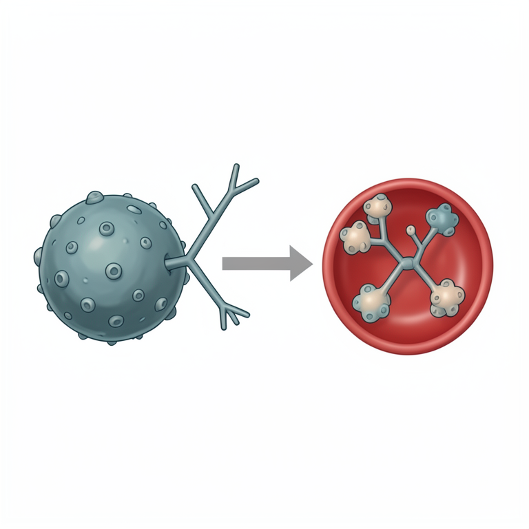
Molecular Mimicry Process
AI-generated conceptual illustration (human red blood cells and a spherical bacterium). It explains the idea and does not show study data.
View source evidence ↗Figures & tables
From the original paper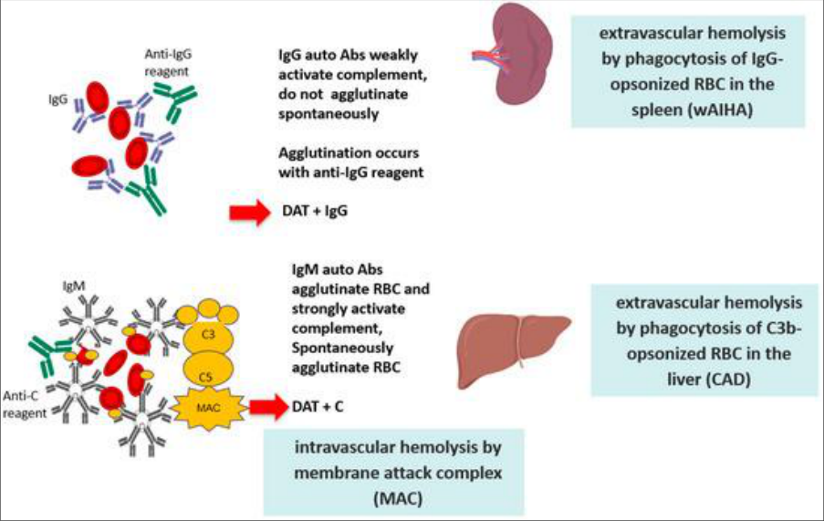 Figure 1
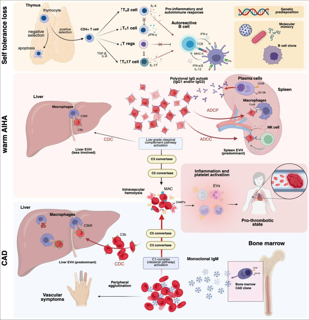 Figure 2
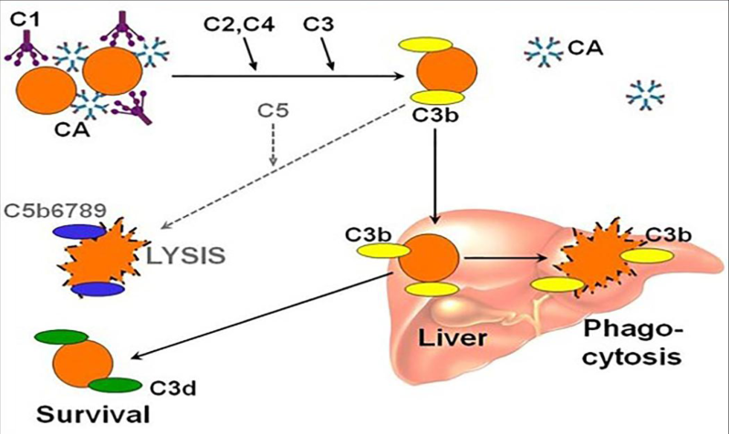 Figure 3
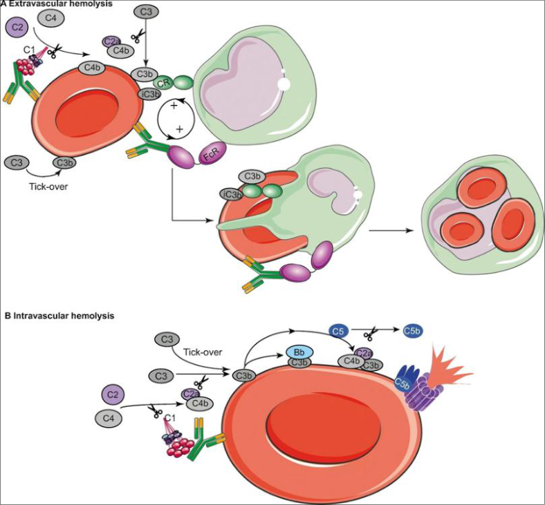 Figure 4
Introduction Scientific definition of autoimmune hemolytic anemia
The AIHA is consider a very rare disorder but clinically relevant hematological condition that is characterized by autoantibodies against self-antigens that cause red blood cells (RBCs) to be destroyed too soon. The prevalence is higher in women, and the global incidence varies between 0.8 and 3 instances per 100,000 people per year, peaking in early childhood and elderly adults (Barcellini & Fattizzo, 2020; Jäger et al., 2020) [1,2] . Immune anemia, also known as autoimmune hemolytic anemia (AIHA), is a rare hematologic disease that affects 1-3 people out of every 100,000 (Böttiger & Westerholm, 1973) [9] . AIHA occurs when autoantibodies attack the patient's own red blood cells (RBCs), causing them to be prematurely destroyed (lysed) and therefore reduced in circulation. In extreme situations, the RBCs' normally 100-120 day lifespan is shortened to a few days (Shoenfield, Cervera, & Gershwin, 2008;) [13] . During RBC lysis, the intracellular contents of RBCs rupture and spill into the blood and tissues, resulting in the majority of the disease's distinctive clinical characteristics. Pathophysiologically, the immune response can initiate the membrane assault complex by activating the conventional and/or alternative complement pathways. The ability of immunoglobulin molecules to activate complement is determined by their structural differences (Hill et al., 2017) [3] . The extramedullary damage caused by the binding of IgG, IgM, or, less frequently, IgA antibodies to RBC surfaces is what defines AIHA (Branch & Parry, 2024) [5] figure 1. [14] Patterns of autoimmune hemolytic anemia (AIHA) • Warm-Antibody Autoimmune Hemolytic Anemia (wAIHA) Warm autoimmune hemolytic anemia (WAIHA) is typified by increased RBC degradation brought on by warm agglutinins, which are essentially all immunoglobulin G (IgG) antibodies that interact best with erythrocyte antigens around 37 °C. According to annual reports, the frequency is roughly 1 to 3 per 100,000 people (Eaton et al., 2007) [4] . Although the disease can strike at any age, the usual time of diagnosis is roughly 52 years, and most studies show a small female predominance. The majority of patients experience a relapsing, chronic illness pattern, while up to 30% of patients may get a durable remission with first medication (Barcellini). Extravascular processes in wAIHA mainly cause hemolysis in the liver and spleen (Barcellini, 2015) [14] . Using Fcγ receptors, splenic macrophages identify IgG-coated erythrocytes and either fully phagocytose the cell or partially remove the cell membrane. During succeeding circulations, the latter become microspherocytes, which are sequestered in the splenic sinusoids and are less deformable than regular RBCs. Additionally, antibody-dependent cellmediated cytotoxicity by T cells with Fc receptors may be included in extravascular hemolysis (Costa et al., 2025) [6] . The complement system can be triggered by specific IgG subclasses, which results in the deposition of C3 fragments on RBC membranes. Liver macrophages with C3 fragment receptors then eliminate these opsonized cells. Severe complement activation causes intravascular hemolysis, which is clinically characterized by hemoglobinuria and significantly high lactate dehydrogenase levels, as well as the building of the membrane assault complex (C5b-9) on the surface of the red blood cell (Brodsky, 2015), (Figure 2). [16] • Cold-Antibody Autoimmune Hemolytic Anemia (CAIHA) A rare form of AIHA known as cold-antibody autoimmune hemolytic anemia (CAIHA), which includes cold agglutinin syndrome (CAS) and cold agglutinin disease (CAD), is defined by elevated levels of cold-reactive autoantibodies, primarily immunoglobulin M (IgM), that are active at temperatures lower than 30 °C (86 °F). Red blood cell (RBC) surface antigens, especially the I and i antigens, are strongly reacted to by these antibodies (Petz & Branch, 1983) [17] , causing complement activation, RBC agglutination, and subsequent intravascular and/or extravascular hemolysis. CAD and CAS, which today constitute two separate clinical entities, were formerly thought to be included in CAD (Nunes et al., 2024) [18] . A. Cold agglutinin syndrome (CAS) is a secondary, transient illness most often seen in the setting of bacterial or viral infections such as Mycoplasma pneumoniae or Epstein-Barr virus-associated mononucleosis (Petz & Garratty, 2004). CAS can also be seen in the setting of certain malignancies or autoimmune illnesses (Swiecicki et al., 2013) [19] . CAS is generally self-limited with resolution after clearance of the inciting infection. B. Cold agglutinin disease (CAD) is a chronic, primary condition where there is a clonal expansion of B lymphocytes producing monoclonal IgM autoantibodies reacting at temperatures ≤ 30 °C, again against the I or i antigens. These monoclonal antibodies activate the complement pathway with resultant potential intravascular hemolysis of scale (Malecka et al., 2023) [16] .
Although the causes of CAS and CAD are different-CAS is caused by polyclonal IgM, whereas CAD is caused by monoclonal IgM-they have similar diagnostic characteristics. IgM antibodies attach to red blood cells and activate complement in both disorders, causing significant intravascular hemolysis (Broome, 2023) ,(Figure 3) . ) [14] .
• Mixed Autoimmune Hemolytic Anemia (mAIHA) Immunoglobulin G (IgG) and immunoglobulin M (IgM) subtypes that are both warm-and cold-reactive autoantibodies coexist in mixed autoimmune hemolytic anemia (mAIHA). Less than 5% of all instances of AIHA are of this kind, and it is significantly less common in children (Mayer et al., 2008). mAIHA may manifest as an idiopathic condition may develop secondary to autoimmune disorders or cancers like lymphoma or systemic lupus erythematosus (SLE). It can be difficult to identify the particular heat range that causes hemolysis as well as the main pathogenic antibody class (IgG or IgM). Patients frequently have both warm and cold antibody components, which can target various red blood cell antigens at the same time) (Rinaldi & Kerr, 2023) [11] .
Causes of autoimmune hemolytic anemia
There is currently no explanation for the pathophysiology of autoimmune anemia. It could be the primary or secondary ailment. Idiopathic primary immunological anemia makes up around half of all cases (Gupta et al., 2011) [22] . Both warm and cold immune anemias can be caused by a variety of different illnesses, the most prevalent of which are as follows: The most prevalent causes of warm secondary immune anemia include autoimmune conditions like lupus, arthritis, scleroderma, and ulcerative colitis, as well as lymphoid disorders like lymphoma and chronic lymphocytic leukemia. Infections and non-lymphoid cancers are less frequent causes of warm secondary immunological anemia.
Although lymphatic diseases can produce secondary cold immune anemia, infections, including mycoplasmosis, viral pneumonia, infectious mononucleosis, and other respiratory infections, are typically the cause. Less frequently, related autoimmune illnesses could be the culprit (Sokol et al., 1981) [8] .
Although uncommon, a variety of medications, such as penicillin and methyldopa, have been known to cause druginduced autoimmune hemolytic anemia. Drug molecules attach themselves to the surface of red blood cells and function as antigens in this type II hypersensitivity reaction.
The complement system is being activated by antibodies directed against red blood cells. While the remaining complement system proteins (C6, C7, C8, and C9) either form the membrane attack complex (MAC) or bind antibodies to aid macrophage phagocytosis (C3b), small complement system components like C3a, C4a, and C5a activate granular leukocytes (such as neutrophils). One kind of penicillin allergy is this one (Figure 4). [12] .
Incidence of Autoimmune Hemolytic Anemia
The size of the population under study, the distribution of ages, and variations in national health care systems all have a substantial impact on the incidence and prevalence of autoimmune hemolytic anemia. Since the majority of current research is conducted in hospitals, primary healthcare providers may not fully recognize AIHA. Therefore, if hospital cases are not selected from trustworthy local community samples, many patients might not be included in epidemiological data (Barcellini & Fattizzo, 2020) [1] . Autoimmune hemolytic anemia (AIHA) is a rare but significant Iraqi hematologic disease, with limited epidemiologic data. An incidence of 2% in patients with ulcerative colitis was documented in one Baghdad Medical City study, underscoring AIHA as an uncommon aftereffect of inflammatory bowel illness (Al-Khazraji, 2008) [23] . While the US Immune Hematology database estimates an incidence of 17.8 cases per million, reported incidence rates of AIHA include roughly 0.8 occurrences per 100,000 person-years in northern France and 3.9 cases per 100,000 in Spain. There are two known occurrence peaks: the first primarily occurs in children under the age of ten, and the second occurs in people 70 years of age and older. With a female-to-male ratio of more than 1.5, AIHA is more prevalent in females than in males (Barcellini & Fattizzo, 2020) [1] .
Clinical Symptoms of Autoimmune Hemolytic Anemia
A vast but wildly varied variety of symptoms are present in AIHA patients. Dizziness, weakness, weariness, and dyspnea (shortness of breath) are the most often reported symptoms, which indicate general anemia and decreased tissue oxygen delivery (Costa et al., 2025) [6] . Hemolysis symptoms typically include pallor, jaundice, and colored urine, which are signs of high bilirubin and accelerated red blood cell breakdown (Loriamini et al., 2024). Physical signs like splenomegaly can also appear in severe or persistent illnesses (Merck Manual, reviewed April 2024). Patients may present with fever, tachycardia, and chest pain after acute or severe hemolysis events, such as acute crises or COVID-associated AIHA, exhibiting more pronounced clinical decompensation (Ayyad et al., 2025) [24] .
Diagnosis of Autoimmune Hemolytic Anemia
The diagnosis of AIHA include different ways for example the Clinical assessment, laboratory or immunological testing, For therapy to be guided and results to improve, an early and precise diagnosis is essential (Hill et al., 2017) [3] .
• Clinical Evaluation: Symptoms including pallor, jaundice, and exhaustion are common when patients first arrive. Potential triggers such autoimmune illnesses, infections, cancers, or drugs should be investigated during a comprehensive history and physical examination (Barcellini & Fattizzo, 2020) [1] . • Laboratory Findings: A complete blood count (CBC) in general recorded a reticulocytosis and or anemia. In some time thehemolysis include decreased haptoglobin or increased indirect bilirubin, in addition to lactate dehydrogenase (LDH) elevation (Jäger et al., 2020) [2] . • Direct Antiglobulin Test (DAT): The Coombs test, in some reference called direct antiglobulin test these test considered a crucial for diagnosing AIHA. in these test the AIHA could be distinguished from other type of blood disease such as anemias by a positive DAT, that mean the presence of autoantibodies or complement protein in the cells (Hill et al., 2017) [3] .
• Immunological Testing: by identifying the type of autoantibodies involved (IgG, IgM) and complementing elements like C3d (Hill et al., 2023) [10] . • Imaging Studies: Abdominal ultrasound figure or CT scans check for splenomegaly or hepatomegaly, that give an idea about the severity of the disease or goose its secondary causes (Barcellini & Fattizzo, 2020) [1] .
References
24 in the original paper- Barcellini W, Giannotta J, Fattizzo B. Autoimmune hemolytic anemia in adults: primary risk factors and diagnostic procedures. Expert Rev Hematol. 2020;13(6):585-597.
- Jäger U, Barcellini W, Broome CM, Gertz MA, Hill A, Hill QA, et al. Diagnosis and treatment of autoimmune hemolytic anemia in adults: recommendations from the First International Consensus Meeting. Blood Rev. 2020;41:100648.
- Hill QA, Stokes M, Massey E, Grainger JD, Provan D, Hill A. The diagnosis and management of primary autoimmune haemolytic anaemia. Br J Haematol. 2017;176(3):395-411.
- Eaton WW, Rose NR, Kalaydjian A, Pedersen MG, Mortensen PB. Epidemiology of autoimmune diseases in Denmark. J Autoimmun. 2007;29(1):1-9.
- Branch DR, Parry R. Autoimmune hemolytic anemias: classifications, pathophysiology and therapeutic insights. Int J Mol Sci. 2024;25(8):4296-4298.
- Costa A, Mulas O, Mereu AM, Schintu M, Greco M, Caocci G. Beneath the surface in autoimmune hemolytic anemia: pathogenetic networks, therapeutic advancements and open questions. Front Immunol. 2025;16:1624667.
- Brodsky RA. Complement in hemolytic anemia. Blood. 2015;126(22):2459-2465.
- Sokol RJ, Hewitt S, Stamps BK. Autoimmune hemolysis: an 18-year study of 865 cases referred to a regional transfusion center. Br Med J. 1981;282(6281):2023-2027.
- Böttiger LE, Westerholm B. Acquired hemolytic anaemia. I. Incidence and aetiology. Acta Med Scand. 1973;193(3):223-226.
- Hill QA, Hill A, Berentsen S, Randen U, Tjønnfjord GE, Barcellini W. The changing landscape of autoimmune hemolytic anemia: diagnosis, classification and epidemiology. Blood Rev. 2023;52:100-130.
- Serrano K, Rinaldi J, Kerr P. Regulatory T-cell dysfunctions are associated with increase in tumor necrosis factor alpha in autoimmune hemolytic anemia and participate in Th17 polarization. Haematologica. 2023;109(2):398-408.
- Patidar P, Jain A, Prakash T. Deciphering the role of molecular mimicry in the etiopathogenesis of autoimmune hemolytic anemia using an immunoinformatics approach. Immunoinformatics. 2025;17:100047.
- Shoenfield Y, Cervera R, Gershwin ME. Diagnostic criteria in autoimmune diseases. Humana Press; 2008.
- Barcellini W. New insights in the pathogenesis of autoimmune hemolytic anemia. Transfus Med Hemother. 2015;42(5):287-293.
- Barcellini W, Fattizzo B, Zaninoni A, Giannotta JA, Lunghi M, Rossi F, et al. Clinical heterogeneity and predictors of outcome in primary autoimmune hemolytic anemia: a GIMEMA study of 308 patients. Blood. 2014;124(19):2930-2936.
- Malecka A, Ostlie I, Troen G, Małecki J, Delabie J, Tierens A, et al. Gene expression analysis revealed downregulation of complement receptor 1 in clonal B cells in cold agglutinin disease. Clin Exp Immunol. 2023;216(1):45-54.
- Petz LD, Branch DR. Serological tests for the diagnosis of immune hemolytic anemias. In: McMillan R, editor. Methods in hematology: immune cytopenias. New York: Churchill Livingstone; 1983. p. 9-48.
- Nunes BS, Gouveia C, Kjollstrom P, Neves JF. Cold agglutinin syndrome and hemophagocytic lymphohistiocytosis: an unusual combination caused by Epstein-Barr virus infection. Cureus. 2024;16(4):e52179.
- Swiecicki PL, Hegerova LT, Gertz MA. Cold agglutinin disease. Blood. 2013;122(7):1114-1121.
- Broome CM. Complement-directed therapy for cold agglutinin disease: sutimlimab. Expert Rev Hematol. 2023;16(6):479-494.
- Mayer B, Yurek S, Kiesewetter H, Salama A. Mixedtype autoimmune hemolytic anemia: differential diagnosis and a critical review of reported cases. Transfusion. 2008;48(10):2229-2234.
- Gupta S, Szerszen A, Nakhl F, et al. Severe refractory autoimmune hemolytic anemia with both warm and cold autoantibodies that responded completely to a single cycle of rituximab: a case report. J Med Case Rep. 2011;5:156.
- Al-Khazraji KA. Prevalence of autoimmune hemolytic anemia in ulcerative colitis. J Fac Med Baghdad. 2008;50(3):273-281.
- Ayyad M, Abu Alya W, Barabrah AM, et al. Autoimmune hemolytic anemia in COVID-19 patients: a systematic review of 105 cases on clinical characteristics and outcomes. Clin Immunol. 2025:277300.