
Research article · 2026 · Full text
International Journal of Biology SciencesDiagnostic evaluation of inappropriate lactation syndrome (Precocious Udder) in a goat kid: A clinicopathological study
Volume 8(5) · Pages 27–29 · DOI: 10.33545/26649926.2026.v8.i5a.823 ↗
Research areasVeterinary Medicine and SurgeryReproductive Physiology in LivestockGrowth Hormone and Insulin-like Growth Factors
Abstract
Author-writtenPrecocious mammary gland development in prepubertal animals is an uncommon clinical finding in veterinary practice. This report describes a rare case of inappropriate lactation in a 10-day-old female goat kid presented with bilateral udder enlargement and milk secretion. Clinical examination revealed normal physiological parameters except for mild hyperthermia (104.7°F). Milk secretion was observed from both teats without any signs of mastitis. Based on history and clinical findings, the condition was diagnosed as precocious udder development. The animal was treated with prednisolone and supportive management was advised. The condition showed gradual regression. This case highlights the importance of recognizing endocrine-mediated abnormalities in neonatal animals and differentiating them from pathological conditions like mastitis.
Summary
Source-checkedPlain-language reading of the full paper, prepared with AI and checked against the text. Each point links to the authors' own words.
Why it matters
- Udder development in goats is controlled by hormones, specifically growth hormone, prolactin, progesterone, and oestrogen. View evidence ↗
- Under normal conditions, significant development of the udder happens during puberty or late pregnancy. View evidence ↗
- Precocious udder, also known as inappropriate lactation syndrome, is a rare condition where mammary development occurs prematurely in prepubertal or neonatal animals. View evidence ↗
The question
- The study aims to describe and evaluate a rare case of inappropriate lactation in a 10-day-old female goat kid exhibiting milk secretion and bilateral udder enlargement. View evidence ↗
What they did
- The subject was a 10-day-old female Salem Black goat kid weighing about 5 kg with a 10-day history of milk secretion and sudden udder enlargement. View evidence ↗
- Clinical examination was performed, which showed normal physiological parameters except for a mild fever of 104.7°F. View evidence ↗
- Ultrasonography was used to confirm secretory activity by identifying anechoic milk content within dilated lacteals. View evidence ↗
- The animal was treated with an intramuscular injection of prednisolone at 1 mg/kg and oral prednisolone tablets of 5 mg for 5 days. View evidence ↗
What they found
- Because there were no inflammatory signs, the authors ruled out mastitis as the cause of the udder enlargement. View evidence ↗
- Following treatment with prednisolone and supportive care, the precocious udder condition gradually regressed. View evidence ↗
What it means
- The authors conclude that careful clinical evaluation is necessary to distinguish this syndrome from mastitis to prevent the use of unnecessary antibiotics. View evidence ↗
- The authors state that the condition typically has a favorable prognosis even with minimal intervention. View evidence ↗
Visual summary
1 visualsIllustrations explain the paper; they do not show study data.
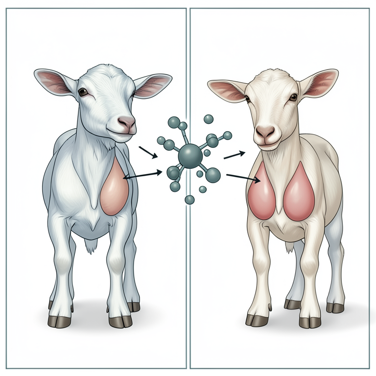
Hormonal Regulation of Mammary Development
AI-generated conceptual illustration (goat kids). It explains the idea and does not show study data.
View source evidence ↗Figures & tables
From the original paper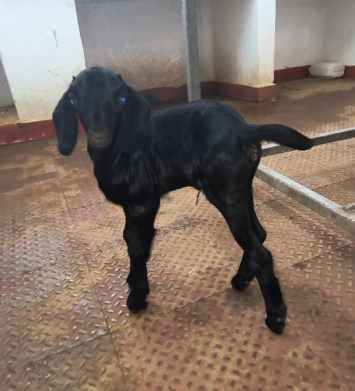 Figure 1
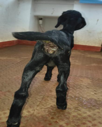 Figure 2
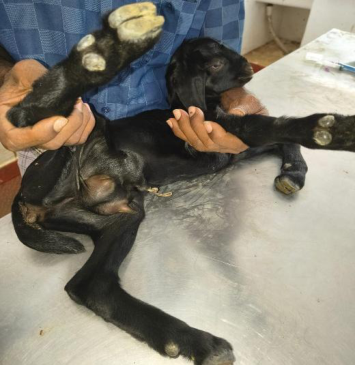 Figure 3
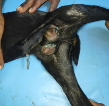 Figure 4
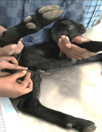 Figure 5
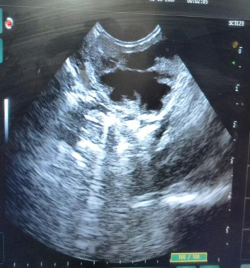 Figure 6
Introduction
Mammary gland development in goats is a hormonally regulated process primarily influenced by oestrogen, progesterone, prolactin, and growth hormone (Smith and Sherman, 2009) [5]. Normally, significant udder development occurs during late pregnancy or puberty (Radostits et al., 2007) [3]. However, premature mammary development in neonatal or prepubertal animals is rare and is often referred to as precocious udder or inappropriate lactation syndrome. Such conditions may arise due to endocrine imbalances (Singh et al., 2020) [4], exposure to exogenous hormones, or ingestion of phytoestrogen-rich feed (Foster, 2016) [2].
Although reported sporadically in calves and goat kids, the exact pathophysiology remains unclear. Early recognition is essential to avoid unnecessary treatment and to differentiate it from mastitis or congenital abnormalities (Radostits et al., 2007) [3].
Materials And Methods
(Case Description) A 10-day-old female Salem Black goat kid weighing approximately 5 kg was presented to the Veterinary Clinical Complex with a history of sudden udder enlargement (Fig 1,2 and 3) and milk secretion from the teats for the past 10 days. Clinical Examination • General appearance: Normal • Behavior: Active • Appetite: Satisfactory • Urination and defecation: Normal • Rectal temperature: 104.7°F Local Examination • Bilateral enlargement of the udder (Fig 3 &4) NAAS Rating (2026): 4.82 IJBS 2026; 8(5): 27-29 K Karthika Assistant Professor, Department of Veterinary Medicine, Veterinary College and Research Institute, Orathanadu, Thanjavur, Tamil Nadu, India S Yogeshpriya Assistant Professor, Department of Veterinary Medicine, Veterinary College and Research Institute, Orathanadu, Thanjavur, Tamil Nadu, India M Veeraselvam Assistant Professor, Department of Veterinary Medicine, Veterinary College and Research Institute, Orathanadu, Thanjavur, Tamil Nadu, India KK Ponnuswamy Professor and Head, Department of Veterinary Medicine, Veterinary College and Research Institute, Orathanadu, Thanjavur, Tamil Nadu, India T Yuvarani UG Scholar, Department of Veterinary Medicine, Veterinary College and Research Institute, Orathanadu, Thanjavur, Tamil Nadu, India M Jeevitha UG Scholar, Department of Veterinary Medicine, Veterinary College and Research Institute, Orathanadu, Thanjavur, Tamil Nadu, India E Tamileniyan UG Scholar, Department of Veterinary Medicine, Veterinary College and Research Institute, Orathanadu, Thanjavur, Tamil Nadu, India K Karthika Assistant Professor, Department of Veterinary Medicine, Veterinary College and Research Institute, Orathanadu, Thanjavur, Tamil Nadu, India https://www.biologyjournal.net
Discussion
Precocious udder development in neonatal goat kids is an uncommon condition likely associated with endocrine disturbances. The presence of milk secretion suggests premature activation of the hypothalamic-pituitary- mammary axis, possibly due to elevated prolactin or estrogenic stimulation (Smith and Sherman, 2009) [5]. Similar cases have been attributed to: • Maternal hormonal influence during gestation • Phytoestrogen ingestion through feed • Idiopathic endocrine dysregulation (Radostits et al., 2007) [3] The absence of inflammatory signs in this case ruled out mastitis, which is a critical differential diagnosis. Mastitis typically presents with pain, heat and altered milk consistency (Radostits et al., 2007) [3].
Mild hyperthermia observed could be incidental or stress-related rather than directly associated with the condition. Administration of corticosteroids such as prednisolone may help modulate hormonal imbalance and reduce mammary gland stimulation. Most reported cases show spontaneous regression (Divers and Peek, 2018) [1], indicating a favourable prognosis (Foster, 2016) [2]. • Non-painful swelling • Milk secretion from both teats (Fig 5) • No signs of inflammation (heat, redness, or pain) Milk samples were collected for further examination.
Results
Clinical findings indicated: • Normal systemic health status • Absence of mastitis signs • Presence of functional milk secretion in a neonatal animal Based on the clinical presentation and absence of inflammatory signs, the condition was diagnosed as precocious udder development (inappropriate lactation syndrome). Ultrasonographic examination showed presence of dilated lacteals with anechoic milk content, confirming the presence of secretory activity within the gland (Foster, 2016) [2] (Fig 6). Treatment • Inj. Prednisolone @ 1 mg/kg IM • Tab.
Prednisolone 5 mg orally for 5 days Supportive care and monitoring were advised. Fig 1: Goat kid presented with abducted hindlimbs Fig 2: Swollen udder noticed from rear view Fig 3: Swollen mammary gland with milk discharge noticed Fig 4: Goat kid prepared for ultrasonographic examination https://www.biologyjournal.net Fig 6: Ultrasonographic examination showed Fig 5: Expression of mammary gland revealed milk content dilated lacteals with anechoic milk content
Conclusion
This case highlights a rare instance of precocious udder development with milk secretion in a neonatal goat kid. Proper clinical evaluation is essential to differentiate it from mastitis and avoid unnecessary antimicrobial therapy. The condition generally carries a good prognosis with minimal intervention.
Acknowledgment
The authors acknowledge the staff of the Veterinary Clinical Complex for their support in managing the case. Conflict of Interest The authors declare no conflict of interest.
References
5 in the original paper- Divers TJ, Peek SF. Rebhun’s diseases of dairy cattle. Elsevier; 2018.
- Foster RA. Female reproductive system. In: Jubb KVF, Kennedy PC, Palmer N, editors. Pathology of domestic animals. Elsevier; 2016.
- Radostits OM, Gay CC, Hinchcliff KW, Constable PD, et al. Veterinary medicine: a textbook of the diseases of cattle, sheep, pigs, goats and horses. Elsevier; 2007.
- Singh K, Marnet PG, Sharma A. Hormonal regulation of mammary gland development and lactation in domestic animals. Animal Reproduction Science. 2020;220:106513.
- Smith MC, Sherman DM. Goat medicine. Wiley- Blackwell; 2009.
Cited by 1
From OpenAlex, OpenCitations and reference lists read by Scinovex
- PRECOCIOUS UDDER DEVELOPMENT ASSOCIATED WITH CYSTIC FOLLICLES IN A 22 DAYS OLD FEMALE CALF ↗
S. Satheshkumar, S Raja, Prabaharan V, Subramaniyan Prakash, P. Jayaganthan · Indian journal of veterinary and animal sciences research. · 2026
A 22 days old Jersey crossbred female calf was reported with the history of udder enlargement and milk secretion for a period of over 10 days after birth. Based on the follicular, udder and endocrine status, the present case was diagnosed as precocious lactogenic udder development associated with cystic follicles and endocrine imbalance. Hyper-oestrogenisim and hyper-prolactinemia were ascribed to be…
❝ 0PDF