
Research article · 2025 · Full text
International Journal of Agriculture and NutritionCharacterization of xanthomonas strains causing bacterial leaf spot in capsicum
Volume 7(1) · Pages 85–88 · DOI: 10.33545/26646064.2025.v7.i1b.376 ↗
Research areasPlant Pathogenic Bacteria StudiesPlant-Microbe Interactions and ImmunityPlant Pathogens and Fungal Diseases
Abstract
Author-writtenBacterial leaf spot (BLS) caused by Xanthomonas spp. destroys 20–60% of capsicum yield annually in western India, yet the pathogen's strain-level diversity in Gujarat remains poorly documented. This research characterised seven Xanthomonas isolates collected from symptomatic capsicum fields across Navsari and Dantiwada districts during the 2022 kharif season. Morphological, biochemical (LOPAT), molecular (16S rDNA sequencing), and pathogenicity tests were performed at Navsari Agricultural University. All seven isolates were confirmed as Xanthomonas euvesicatoria. Disease severity on five capsicum cultivars ranged from 1.1 to 4.9 on a 1–5 scale. Strains XCV-02 and XCV-05 were the most aggressive, producing disease scores above 4.0 on susceptible cultivars California Wonder and Arka Mohini. Antibiotic sensitivity tests showed that streptomycin sulphate at 500 ppm gave the widest inhibition zones. The findings provide a strain-level baseline for designing targeted resistance breeding and chemical management strategies against BLS in Gujarat.
Summary
Source-checkedPlain-language reading of the full paper, prepared with AI and checked against the text. Each point links to the authors' own words.
Why it matters
- Bacterial leaf spot (BLS), caused by Xanthomonas species, is a significant agricultural problem in western India, reducing annual capsicum yields by 20% to 60%. View evidence ↗
- Identifying the specific strain of Xanthomonas is critical because multiple species cause similar symptoms but vary in their virulence and how they respond to fungicides. View evidence ↗
- Despite Gujarat being a major producer of capsicum, there has been no published research characterizing the local Xanthomonas population at the strain level. View evidence ↗
The question
- The study aimed to isolate and identify Xanthomonas strains from Gujarat, evaluate their virulence across five commercial cultivars, and determine their sensitivity to antibiotics. View evidence ↗
What they did
- Seven Xanthomonas isolates were collected from symptomatic capsicum leaves in the Navsari and Dantiwada districts of Gujarat during the 2022 kharif season. View evidence ↗
- The researchers used five commercial capsicum cultivars, California Wonder, Pusa Jwala, Arka Mohini, Kashi Anmol, and Punjab Lal, to test for virulence. View evidence ↗
- Isolates were identified using 16S rDNA sequencing and compared against the NCBI GenBank database. View evidence ↗
- Pathogenicity was tested by infiltrating a bacterial suspension into 45-day-old plants, with disease severity scored on a scale of 1 to 5 after 14 days. View evidence ↗
- Antibiotic sensitivity was tested using discs of streptomycin sulphate (100, 300, 500 ppm), copper oxychloride (0.2% and 0.3%), and kasugamycin (200 ppm). View evidence ↗
What they found
- All seven isolates were identified as Xanthomonas euvesicatoria, with 16S rDNA identity scores of 98.7% or higher. View evidence ↗
- The disease severity across the five tested cultivars varied significantly, ranging from 1.1 to 4.9 on a 5-point scale. View evidence ↗
- Strains XCV-02 and XCV-05 were the most aggressive, scoring between 4.5 and 4.9 on California Wonder and Arka Mohini, while XCV-06 was the least virulent with a maximum score of 2.2. View evidence ↗
- The Pusa Jwala cultivar exhibited the lowest disease scores across all tested strains, indicating it has partial resistance to the pathogen. View evidence ↗
- Streptomycin sulphate at 500 ppm was the most effective treatment with a mean inhibition zone of 23.4 mm, followed by kasugamycin (15.3 mm), while 0.2% copper oxychloride was the least effective (7.8 mm). View evidence ↗
What it means
- The authors conclude that these results establish a necessary baseline for developing targeted breeding for resistance and improving chemical management of BLS in Gujarat. View evidence ↗
- The authors state that broader surveys across more seasons and districts are required to confirm if other Xanthomonas species are truly absent from the region. View evidence ↗
Visual summary
2 visualsIllustrations explain the paper; charts are replotted from the paper's own table values.
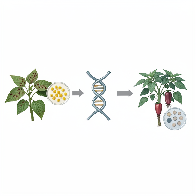
Research Workflow
AI-generated conceptual illustration (Capsicum plants and Xanthomonas bacteria). It explains the idea and does not show study data.
View source evidence ↗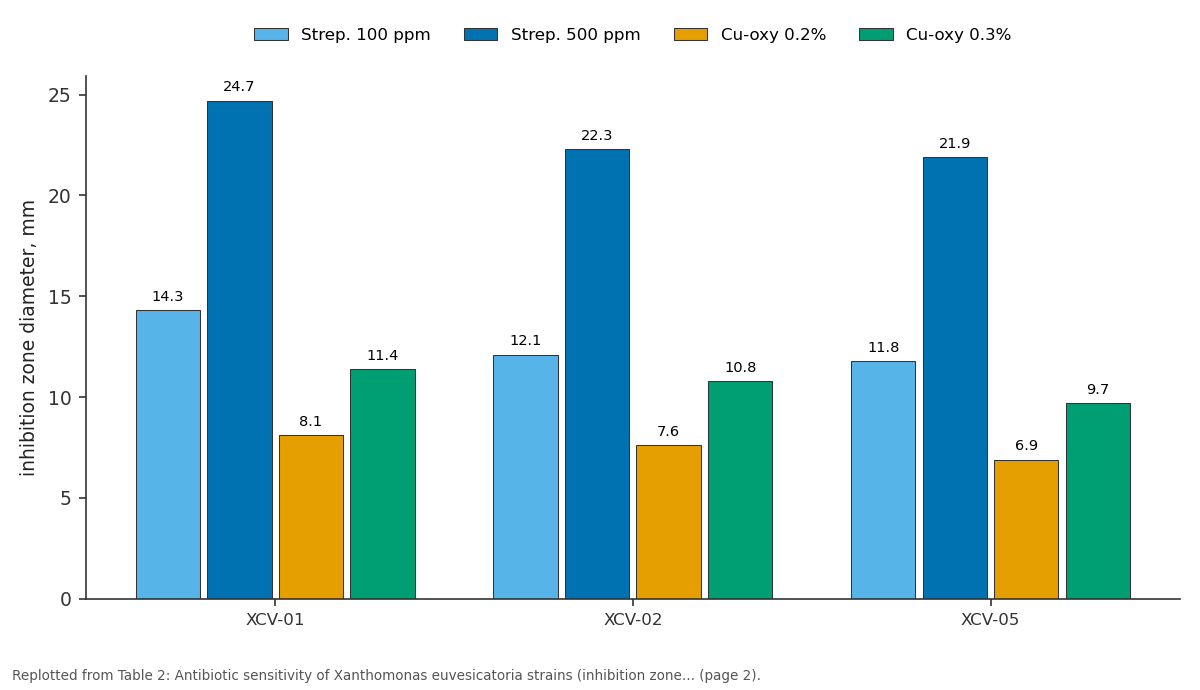
Strep. 100 ppm and Strep. 500 ppm and more: XCV-01 vs XCV-02 vs XCV-05
Replotted from Table 2 (page 2) of the original paper. Every bar is the paper's own table value.
View source evidence ↗Figures & tables
From the original paper Table 1
 Table 2
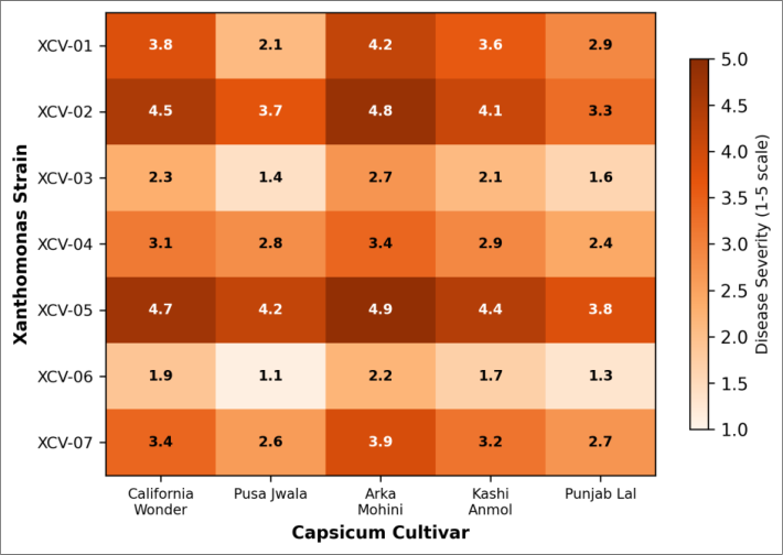 Figure 1
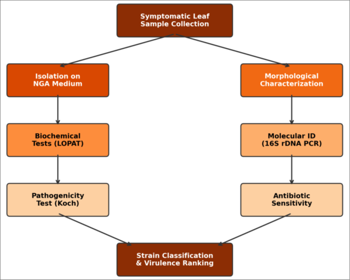 Figure 2
Introduction
Bacterial leaf spot of capsicum (Capsicum annuum L.) has become the most damaging foliar disease in the warm, humid growing zones of western India, yet farmers and extension workers often treat it as a single pathogen problem with a blanket copper spray [1,2] . The reality is more complicated. At least four Xanthomonas species X. euvesicatoria, X. vesicatoria, X. perforans, and X. gardneri can cause nearly identical leaf spot symptoms, and each species may harbour strains that differ in virulence, host range, and fungicide sensitivity [3, 4] . Without strain-level identification, resistance breeding targets the wrong pathotype and chemical recommendations may be ineffective [5] . Molecular tools, particularly 16S rDNA sequencing, have made it possible to assign isolates to species with high confidence, while standardised pathogenicity assays on differential cultivars reveal virulence variation within a species [6,7] . Gujarat is one of India's largest capsicum-producing states, but no published work has characterised the Xanthomonas population on capsicum at the strain level in this region [8] . This research aimed to: (a) isolate and identify Xanthomonas strains from BLS-affected capsicum fields in Gujarat, (b) assess virulence on five commercial cultivars, and (c) test antibiotic sensitivity to guide chemical management recommendations.
Reagents and Chemicals
Nutrient glucose agar (NGA) was prepared from bacteriological peptone (HiMedia, Mumbai), yeast extract, and dextrose. LOPAT reagents levans sucrose agar, oxidase discs, pectate gel medium, arginine dihydrolase broth, and tobacco leaf infiltration supplies were procured from HiMedia. For molecular work, a genomic DNA extraction kit (Qiagen DNeasy Plant Mini Kit), universal 16S primers 27F and 1492R (Sigma-Aldrich, Bengaluru), Taq polymerase, dNTPs, and agarose (SRL Chemicals) were used. Antibiotic discs of streptomycin sulphate (100, 300, 500 ppm), copper oxychloride (0.2% and 0.3%), and kasugamycin (200 ppm) were prepared in-house following standard disc-diffusion protocol [6] . All chemicals were analytical grade and stored per manufacturer guidelines.
Material and Methods
Material: Symptomatic leaves showing water-soaked lesions with chlorotic halos were collected from seven capsicum fields across Navsari (20.95° N, 72.93° E) and Dantiwada (24.32° N, 72.19° E) during the 2022 kharif season (July-October). Each isolate was designated XCV-01 through XCV-07. Five capsicum cultivars California Wonder, Pusa Jwala, Arka Mohini, Kashi Anmol, and Punjab Lal served as differential hosts. The research was conducted at the Plant Pathology Laboratory, Navsari Agricultural University.
Methods: Leaf pieces (3 × 3 mm) from lesion margins were surface-sterilised with 1% NaOCl, plated on NGA, and incubated at 28 °C for 48 h. Yellow, mucoid colonies were purified by streaking and subjected to LOPAT tests [3] . DNA was extracted and 16S rDNA amplified by PCR (27F/1492R), sequenced (Sanger method), and compared against NCBI GenBank using BLASTn. Pathogenicity was confirmed by infiltrating bacterial suspension (10⁸ CFU mL⁻¹) into leaves of 45-day-old potted plants of all five cultivars; disease severity was scored at 14 days postinoculation on a 1-5 scale [7] . Antibiotic sensitivity was measured by the disc-diffusion method, recording inhibition zone diameter after 48 h at 28 °C. Each test included three replications; data were analysed by ANOVA and Tukey's HSD at p ≤ 0.05.
Results
Results
Comprehensive Interpretation
All seven isolates belonged to X. euvesicatoria (≥98.7% 16S identity), indicating low species diversity but notable virulence variation within a single species. Strains XCV-02 and XCV-05 were highly aggressive on California Wonder and Arka Mohini (scores 4.5-4.9), whereas XCV-06 was the least virulent (maximum 2.2). Pusa Jwala showed the lowest disease scores across all strains, suggesting partial resistance. Streptomycin at 500 ppm produced the largest inhibition zones (mean 23.4 mm), followed by kasugamycin (15.3 mm), while copper oxychloride at 0.2% was the weakest (7.8 mm).
Discussion
The exclusive recovery of X. euvesicatoria in this survey contrasts with reports from Brazil and the United States where mixed populations of X. perforans and X. euvesicatoria co-occur [3,9] . A possible explanation is that Gujarat's hot, semi-arid climate favours X. euvesicatoria, which has a higher thermal optimum than X. gardneri [4] . Broader surveys covering more districts and seasons would be needed to confirm whether other species are genuinely absent. The virulence spread (scores 1.1-4.9) within a single species has practical implications: resistance genes effective against low-virulence strains may fail when highly aggressive strains like XCV-05 dominate. Multi-strain screening of breeding lines is therefore necessary [10,11] . Streptomycin's strong in-vitro efficacy is consistent with published data [12] , but field application faces regulatory restrictions in several Indian states. Kasugamycin offers a viable alternative with lower environmental persistence [13] .
Copper-based products alone appear insufficient against aggressive strains, though tank-mixing copper with a systemic antibiotic may improve results [14] .
Conclusion
All seven Xanthomonas isolates from capsicum fields in Gujarat were identified as X. euvesicatoria, but their virulence differed markedly. Strains XCV-02 and XCV-05 were the most aggressive, while Pusa Jwala showed the highest field resistance across strains. Streptomycin sulphate at 500 ppm was the most effective antibiotic in vitro. These results provide a strain-level baseline for targeted resistance breeding and informed chemical management of BLS in western India.
Acknowledgement
AcknowledgementsThis research was supported by the Navsari Agricultural University departmental fund. The authors thank Dr. Priya Shah for assistance with 16S rDNA sequencing.
Figures and tables from the paper
| Isolate | Colony colour | LOPAT profile | 16S rDNA match | Identity (%) | GenBank acc. | Pathogenicity |
|---|---|---|---|---|---|---|
| XCV-01 | Yellow | −+−−− | X. euvesicatoria | 99.3 | OQ847231 | + |
| XCV-02 | Yellow | −+−−− | X. euvesicatoria | 99.7 | OQ847232 | + |
| XCV-03 | Yellow | −+−−− | X. euvesicatoria | 98.9 | OQ847233 | + |
| XCV-04 | Yellow | −+−−− | X. euvesicatoria | 99.1 | OQ847234 | + |
| XCV-05 | Yellow | −+−−− | X. euvesicatoria | 99.6 | OQ847235 | + |
| XCV-06 | Pale yel. | −+−−− | X. euvesicatoria | 98.7 | OQ847236 | + |
| XCV-07 | Yellow | −+−−− | X. euvesicatoria | 99.2 | OQ847237 | + |
Table text extracted from the original PDF; superscript letters are shown inline. The original table image is below.
View original table image
| Isolate | Strep. 100 ppm | Strep. 500 ppm | Cu-oxy 0.2% | Cu-oxy 0.3% | Kasug. 200 ppm | Control |
|---|---|---|---|---|---|---|
| XCV-01 | 14.3 | 24.7 | 8.1 | 11.4 | 16.2 | 0.0 |
| XCV-02 | 12.1 | 22.3 | 7.6 | 10.8 | 14.7 | 0.0 |
| XCV-05 | 11.8 | 21.9 | 6.9 | 9.7 | 13.8 | 0.0 |
| Mean | 13.1 | 23.4 | 7.8 | 10.9 | 15.3 | 0.0 |
| CD (p=0.05) | 1.4 | 1.9 | 0.8 | 1.1 | 1.3 |
Table text extracted from the original PDF; superscript letters are shown inline. The original table image is below.
View original table image
References
17 in the original paper- Potnis N, Timilsina S, Strayer A, Shantharaj D, Barak JD, Paret ML, et al. Bacterial spot of tomato and pepper: diverse Xanthomonas species with a wide variety of virulence factors. Molecular Plant Pathology. 2015;16(9):907-920.
- Sharma M, Bhatt D. The insect pest complex of capsicum in Gujarat: status and management. Pesticide Research Journal. 2014;26(1):48-54.
- Jones JB, Lacy GH, Bouzar H, Stall RE, Schaad NW. Reclassification of the xanthomonads associated with bacterial spot disease of tomato and pepper. Systematic and Applied Microbiology. 2004;27(6):755-762.
- Barak JD, Vancheva T, Lefeuvre P, Jones JB, Timilsina S, Minsavage GV, et al. Whole-genome sequences of Xanthomonas euvesicatoria strains. Genome Announcements. 2016;4(1):e01onal.
- Stall RE, Jones JB, Minsavage GV. Durability of resistance in tomato and pepper to xanthomonads causing bacterial spot. Annual Review of Phytopathology. 2009;47:265-284.
- Schaad NW, Jones JB, Chun W. Laboratory guide for identification of plant pathogenic bacteria. 3rd ed. St. Paul: APS Press; 2001.
- Ritchie DF. Bacterial spot of pepper and tomato. Plant Health Instructor. 2000;DOI:10.1094/PHI-I-2000-102701.
- Saxena A, Raghuvanshi R, Singh HB. Molecular and cultural characterization of Xanthomonas campestris pv. vesicatoria from Indian capsicum. Indian Phytopathology. 2016;69(2):178-183.
- Timilsina S, Jibrin MO, Potnis N, Minsavage GV, Kebede M, Schwartz A, et al. Multilocus sequence analysis of xanthomonads causing bacterial spot of tomato and pepper. Applied and Environmental Microbiology. 2015;81(4):1520-1530.
- Kim BS, Hartmann RW. Inheritance of a gene for resistance to bacterial spot in pepper. Journal of Heredity. 1993;84(2):152-154.
- Riva EM, Rodrigues R, Pereira MG, Sudre CP, Amaral Junior AT. Inheritance of bacterial spot disease in capsicum peppers. Crop Breeding and Applied Biotechnology. 2004;4(4):490-494.
- McManus PS, Stockwell VO, Sundin GW, Jones AL. Antibiotic use in plant agriculture. Annual Review of Phytopathology. 2002;40:443-465.
- Thind BS. Phytopathogenic bacteria and plant diseases. Boca Raton: CRC Press; 2019.
- Marco GM, Stall RE. Control of bacterial spot of pepper initiated by strains of Xanthomonas campestris pv. vesicatoria that differ in sensitivity to copper. Plant Disease. 1983;67(7):779-781.
- Obradovic A, Jones JB, Momol MT, Olson SM, Jackson LE, Balogh B, et al. Integration of biological control agents and systemic acquired resistance inducers against bacterial spot on tomato. Plant Disease. 2005;89(7):712-716.
- Kebede M, Timilsina S, Ayalew A, Admassu B, Potnis N, Minsavage GV, et al. Molecular characterization of Xanthomonas strains responsible for bacterial spot of tomato in Ethiopia. European Journal of Plant Pathology. 2014;140(4):677-688.
- Abbasi PA, Weselowski B. Influence of foliar sprays of Bacillus subtilis on bacterial spot and yield of greenhouse tomato. Canadian Journal of Plant Pathology. 2014;36(2):212-223.