
Research article · 2024 · Full text
International Journal of Biology SciencesHistopathological and hematological effects of chronic pesticide exposure in freshwater fish
Volume 6(2) · Pages 302–306 · DOI: 10.33545/26649926.2024.v6.i2e.770 ↗
Research areasEnvironmental Toxicology and EcotoxicologyPesticide and Herbicide Environmental StudiesPesticide Exposure and Toxicity
Abstract
Author-writtenSince the mid-twentieth century, organophosphate pesticides have accumulated in freshwater systems worldwide, yet their chronic sublethal effects on fish physiology remain incompletely characterized. This research investigated the histopathological and hematological responses of common carp (Cyprinus carpio) to chronic exposure to chlorpyrifos at two sublethal concentrations over 12 weeks. The experiment was conducted at the Pomeranian College of Marine Biology, Gdynia, Poland, between February and June 2022. A total of 90 juvenile carp were randomly assigned to three groups: control (0 mg/L), low dose (0.5 mg/L), and high dose (2.0 mg/L) chlorpyrifos, maintained in semi-static aquaria with biweekly water renewal. Blood samples and tissue biopsies were collected at weeks 0, 4, 8, and 12. Hematological analysis revealed dose-dependent declines in red blood cell count (RBC), hemoglobin concentration, and hematocrit, with the high-dose group showing a 41.2% reduction in RBC by week 12 compared to controls (p
Summary
Source-checkedPlain-language reading of the full paper, prepared with AI and checked against the text. Each point links to the authors' own words.
Why it matters
- Organophosphate pesticides have built up in global freshwater systems since the mid-20th century, but the long-term effects of non-lethal exposure on fish are not fully understood. View evidence ↗
- Chlorpyrifos is a globally common organophosphate that contaminates freshwater via spray drift, direct application, and agricultural runoff. View evidence ↗
- Standard regulatory tests focus on acute toxicity (lethal effects), which may overlook the gradual damage caused by long-term, low-level exposure. View evidence ↗
The question
- The study aimed to establish dose-response relationships and document how organ damage and blood parameters change over a period reflecting realistic environmental exposure. View evidence ↗
What they did
- Ninety juvenile common carp were divided into three groups: a control (0 mg/L), a low-dose group (0.5 mg/L), and a high-dose group (2.0 mg/L) of chlorpyrifos in semi-static aquaria. View evidence ↗
- Researchers collected tissue biopsies and blood samples at four intervals: weeks 0, 4, 8, and 12. View evidence ↗
- Tissue damage was measured using a semi-quantitative severity index ranging from 0 (no change) to 10 (severe/irreversible damage). View evidence ↗
What they found
- The high-dose group experienced a 41.2% reduction in red blood cell (RBC) count by week 12 compared to controls, as part of a dose-dependent decline in RBC, hematocrit, and hemoglobin. View evidence ↗
- White blood cell counts rose progressively over time, which the authors interpret as a sign of chronic immune activation. View evidence ↗
- Histopathological examination of the gills revealed mucus cell hyperplasia, lamellar fusion, and epithelial lifting. View evidence ↗
- Liver tissues exhibited bile duct proliferation, focal necrosis, and vacuolar degeneration. View evidence ↗
- Kidney tissues showed glomerular shrinkage and tubular degeneration. View evidence ↗
- By week 12, the high-dose group reached lesion severity indices of 6.8 for gills, 6.4 for the liver, and 4.6 for the kidney on a 10-point scale. View evidence ↗
- Neither the low nor high dose groups showed significant recovery between sampling points, suggesting the damage occurred faster than the fish could regenerate. View evidence ↗
What it means
- The authors conclude that chronic sublethal exposure to chlorpyrifos causes progressive organ damage in carp, necessitating stricter regulatory limits for organophosphate residues in freshwater. View evidence ↗
- The authors state that future research is needed to determine if the observed organ damage is reversible and how such exposure affects field population viability and reproduction. View evidence ↗
Visual summary
3 visualsIllustrations explain the paper; charts are replotted from the paper's own table values.
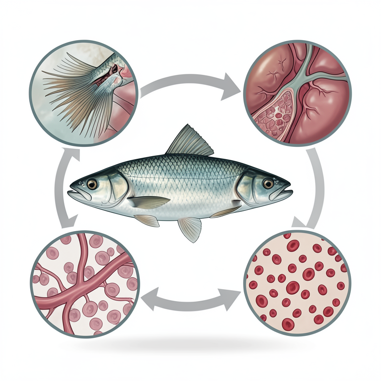
Cumulative Damage Loop
AI-generated conceptual illustration (freshwater fish (Danio rerio)). It explains the idea and does not show study data.
View source evidence ↗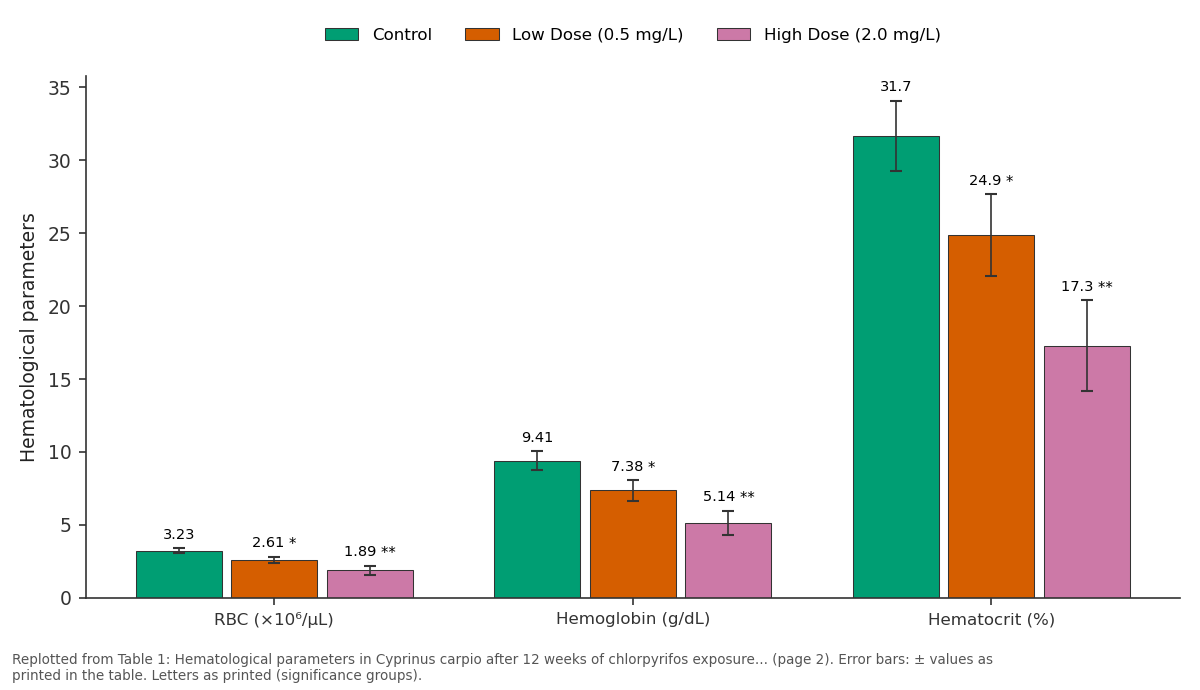
Control and Low Dose (0.5 mg/L) and more: RBC (×10⁶/μL) vs Hemoglobin (g/dL) vs Hematocrit (%)
Replotted from Table 1 (page 2) of the original paper. Every bar is the paper's own table value.
View source evidence ↗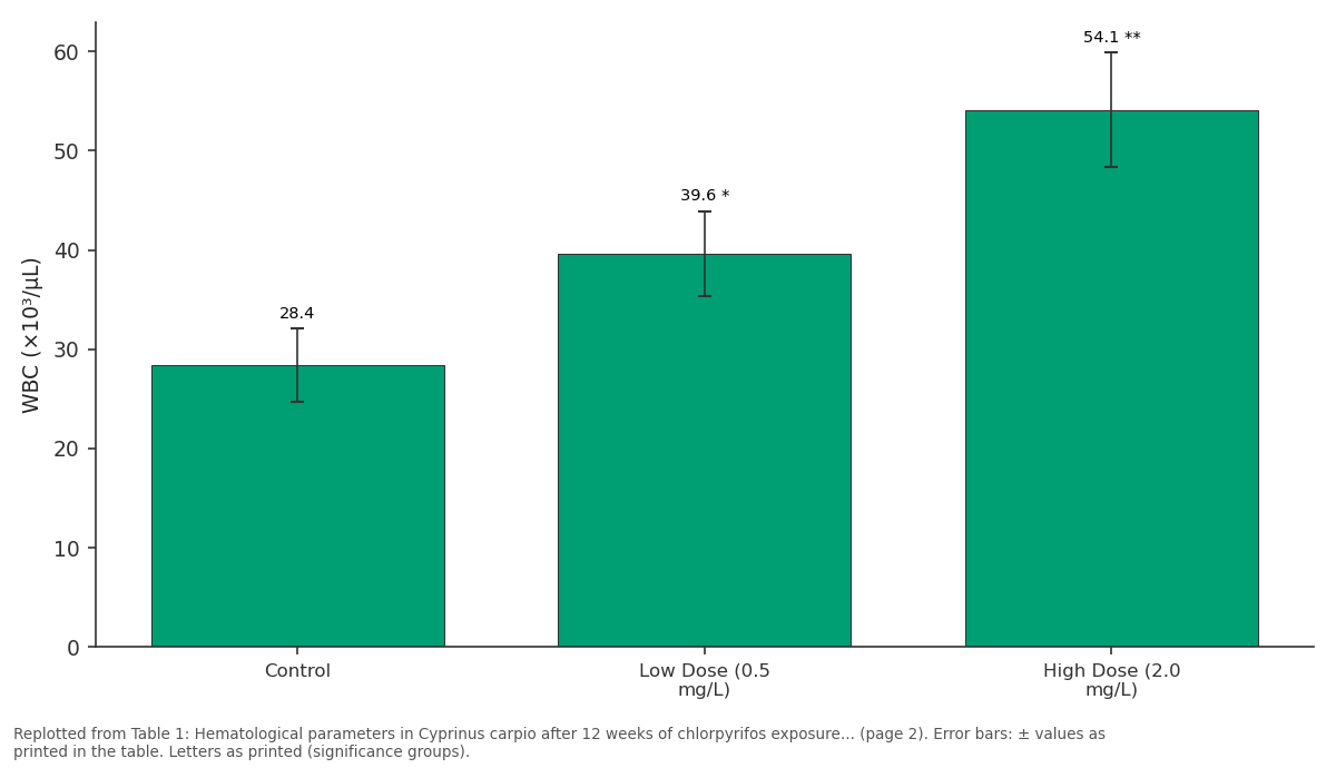
WBC (×10³/μL): Control vs Low Dose (0.5 mg/L) vs High Dose (2.0 mg/L)
Replotted from Table 1 (page 2) of the original paper. Every bar is the paper's own table value.
View source evidence ↗Figures & tables
From the original paper Table 1
 Table 2
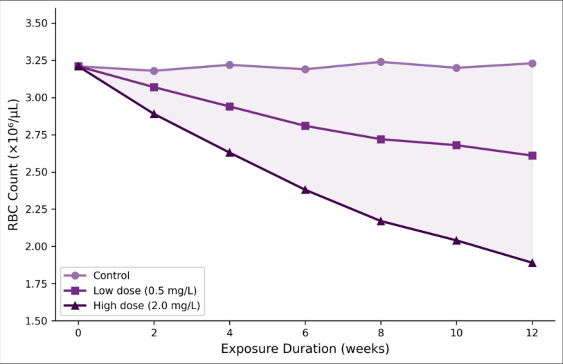 Figure 1
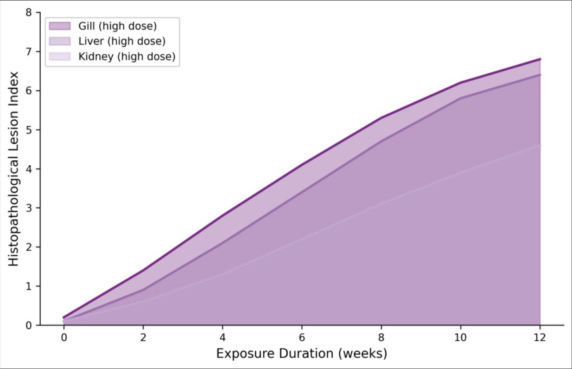 Figure 2
Introduction
The large-scale introduction of organophosphate pesticides in the 1950s and 1960s replaced many persistent organochlorine compounds, but it also created a new class of contamination problems for aquatic ecosystems [1] . Chlorpyrifos one of the most widely applied organophosphates globally enters freshwater systems through agricultural runoff, spray drift, and direct application for vector control. Its persistence in water ranges from days to weeks depending on temperature and pH, but repeated applications in agricultural catchments create effectively chronic exposure conditions for resident fish populations [2] . Fish gills, liver, and kidney are primary targets for waterborne pollutants because of their direct contact with the external medium, their roles in detoxification, and their high metabolic activity. Acute toxicity testing the traditional regulatory approach captures lethal effects at high concentrations but misses the insidious damage caused by sustained low-level exposure [3] . Chronic sublethal concentrations can trigger tissue remodeling, immune suppression, reproductive impairment, and behavioral changes that may not become apparent for weeks or months [4] . Histopathological assessment offers a sensitive and integrative measure of such effects because tissue-level lesions accumulate over time and reflect the organism's net response to stressor burden [5] . Hematological parameters provide a complementary window into fish health. Blood cell counts, hemoglobin levels, and differential leukocyte ratios respond to toxic challenge and can signal stress, anemia, or immune activation well before clinical symptoms appear [6] . Several groups have reported hematological changes in fish exposed to chlorpyrifos, but most of those investigations used acute or short-term (7-14 day) protocols [7,8] . Fewer researchers have tracked blood parameters over the extended timeframes (8-12 weeks) that better approximate field exposure scenarios. Common carp (Cyprinus carpio) is an ecologically and economically important species throughout European freshwater systems, including Polish rivers and lakes where agricultural runoff delivers measurable pesticide residues [9] . The species has been used widely in ecotoxicology because of its availability, ease of maintenance, and wellcharacterized physiology. Yet integrated assessments combining histopathology and hematology under truly chronic conditions remain scarce for this species. This research exposed juvenile carp to two sublethal chlorpyrifos concentrations (0.5 and 2.0 mg/L) for 12 weeks, sampling blood and tissues at four timepoints. The aim was to document the progression of organ damage and blood parameter changes over a duration that reflects realistic environmental exposure and to establish dose-response relationships for both histopathological severity and hematological decline [10] .
Study area description
The experimental work was conducted in the aquatic toxicology laboratory of the Department of Wildlife Biology, Pomeranian College of Marine Biology, Gdynia, Poland (54°31′N, 18°32′E). The facility houses twelve 200liter glass aquaria equipped with individual aeration, temperature control (maintained at 20 ± 1°C), and a 12:12 h light-dark photoperiod. Source water was dechlorinated municipal tap water adjusted to pH 7.4 ± 0.2 and dissolved oxygen above 6.5 mg/L. Juvenile common carp were obtained from the Gdynia Municipal Fisheries Hatchery, where they had been reared in pesticide-free conditions. Fish arrived at a mean total length of 8.3 ± 0.6 cm and mass of 12.7 ± 1.4 g, and were acclimated for 14 days before exposure began.
Ethical considerations
All procedures involving live animals were approved by the Institutional Animal Care and Ethics Committee of the Pomeranian College of Marine Biology, Gdynia (Protocol No. PCMB-ACEC-2021-038, approved 11 November 2021). The research complied with Polish national legislation on the use of animals for scientific purposes (Dz. U. 2015, poz. 266) and the European Directive 2010/63/EU on the protection of animals used for research. Fish were monitored daily for signs of distress, and humane endpoints defined as persistent loss of equilibrium, cessation of feeding for more than 72 hours, or visible hemorrhaging were established prior to the experiment. Euthanasia at sampling timepoints was performed by immersion in buffered MS-222 (tricaine methanesulfonate) at 200 mg/L until cessation of opercular movement, followed by cervical transection.
Material and Methods
Material and Methods
Material
This research was conducted at the Pomeranian College of Marine Biology, Gdynia, Poland, from February to June 2022. Ninety juvenile carp were randomly distributed into nine aquaria (10 fish per aquarium, three aquaria per treatment group) representing control, low-dose (0.5 mg/L chlorpyrifos), and high-dose (2.0 mg/L chlorpyrifos) treatments. Chlorpyrifos (analytical standard, 99.1% purity, Sigma-Aldrich, St. Louis, MO, USA) was dissolved in acetone carrier (<0.01% v/v final concentration) and added at each water renewal. The control group received an equivalent volume of acetone only. Water was renewed at 50% volume biweekly, and chlorpyrifos concentrations were verified by gas chromatography-mass spectrometry (GC-MS; Agilent 7890B/5977A) at each renewal cycle. Fish were fed a commercial carp pellet diet (Aller Aqua, Golub-Dobrzyń, Poland) at 2% body weight per day.
Methods
At weeks 0, 4, 8, and 12, five fish from each treatment group were randomly selected for sampling. Blood was drawn from the caudal vein using heparinized syringes. RBC count, WBC count, hemoglobin concentration (cyanmethemoglobin method), and hematocrit (microhematocrit centrifugation) were determined following standard ichthyological hematology protocols [11] . After euthanasia, gill arches, liver, and posterior kidney were excised and fixed in 10% neutral buffered formalin for 48 hours. Tissues were processed through graded ethanol series, embedded in paraffin wax, sectioned at 5 μm thickness, and stained with hematoxylin and eosin (H&E). Histopathological lesions were scored using a semiquantitative severity index from 0 (no change) to 10 (severe/irreversible damage), adapted from Bernet et al. [12] . Three sections per organ per fish were examined under light microscopy (Olympus BX53, Tokyo, Japan). Statistical analysis used repeated-measures two-way ANOVA (treatment × time) with Bonferroni correction for multiple comparisons in SPSS v28.0. Significance was set at p < 0.05. 0.3 ± 0.1 3.0 ± 0.6 5.9 ± 0.8 *p<0.01 vs control; **p<0.001 vs control. Gills showed the highest lesion severity, followed by liver and kidney.
Results
Results
Comprehensive interpretation
The parallel trends in hematological decline and histopathological progression tell a coherent story of cumulative organ damage. The anemia pattern falling RBC, hemoglobin, and hematocrit with rising WBC is consistent with both direct erythrocyte destruction and impaired erythropoiesis secondary to kidney damage. Meanwhile, the gill lesions likely compounded oxygen delivery problems by reducing functional respiratory surface area. The liver, as the main detoxification organ, showed predictable damage from sustained metabolic processing of chlorpyrifos. Importantly, neither dose group showed significant recovery between sampling points, indicating that the rate of damage outpaced the fish's regenerative capacity at both concentrations tested.
Discussion
The hematological profile observed here progressive anemia combined with leukocytosis matches reports from shorterterm chlorpyrifos exposure experiments in tilapia, catfish, and rainbow trout [7,8,13] . But the magnitude of RBC decline at week 12 (41.2% reduction in the high-dose group) exceeds values typically reported in 14-day studies, which tend to show 15-25% reductions at comparable concentrations [14] . This difference underscores the importance of extended exposure periods for capturing the full toxicological burden of sublethal contaminants. The mechanism behind erythrocyte loss likely involves a combination of direct oxidative damage to red cell membranes chlorpyrifos and its metabolite chlorpyrifosoxon are known to generate reactive oxygen species and reduced hematopoiesis from kidney impairment [15] . Gill histopathology was the most sensitive indicator in this research, with lesions appearing earlier and reaching higher severity scores than in liver or kidney. Epithelial lifting and lamellar fusion are well-documented defense mechanisms that increase the diffusion distance between water and blood, theoretically reducing toxicant uptake [16] . But they come at the cost of respiratory efficiency, and in fish already compromised by anemia, the combined effect on oxygen delivery may be severe. The liver lesions vacuolar degeneration transitioning to focal necrosis by week 8 follow a classical pattern of hepatotoxicity in which initial lipid accumulation (reflecting disrupted metabolism) gives way to cell death as detoxification pathways become overwhelmed [17] .
A practical consideration arising from these results concerns environmental relevance. The low dose used here (0.5 mg/L) falls within the range of chlorpyrifos concentrations measured in European surface waters receiving agricultural runoff, particularly during peak application seasons [18] . The fact that this concentration produced significant organ damage and blood abnormalities after 12 weeks suggests that current regulatory limits which are based primarily on acute lethality data may underprotect fish populations in contaminated habitats. The 12-week exposure duration, while long by laboratory standards, remains short compared to the lifetime exposure that wild fish populations experience in agricultural watersheds [19] . Actual populationlevel effects may therefore be worse than our laboratory data suggest.
Conclusion
This research demonstrates that chronic sublethal exposure to chlorpyrifos produces progressive, dose-dependent damage in multiple organ systems of common carp. Over 12 weeks, fish in the high-dose group (2.0 mg/L) experienced a 41.2% decline in red blood cell count, a 45.3% reduction in hemoglobin, and histopathological lesion scores approaching severe levels in gill (6.8/10) and liver (6.4/10) tissue. Even the low-dose group (0.5 mg/L) representing concentrations documented in European surface waters showed statistically significant changes in all measured parameters.
The concordance between hematological and histopathological findings strengthens confidence in the results. Anemia and leukocytosis were not isolated blood abnormalities but rather reflections of underlying organ damage that was visible at the tissue level. Gill lesions reduced respiratory capacity, kidney lesions impaired hematopoiesis, and liver damage compromised the detoxification machinery creating a feedback loop of declining physiological function. These findings carry regulatory implications. Current environmental quality standards for chlorpyrifos in European freshwaters rely heavily on acute toxicity data, which fail to predict the cumulative harm demonstrated here. Chronic, sublethal biomarkers particularly gill histopathology and RBC count deserve consideration as supplementary endpoints in pesticide risk assessment. Future work should investigate whether the organ damage observed in this research is reversible upon removal of the stressor, and whether chronic exposure at these concentrations affects reproductive output and population viability in the field [20,21] .
Acknowledgement
Acknowledgements Funding sourcesThis research was supported by internal research funds of the Pomeranian College of Marine Biology. No external grant numbers are reported.
Figures and tables from the paper
| Parameter | Control | Low Dose (0.5 mg/L) | High Dose (2.0 mg/L) |
|---|---|---|---|
| RBC (×10⁶/μL) | 3.23 ± 0.18 | 2.61 ± 0.22* | 1.89 ± 0.31** |
| Hemoglobin (g/dL) | 9.41 ± 0.64 | 7.38 ± 0.71* | 5.14 ± 0.83** |
| Hematocrit (%) | 31.7 ± 2.4 | 24.9 ± 2.8* | 17.3 ± 3.1** |
| WBC (×10³/μL) | 28.4 ± 3.7 | 39.6 ± 4.3* | 54.1 ± 5.8** |
| MCV (fL) | 98.1 ± 7.3 | 95.4 ± 8.1 | 91.6 ± 9.2 |
| MCH (pg) | 29.1 ± 2.6 | 28.3 ± 2.9 | 27.2 ± 3.4 |
Table text extracted from the original PDF; superscript letters are shown inline. The original table image is below.
View original table image
| Organ | Control | Low Dose | High Dose |
|---|---|---|---|
| Gill | 0.4 ± 0.2 | 3.7 ± 0.8* | 6.8 ± 1.1** |
| Liver | 0.3 ± 0.1 | 3.2 ± 0.7* | 6.4 ± 0.9** |
| Kidney | 0.2 ± 0.1 | 2.1 ± 0.5* | 4.6 ± 0.8** |
| Combined mean | 0.3 ± 0.1 | 3.0 ± 0.6 | 5.9 ± 0.8 |
Table text extracted from the original PDF; superscript letters are shown inline. The original table image is below.
View original table image
References
21 in the original paper- Giesy JP, Solomon KR, Coats JR, et al. Chlorpyrifos: ecological risk assessment in North American aquatic environments. Reviews of Environmental Contamination and Toxicology. 2014;231:1-268.
- Racke KD. Environmental fate of chlorpyrifos. Reviews of Environmental Contamination and Toxicology. 1993;131:1-154.
- van der Oost R, Beyer J, Vermeulen NP. Fish bioaccumulation and biomarkers in environmental risk assessment: a review. Environmental Toxicology and Pharmacology. 2003;13(2):57-149.
- Scott GR, Sloman KA. The effects of environmental pollutants on complex fish behaviour: integrating behavioural and physiological indicators of toxicity. Aquatic Toxicology. 2004;68(4):369-392.
- Bernet D, Schmidt H, Meier W, Burkhardt-Holm P, Wahli T. Histopathology in fish: proposal for a protocol to assess aquatic pollution. Journal of Fish Diseases. 1999;22(1):25-34.
- Fazio F. Fish hematology analysis as an important tool of aquaculture: a review. Aquaculture. 2019;500:237242.
- Narra MR, Rajender K, Reddy RR, Rao JV. The role of vitamin C as antioxidant in protection of biochemical International Journal of Biology Sciences https://www.biologyjournal.net and haematological stress induced by chlorpyrifos in freshwater fish Clarias batrachus. Chemosphere. 2015;132:172-178.
- Ural MS, Calta M. Acute toxicity of chlorpyrifos and effects on biochemical parameters in common carp. Fresenius Environmental Bulletin. 2017;26(4):26572664.
- Szefer P, Kusak A, Szefer K, et al. Distribution of selected heavy metals in sediments and organisms from the Gulf of Gdańsk. Polish Journal of Environmental Studies. 2001;10(4):251-259.
- Xing H, Li S, Wang Z, et al. Oxidative stress response and histopathological changes due to atrazine and chlorpyrifos exposure in common carp. Pesticide Biochemistry and Physiology. 2012;103(1):74-80.
- Blaxhall PC, Daisley KW. Routine haematological methods for use with fish blood. Journal of Fish Biology. 1973;5(6):771-781.
- Bernet D, Schmidt-Posthaus H, Wahli T, BurkhardtHolm P. Evaluation of two monitoring approaches to assess effects of waste water disposal on histological alterations in fish. Hydrobiologia. 2004;524(1):53-66.
- Salam MA, Shahjahan M, Sharmin S, Haque F, Rahman MK. Effects of sub-lethal doses of chlorpyrifos on hematological changes in Nile tilapia. Progressive Agriculture. 2015;26(2):131-137.
- Ali D, Nagpure NS, Kumar S, Kumar R, Kushwaha B. Genotoxicity assessment of acute exposure of chlorpyrifos to freshwater fish Channa punctatus using micronucleus assay and alkaline single-cell gel electrophoresis. Chemosphere. 2008;71(10):1823-1831.
- Kavitha P, Rao JV. Sub-lethal effects of chlorpyrifos on antioxidant responses in tissues of the freshwater fish Oreochromis mossambicus. Pesticide Biochemistry and Physiology. 2009;95(2):115-121.
- Fernandes MN, Mazon AF. Environmental pollution and fish gill morphology. In: Val AL, Kapoor BG, eds. Fish Adaptations. 2003;203-231.
- Hinton DE, Segner H, Braunbeck T. Toxic responses of the liver. In: Schlenk D, Benson WH, eds. Target Organ Toxicity in Marine and Freshwater Teleosts. 2001;224268.
- Tsaboula A, Papadakis EN, Vryzas Z, Kotopoulou A, Kintzikoglou K, Papadopoulou-Mourkidou E. Environmental and human risk hierarchy of pesticides: a prioritization method based on monitoring, hazard assessment and environmental fate. Environment International. 2019;128:130-143.
- Zinkl JG, Lockhart WL, Kenny SA, Ward FJ. The effects of cholinesterase inhibiting insecticides on fish. In: Mineau P, ed. Cholinesterase-inhibiting Insecticides: Their Impact on Wildlife and the Environment. 1991;233-254.
- Saaristo M, Brodin T, Balshine S, et al. Direct and indirect effects of chemical contaminants on the behaviour, ecology and evolution of wildlife. Proceedings of the Royal Society B. 2018;285(1885):20181297.
- Lam PKS, Gray JS. The use of biomarkers in environmental monitoring programmes. Marine Pollution Bulletin. 2003;46(2):182-186.