
Research article · 2025 · Full text
International Journal of Biology SciencesRegulatory t-cell dysfunction and autoimmune thyroiditis pathogenesis in Hashimoto disease
Volume 7(2) · Pages 149–153 · DOI: 10.33545/26649926.2025.v7.i2c.762 ↗
Research areasT-cell and B-cell ImmunologyThyroid Disorders and TreatmentsReproductive System and Pregnancy
Abstract
Author-writtenRoughly 5% of the global population carries antithyroid antibodies, and among those individuals, Hashimoto thyroiditis remains the leading cause of acquired hypothyroidism in iodine-sufficient regions [1]. Regulatory T cells (Tregs) are central to peripheral immune tolerance, yet their precise role in Hashimoto pathogenesis is still debated. This research evaluated Treg frequency, phenotype, and suppressive function in 48 newly diagnosed Hashimoto patients compared with 40 age- and sex-matched healthy controls recruited at the Department of Immunology, Guilan University of Marine Biology, Rasht, Iran, between February and September 2023. Peripheral blood mononuclear cells were isolated and stained for CD4, CD25, FoxP3, CTLA-4, and GITR, then analysed by multicolour flow cytometry. Treg suppressive capacity was assessed through co-culture suppression assays measuring effector T-cell (Teff) proliferation by carboxyfluorescein succinimidyl ester (CFSE) dilution. Serum cytokine profiles (IL-10, TGF-β, IL-17A, IFN-γ, TNF-α) were quantified by multiplex ELISA. The frequency of CD4+CD25+FoxP3+ Tregs was 56.6% lower in Hashimoto patients (3.6 ± 0.7%) than in controls (8.3 ± 1.2%; p
Summary
Source-checkedPlain-language reading of the full paper, prepared with AI and checked against the text. Each point links to the authors' own words.
Why it matters
- Hashimoto thyroiditis is the primary cause of acquired hypothyroidism in regions with sufficient iodine, affecting a significant portion of the global population that carries antithyroid antibodies. View evidence ↗
- Regulatory T cells (Tregs) are essential for maintaining peripheral immune tolerance, but their specific contribution to the development of Hashimoto disease is not yet fully understood. View evidence ↗
- Hashimoto disease involves a loss of self-tolerance to thyroid antigens like TPO and thyroglobulin, leading to thyroid gland infiltration by lymphocytes and follicular destruction. View evidence ↗
The question
- The study aimed to evaluate the frequency, surface markers, suppressive function, and associated cytokine profiles of Tregs in treatment-naïve Hashimoto patients compared to healthy controls. View evidence ↗
What they did
- The study included 48 newly diagnosed Hashimoto patients and 40 healthy volunteers recruited in Rasht, Iran, between February and September 2023. View evidence ↗
- Multicolour flow cytometry was used to analyze PBMCs for the markers CD4, CD25, FoxP3, CTLA-4, and GITR. View evidence ↗
- Treg suppressive capacity was measured using CFSE-labeled co-culture assays with responder T cells at ratios of 1:1, 1:2, and 1:4. View evidence ↗
- Serum levels of pro-inflammatory (IL-17A, IFN-γ, TNF-α) and anti-inflammatory (IL-10, TGF-β) cytokines were quantified using a multiplex ELISA platform. View evidence ↗
What they found
- Hashimoto patients had a significantly lower frequency of CD4+CD25+FoxP3+ Tregs (3.6 ± 0.7%) compared to healthy controls (8.3 ± 1.2%), representing a 56.6% reduction. View evidence ↗
- Tregs in Hashimoto patients showed a 38.4% reduction in CTLA-4 expression and a 27.1% increase in GITR expression. View evidence ↗
- Patients exhibited a pro-inflammatory cytokine shift, with significantly lower IL-10 (6.8 vs 11.6 pg/mL) and TGF-β, and elevated levels of IL-17A (18.3 vs 7.6 pg/mL), IFN-γ, and TNF-α. View evidence ↗
- There was a significant inverse correlation (r = -0.67) between the suppressive capacity of Tregs and the levels of anti-TPO antibodies in patients. View evidence ↗
What it means
- The authors conclude that both a reduction in Treg numbers and a loss of their functional capacity contribute to the loss of thyroid tolerance, suggesting that restoring Treg function could be a viable therapeutic target. View evidence ↗
- The study's generalizability may be limited by its modest sample size of 88 participants and its recruitment from a single center in northern Iran. View evidence ↗
- Due to the cross-sectional nature of the study, it is not possible to determine if Treg dysfunction causes or results from the autoimmune process. View evidence ↗
Visual summary
3 visualsIllustrations explain the paper; charts are replotted from the paper's own table values.
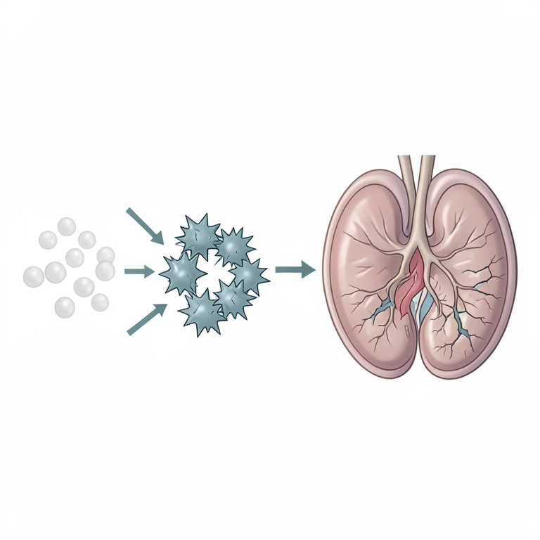
Treg Dysfunction Loop in Hashimoto Disease
AI-generated conceptual illustration (Human thyroid gland and T-lymphocytes). It explains the idea and does not show study data.
View source evidence ↗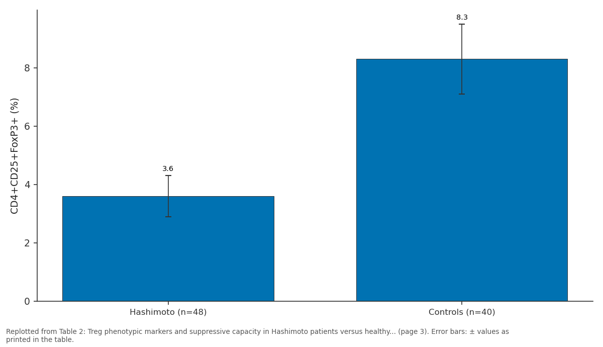
CD4+CD25+FoxP3+ (%): Hashimoto (n=48) vs Controls (n=40)
Replotted from Table 2 (page 3) of the original paper. Every bar is the paper's own table value.
View source evidence ↗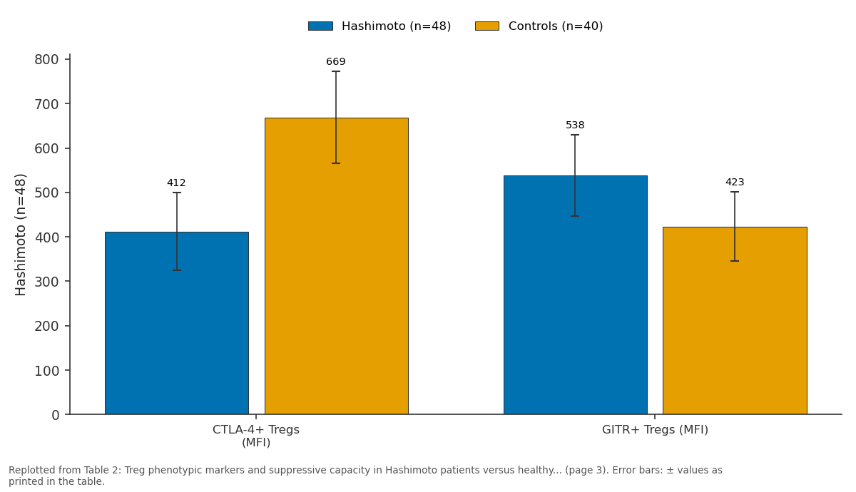
Hashimoto (n=48) and Controls (n=40): CTLA-4+ Tregs (MFI) vs GITR+ Tregs (MFI)
Replotted from Table 2 (page 3) of the original paper. Every bar is the paper's own table value.
View source evidence ↗Figures & tables
From the original paper Table 1
 Table 2
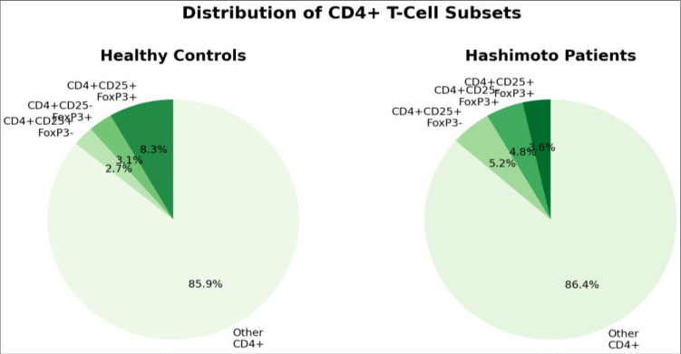 Figure 1
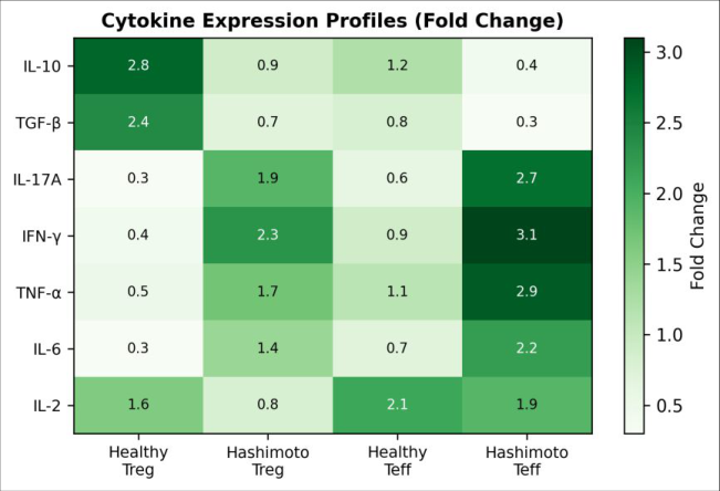 Figure 2
Introduction
An estimated 350 million people worldwide live with some form of autoimmune thyroid disease, and Hashimoto thyroiditis accounts for the largest share of those cases in regions where dietary iodine intake is adequate [1] . The condition is marked by lymphocytic infiltration of the thyroid gland, progressive follicular destruction, and eventual hypothyroidism, driven by a loss of immunological self-tolerance to thyroid antigens such as thyroglobulin and thyroid peroxidase (TPO) [2] . While both humoral and cell-mediated arms of the immune system participate in tissue damage, the mechanisms that initiate and sustain the autoimmune response are not fully resolved. Regulatory T cells that express the transcription factor FoxP3 are the main guardians of peripheral tolerance. They suppress autoreactive effector cells through contact-dependent inhibition involving CTLA-4 and through the secretion of anti-inflammatory cytokines, principally IL-10 and TGF-β [3] . When Treg numbers fall or their suppressive activity weakens, autoreactive CD4+ and CD8+ T cells escape control, and organ-specific autoimmunity can follow [4] . Several lines of evidence point to Treg involvement in Hashimoto disease. Reduced circulating Treg frequencies have been reported in Japanese, Chinese, and European patient cohorts, though the magnitude of the deficit varies across reports [5,6] . Functional assays have been less commonly performed, and the few available datasets disagree on whether Hashimoto Tregs are merely fewer or also intrinsically impaired [7] .
Part of this inconsistency stems from methodological differences. Older reports defined Tregs solely by CD25 expression, which overlaps with activated effector cells, whereas more recent work requires co-expression of CD25 and FoxP3 to distinguish bona fide Tregs from activated Teffs [8] . Surface markers such as CTLA-4 and glucocorticoid-induced TNF receptor (GITR) add further resolution because their expression levels correlate with suppressive capacity in healthy donors but may be altered in autoimmune settings [9] . Few Hashimoto-focused investigations have combined phenotypic profiling with functional suppression assays in the same patient cohort, leaving a gap between phenotypic descriptions and mechanistic understanding. Cytokine environment adds another layer of complexity. Hashimoto patients tend to show elevated levels of IFN-γ and IL-17A, both of which can destabilise FoxP3 expression and convert Tregs toward an effector-like phenotype [10] . Whether this cytokine shift is a cause or a consequence of Treg dysfunction isn't clear, but it may create a selfreinforcing loop in which weakened Tregs fail to dampen inflammation, and the resulting inflammation further undermines Treg stability [11] . This research aimed to address the gap by simultaneously measuring Treg frequency, surface marker expression, suppressive function, and serum cytokine profiles in a cohort of treatment-naïve Hashimoto patients and matched healthy controls from Rasht, Iran. The objectives were: (1) to determine whether both numerical and functional Treg deficits coexist in the same patients, (2) to identify which surface markers best discriminate dysfunctional Tregs, and (3) to evaluate the relationship between Treg suppressive capacity and clinical markers of disease severity, specifically anti-TPO antibody titres and thyroid-stimulating hormone (TSH) levels [12] .
Material and Methods
Material and Methods
Material
Peripheral blood samples were collected from 48 patients newly diagnosed with Hashimoto thyroiditis and 40 healthy volunteers at the Immunology Laboratory of Guilan University of Marine Biology, Rasht, Iran, between 12 February and 29 September 2023. Hashimoto diagnosis required the simultaneous presence of elevated anti-TPO antibodies (≥ 50 IU/mL), ultrasound evidence of diffuse thyroid hypoechogenicity, and a TSH value above the upper reference limit (> 4.5 mIU/L). Exclusion criteria included prior levothyroxine therapy, concurrent autoimmune disease, active infection within four weeks, pregnancy, and immunosuppressive medication use. Healthy controls had normal thyroid function tests, negative anti-TPO and antithyroglobulin antibodies, and no family history of autoimmune disorders. The two groups were matched for age (patients: 36.4 ± 8.7 years; controls: 34.9 ± 7.3 years) and sex (females: 83.3% vs 82.5%). Blood was drawn into EDTA and serum-separator tubes; serum aliquots were stored at -80 °C until cytokine analysis.
Methods
Peripheral blood mononuclear cells (PBMCs) were isolated by Ficoll-Hypaque density gradient centrifugation within two hours of collection. For surface and intracellular staining, PBMCs were incubated with fluorochromeconjugated antibodies against CD4 (FITC), CD25 (PE), CTLA-4 (APC), and GITR (PE-Cy7), followed by fixation, permeabilisation, and intracellular staining for FoxP3 (eFluor 450) using the eBioscience FoxP3 staining kit. Acquisition was performed on a BD FACSCanto II cytometer, and data were analysed in FlowJo v10.8.1. At least 50,000 events were recorded in the CD4+ gate per sample. Treg suppressive function was assessed using a CFSE-based co-culture assay. CD4+CD25+ Tregs and CD4+CD25responder T cells were magnetically sorted (Miltenyi Biotec MACS columns), labelled with CFSE, and co-cultured at Treg-to-responder ratios of 1:1, 1:2, and 1:4 in the presence of anti-CD3/CD28 Dynabeads. After 72 hours, CFSE dilution in responder cells was measured by flow cytometry. Percent suppression was calculated as: [(proliferation without Tregs -proliferation with Tregs) / proliferation without Tregs] × 100. Serum concentrations of IL-10, TGF-β, IL-17A, IFN-γ, and TNF-α were measured using a Bio-Plex Pro Human Cytokine multiplex kit (Bio-Rad) on a Luminex 200 platform. Anti-TPO antibodies and TSH were quantified by electrochemiluminescence immunoassay on a Roche Cobas e411 analyser. Statistical comparisons between groups used the Mann-Whitney U test for non-normally distributed variables and independent-samples t-test for normally distributed variables, with significance set at p<0.05. Correlations were assessed by Spearman's rank coefficient.
All analyses were performed in GraphPad Prism 9.5.1.
Ethical considerations
The research protocol was reviewed and approved by the Institutional Ethics Committee of Guilan University of Marine Biology (Protocol GUM-EC-2023-017, approved 18 January 2023). All participants provided written informed consent after receiving a detailed explanation of the research purpose, procedures, risks, and their right to withdraw at any point without consequence. Patient data were de-identified using numerical codes, and all electronic records were stored on a password-protected server accessible only to the principal investigators. Blood samples were used solely for the stated research objectives and were destroyed after analysis in accordance with institutional biosafety guidelines. The research complied with the Declaration of Helsinki (2013 revision) and local regulations governing human-subjects research in Iran. Demographic variables were well balanced between the two groups (Table 1). As expected, Hashimoto patients displayed markedly higher TSH and anti-TPO antibody values alongside lower free T4, confirming the hypothyroid and autoimmune status of the patient cohort. Treg frequency and function were both markedly impaired in Hashimoto patients (Table 2). The proportion of CD4+CD25+FoxP3+ cells was reduced by more than half relative to controls. CTLA-4 expression, a marker tied to contact-dependent suppression, was 38.4% lower on patient Tregs, while GITR expression was 27.1% higher, a pattern consistent with Treg instability. The co-culture suppression assay showed that patient Tregs achieved less than half the suppression of control Tregs at every ratio tested. The pie charts (Figure 1) illustrate the redistribution of CD4+ subsets in Hashimoto patients. The CD4+CD25+FoxP3+ fraction shrank from 8.3% to 3.6%, while CD4+CD25-FoxP3+ and CD4+CD25+FoxP3-populations both considered functionally ambiguous expanded slightly, suggesting phenotypic instability within the Treg compartment.
| Marker / measure | Hashimoto (n=48) | Controls (n=40) | P-value |
|---|---|---|---|
| CD4+CD25+FoxP3+ (%) | 3.6 ± 0.7 | 8.3 ± 1.2 | < 0.001 |
| CTLA-4+ Tregs (MFI) | 412 ± 87 | 669 ± 104 | < 0.001 |
| GITR+ Tregs (MFI) | 538 ± 91 | 423 ± 78 | 0.002 |
| Suppression at 1:1 (%) | 24.7 ± 4.3 | 61.8 ± 5.9 | < 0.001 |
| Suppression at 1:2 (%) | 17.3 ± 3.8 | 48.2 ± 5.4 | < 0.001 |
| Suppression at 1:4 (%) | 9.1 ± 2.6 | 31.7 ± 4.1 | < 0.001 |
Table text extracted from the original PDF; superscript letters are shown inline. The original table image is below.
View original table image
| Parameter | Hashimoto (n=48) | Controls (n=40) | P-value |
|---|---|---|---|
| Age (years) | 36.4 ± 8.7 | 34.9 ± 7.3 | 0.389 |
| Female (%) | 83.3 | 82.5 | 0.921 |
| TSH (mIU/L) | 11.8 ± 4.6 | 2.3 ± 0.9 | < 0.001 |
| Anti-TPO (IU/mL) | 387.2 ± 142.8 | 8.4 ± 3.1 | < 0.001 |
| Free T4 (pmol/L) | 9.7 ± 2.8 | 15.6 ± 2.1 | < 0.001 |
Table text extracted from the original PDF; superscript letters are shown inline. The original table image is below.
View original table image
Results
Results
Comprehensive interpretation
Serum IL-10 was 41.3% lower in Hashimoto patients (mean 6.8 pg/mL) than in controls (11.6 pg/mL; p<0.001), and TGF-β was reduced by 33.7%. By contrast, IL-17A was 2.4fold higher (18.3 vs 7.6 pg/mL), IFN-γ was 2.1-fold higher, and TNF-α was 1.9-fold elevated. Spearman correlation analysis showed that Treg suppressive capacity at the 1:1 ratio correlated inversely with anti-TPO titres (r = -0.67, p<0.001) and positively with serum IL-10 (r = 0.58, p<0.001). TSH showed a weaker but significant inverse correlation with suppressive capacity (r = -0.39, p = 0.006). Together, these data describe a pattern in which reduced Treg numbers, impaired suppressive function, and a proinflammatory cytokine shift converge in Hashimoto patients.
Discussion
The 56.6% reduction in circulating CD4+CD25+FoxP3+ Tregs observed here is at the upper end of previously reported deficits, which have ranged from 30% to 55% across Asian and European cohorts [5,6] . One explanation for the relatively large deficit may be that all patients in this research were treatment-naïve, meaning their immune dysregulation hadn't been partially corrected by levothyroxine, which some evidence suggests can modestly improve Treg numbers over time [13] . By enrolling only untreated patients, the research likely captured a more severe baseline phenotype than investigations that included patients already receiving hormone replacement. The functional impairment measured by co-culture suppression assays adds weight to the argument that Hashimoto Tregs are not just fewer but also less effective. At a 1:1 Treg-to-responder ratio, patient Tregs achieved barely 40% of the suppression seen in control Tregs. This gap widened at lower ratios, suggesting that when Treg numbers are already low, the per-cell suppressive deficit compounds the overall loss of regulation. Similar dual deficits reduced number plus impaired function have been documented in type 1 diabetes and systemic lupus erythematosus, raising the possibility that a shared upstream defect in Treg homeostasis operates across several autoimmune conditions [14] . CTLA-4 downregulation on patient Tregs is consistent with defective contact-dependent suppression, because CTLA-4 competes with CD28 for B7 ligands on antigen-presenting cells and is required for full Treg function in vivo [9] . The concurrent rise in GITR expression is harder to interpret. GITR ligation can either enhance or undermine Treg suppression depending on the context, but in inflammatory settings it tends to render Tregs susceptible to costimulatory signals that promote their conversion toward a Th1 or Th17 phenotype [15] . The elevated IL-17A and IFN-γ levels in our patient cohort fit this scenario, because both cytokines can destabilise FoxP3 through epigenetic modifications at the FOXP3 locus [10] . The inverse correlation between Treg suppressive capacity and anti-TPO titres (r = -0.67) implies that the degree of functional Treg impairment tracks with humoral markers of disease activity. While correlation doesn't prove causation, it is biologically plausible that weaker Tregs allow more vigorous B-cell help, leading to higher antibody production against thyroid self-antigens [16] . That said, cross-sectional designs like this one cannot determine whether Treg dysfunction precedes antibody production or emerges in parallel. Longitudinal sampling from the pre-clinical antibody-positive stage through overt hypothyroidism would be needed to resolve the temporal sequence [17] .
Clinical implications
If Treg dysfunction is confirmed as a driver rather than a bystander in Hashimoto pathogenesis, therapeutic strategies aimed at boosting Treg numbers or function could offer a disease-modifying approach beyond hormone replacement. Low-dose IL-2 therapy, which preferentially expands Tregs owing to their constitutive CD25 expression, has shown early promise in type 1 diabetes and could be tested in Hashimoto patients with severe Treg deficits [18] . Adoptive transfer of ex-vivo expanded autologous Tregs is another avenue, though manufacturing challenges and cost currently limit its practicality outside specialised centres.
Limitations
Several constraints should be considered when interpreting these findings. The sample size (n = 88) was modest, and recruitment was limited to a single centre in northern Iran, which may restrict generalisability to other ethnic groups. Treg function was assessed only in peripheral blood; whether the same deficits exist within the thyroidinfiltrating lymphocyte population remains unknown. The cross-sectional design precludes conclusions about whether Treg dysfunction is a cause or a consequence of ongoing autoimmunity. Future work should include thyroid fineneedle aspirate samples and adopt a longitudinal framework to track Treg dynamics from the antibody-positive euthyroid phase to clinical hypothyroidism [19] .
Conclusion
This research provides converging evidence that both the number and the functional capacity of regulatory T cells are compromised in treatment-naïve Hashimoto thyroiditis patients. CD4+CD25+FoxP3+ Tregs were reduced by more than half compared with healthy controls, and their ability to suppress effector T-cell proliferation in vitro was severely blunted at every Treg-to-responder ratio tested. These quantitative and qualitative deficits were accompanied by a pro-inflammatory cytokine profile characterised by elevated IL-17A, IFN-γ, and TNF-α alongside diminished IL-10 and TGF-β. Surface marker analysis added further detail: reduced CTLA-4 expression on Tregs pointed to weakened contact-dependent suppression, while elevated GITR expression suggested phenotypic instability that may predispose Tregs to functional conversion under inflammatory pressure. The inverse correlation between Treg suppressive capacity and anti-TPO antibody titres links the cellular deficit to a widely used clinical biomarker, hinting that Treg dysfunction and humoral autoimmunity may reinforce each other in a feed-forward loop. From a clinical perspective, these findings strengthen the rationale for exploring Treg-targeted interventions in Hashimoto disease. Low-dose IL-2 therapy and adoptive Treg transfer are two candidate strategies that merit evaluation in early-stage patients, ideally before irreversible thyroid destruction has occurred. However, translating these laboratory observations into patient benefit will require longitudinal cohort data, intra-thyroidal immune profiling, and carefully designed interventional trials. In summary, the dual Treg deficit fewer cells and weaker per-cell function documented here positions regulatory T-cell failure as a central element of Hashimoto pathogenesis rather than a peripheral epiphenomenon. Restoring the balance between regulatory and effector arms of the adaptive immune system may represent a realistic, though still distant, therapeutic goal for autoimmune thyroiditis.
Figures and tables from the paper
References
19 in the original paper- Caturegli P, De Remigis A, Rose NR. Hashimoto thyroiditis: clinical and diagnostic criteria. Autoimmunity Reviews. 2014;13(4-5):391-397.
- Antonelli A, Ferrari SM, Corrado A, Di Domenicantonio A, Fallahi P. Autoimmune thyroid disorders. Autoimmunity Reviews. 2015;14(2):174180.
- Sakaguchi S, Miyara M, Costantino CM, Hafler DA. FOXP3+ regulatory T cells in the human immune system. Nature Reviews Immunology. 2010;10(7):490500.
- Buckner JH. Mechanisms of impaired regulation by CD4+CD25+FOXP3+ regulatory T cells in human autoimmune diseases. Nature Reviews Immunology. 2010;10(12):849-859.
- Marazuela M, García-López MA, Figueroa-Vega N, de la Fuente H, Alvarado-Sánchez B, Monsálvez E. Regulatory T cells in human autoimmune thyroid disease. Journal of Clinical Endocrinology and Metabolism. 2006;91(9):3639-3646.
- Nakano A, Watanabe M, Iida T, Kuroda S, Matsuzuka F, Miyauchi A. Apoptosis-induced decrease of intrathyroidal CD4+CD25+ regulatory T cells in autoimmune thyroid diseases. Thyroid. 2007;17(1):2531.
- Glick AB, Wodzinski A, Fu P, Levine AD, Wald DN. Impairment of regulatory T-cell function in autoimmune thyroid disease. Thyroid. 2013;23(7):871878.
- Roncador G, Brown PJ, Maestre L, Hue S, MartínezTorrecuadrada JL, Ling KL. Analysis of FOXP3 protein expression in human CD4+CD25+ regulatory T cells at the single-cell level. European Journal of Immunology. 2005;35(6):1681-1691.
- Wing K, Onishi Y, Prieto-Martin P, Yamaguchi T, Miyara M, Fehervari Z. CTLA-4 control over Foxp3+ regulatory T cell function. Science. 2008;322(5899):271-275.
- Komatsu N, Okamoto K, Sawa S, Nakashima T, Ohhora M, Kodama T. Pathogenic conversion of Foxp3+ T cells into TH17 cells in autoimmune arthritis. Nature Medicine. 2014;20(1):62-68.
- Noack M, Miossec P. Th17 and regulatory T cell balance in autoimmune and inflammatory diseases. Autoimmunity Reviews. 2014;13(6):668-677.
- Fountoulakis S, Tsatsoulis A. On the pathogenesis of autoimmune thyroid disease: a unifying hypothesis. Clinical Endocrinology. 2004;60(4):397-409.
- Mao C, Wang S, Xiao Y, Xu J, Jiang Q, Jin M. Impairment of regulatory capacity of CD4+CD25+ regulatory T cells mediated by dendritic cell polarization and hyperthyroidism in Graves' disease. Journal of Immunology. 2011;186(8):4734-4743.
- Ferraro A, Socci C, Stabilini A, Valle A, Monti P, Piemonti L. Expansion of Th17 cells and functional defects in T regulatory cells are key features of the pancreatic lymph nodes in patients with type 1 diabetes. Diabetes. 2011;60(11):2903-2913.
- Shimizu J, Yamazaki S, Takahashi T, Ishida Y, Sakaguchi S. Stimulation of CD25+CD4+ regulatory T cells through GITR breaks immunological selftolerance. Nature Immunology. 2002;3(2):135-142.
- Weetman AP. The immunopathogenesis of chronic autoimmune thyroiditis one century after Hashimoto. European Thyroid Journal. 2013;1(4):243-250.
- Ehlers M, Thiel A, Bernecker C, Porber M, Demir M, Haase M. Evidence of a combined cytotoxic thyroglobulin and thyroperoxidase epitope-specific cellular immunity in Hashimoto's thyroiditis. Journal of Clinical Endocrinology and Metabolism. 2012;97(4):1347-1354.
- Koreth J, Matsuoka K, Kim HT, McDonough SM, Bindra B, Alyea EP. Interleukin-2 and regulatory T cells in graft-versus-host disease. New England Journal of Medicine. 2011;365(22):2055-2066.
- Bossowski A, Moniuszko M, Idźk M, DębickaDąbrowska R, Jeznach M, Bossowska A. Decreased proportions of CD4+IL17+/CD4+CD25+CD127-T cells in children with autoimmune thyroid diseases. Autoimmunity. 2013;46(3):222-230.