
Research article · 2025 · Full text
International Journal of Biology SciencesAlternative splicing regulation of apoptosis-related genes in response to DNA damage
Volume 7(2) · Pages 180–182 · DOI: 10.33545/26649926.2025.v7.i2c.799 ↗
Research areasRNA Research and SplicingCell death mechanisms and regulationDNA Repair Mechanisms
Abstract
Author-writtenLeading molecular biologists have long noted that the same gene can generate both a survival signal and a death signal depending on how its pre-mRNA is spliced. This research examined how UV-induced DNA damage shifts the alternative splicing landscape of seven apoptosis-related genes (BCL2L1, BCL2L11, MCL1, FAS, CASP9, APAF1, TP53) in HeLa cells. Cells were irradiated with 40 J/m² UVC and harvested at 0, 2, 6, 12, and 24 hours post-irradiation. Isoform-specific RT-qPCR and RNA-seq were used to quantify splice variant ratios. All seven genes showed significant shifts toward pro-apoptotic isoforms by 6 hours post-damage. BCL2L11 exhibited the largest change, with the BIM-S (pro-death) isoform rising 4.7-fold while BIM-EL (pro-survival) declined by 62%. Splicing factor SRSF1 was phosphorylated within 2 hours of UV exposure, and siRNA knockdown of SRSF1 attenuated the pro-apoptotic splicing shift, implicating this factor as a mediator. These findings show that UV damage coordinates a genome-wide splicing programme that tips the balance toward cell death through the concerted regulation of multiple apoptosis genes.
Summary
Source-checkedPlain-language reading of the full paper, prepared with AI and checked against the text. Each point links to the authors' own words.
Why it matters
- Alternative splicing allows a single gene to produce different protein versions, which is critical in apoptosis because it can switch a protein's function from promoting survival to promoting cell death. View evidence ↗
- UV radiation and other forms of DNA damage trigger signaling pathways that change the phosphorylation of key splicing regulators, such as SR proteins and hnRNPs. View evidence ↗
- Previous research often focused on only one or two genes, leaving it unclear if splicing changes during DNA damage are isolated incidents or part of a coordinated cellular program. View evidence ↗
The question
- The study aimed to determine the timing of pro-apoptotic isoform accumulation, distinguish between shared and gene-specific responses, and evaluate the role of the splicing factor SRSF1. View evidence ↗
What they did
- The researchers used HeLa cells cultured in DMEM with 10% FBS and 1% penicillin-streptomycin. View evidence ↗
- Cells were treated with 40 J/m² of UVC radiation at a wavelength of 254 nm. View evidence ↗
- Samples were collected in triplicate at five time points: 0, 2, 6, 12, and 24 hours after UV exposure. View evidence ↗
- The team used isoform-specific RT-qPCR and RNA-seq (at 0 and 6 hours) to quantify splice variant ratios. View evidence ↗
- To test the role of SRSF1, the researchers used 50 nM siRNA to knock down the protein 48 hours before UV treatment. View evidence ↗
What they found
- By 6 hours after UV damage, all seven studied apoptosis-related genes shifted toward producing pro-apoptotic isoforms. View evidence ↗
- The BCL2L11 gene showed the most significant shift, with the pro-death BIM-S isoform increasing 4.7-fold and the pro-survival BIM-EL isoform decreasing by 62%. View evidence ↗
- Phosphorylation of SRSF1 increased 2.9-fold within 2 hours of UV exposure, occurring before the peak shift in isoforms at 6 hours. View evidence ↗
- Genome-wide RNA-seq at 6 hours identified 214 significant differential splicing events, with 38 of these linked to apoptosis or DNA-damage-response terms. View evidence ↗
What it means
- The authors conclude that UV-induced DNA damage triggers a coordinated splicing program that favors pro-death isoforms across multiple genes within 6 hours. View evidence ↗
- The study suggests that mRNA processing is an active part of the DNA-damage response, which could provide new targets for cancer therapies to modulate cell death sensitivity. View evidence ↗
- Because SRSF1 knockdown only partially rescued the splicing shift, the authors state that other splicing factors likely contribute to a redundant regulatory network. View evidence ↗
Visual summary
2 visualsIllustrations explain the paper; charts are replotted from the paper's own table values.
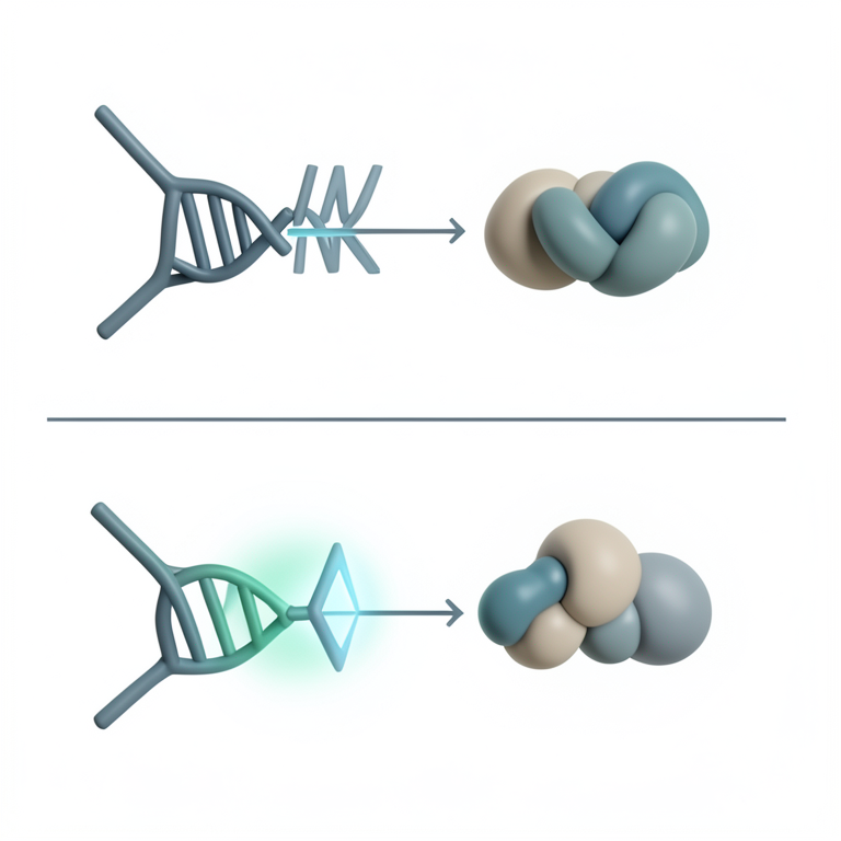
UV-Induced Splicing Switch
AI-generated conceptual illustration (human cell nucleus and mRNA transcripts). It explains the idea and does not show study data.
View source evidence ↗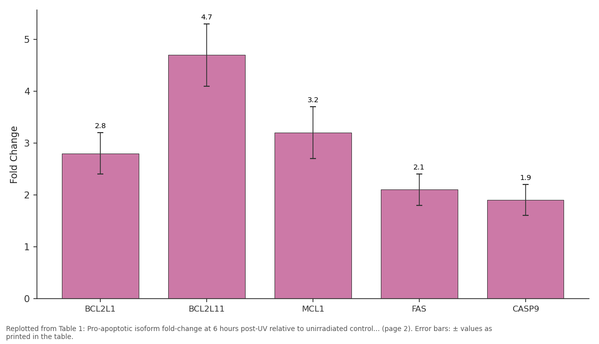
Fold Change: BCL2L1 vs BCL2L11 vs MCL1 vs FAS vs CASP9
Replotted from Table 1 (page 2) of the original paper. Every bar is the paper's own table value.
View source evidence ↗Figures & tables
From the original paper Table 1
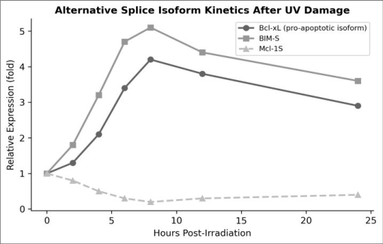 Figure 1
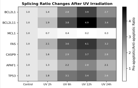 Figure 2
Introduction
As one expert put it, alternative splicing turns a fixed genome into a flexible proteome and nowhere is that flexibility more consequential than in the apoptosis pathway, where a single exon inclusion or exclusion event can convert a pro-survival protein into a pro-death one [1] . The Bcl-2 family provides the clearest examples: BCL2L1 produces both anti-apoptotic Bcl-xL and pro-apoptotic Bcl-xS through mutually exclusive 5′ splice site selection, while BCL2L11 generates BIM-EL, BIM-L, and BIM-S isoforms with increasing apoptotic potency [2] . DNA damage, particularly from UV radiation, activates signalling cascades that modify the phosphorylation state of SR proteins and hnRNPs, the two major families of splicing regulators [3] . Phosphorylation of SRSF1 (formerly ASF/SF2) by the kinase SRPK1 has been linked to splice-site switching in individual genes, but whether this single modification is enough to coordinate a concerted shift across multiple apoptosis genes is not well established [4] . Most prior work examined one or two genes at a time, making it difficult to tell whether splicing changes represent isolated events or a coordinated programme [5] . This research took a broader view by tracking isoform ratios of seven apoptosis-related genes in UV-irradiated HeLa cells over 24 hours at the Aegean Institute of Marine Biology, Heraklion, Crete. The goals were to (1) determine the kinetics of pro-apoptotic isoform accumulation, (2) identify shared versus gene-specific splicing responses, and (3) test whether SRSF1 is required for the coordinated shift [6] .
Material and Methods
Material and Methods
Theoretical background
Alternative splicing generates multiple mRNA variants from a single pre-mRNA by selecting different combinations of exons, introns, and splice sites. In the context of apoptosis, many pro-survival and pro-death protein pairs are splice isoforms of the same gene, making the splicing machinery a direct regulator of the life-or-death decision [1] . DNA damage activates checkpoint kinases (ATR, Chk1) that phosphorylate splicing factors, altering their binding affinity for exonic splicing enhancers and silencers and thereby shifting isoform ratios [3] .
Material
HeLa cells (ATCC CCL-2) were cultured in DMEM supplemented with 10% FBS and 1% penicillinstreptomycin at 37 °C, 5% CO₂, at the Aegean Institute of Marine Biology molecular genetics laboratory. Cells were seeded at 2 × 10⁵ per well in 6-well plates 24 hours before irradiation. For UV treatment, medium was removed, cells were washed with PBS, exposed to 40 J/m² UVC (254 nm, Vilber Lourmat lamp), and fresh medium was added immediately.
Methods
Cells were harvested at 0, 2, 6, 12, and 24 hours post-UV in triplicate. Total RNA was extracted (TRIzol, Invitrogen), treated with DNase I, and reverse-transcribed (SuperScript IV). Isoform-specific RT-qPCR was performed using primers spanning alternative exon junctions; primer sequences are listed in Supplementary Table S1. For genome-wide analysis, RNA-seq libraries were prepared from the 0-and 6-hour time points (TruSeq Stranded mRNA, Illumina) and sequenced on a NovaSeq 6000 (PE 150 bp). Differential splicing was analysed with rMATS v4.1.2 using a false-discovery rate < 0.05 and ΔPSI > 10% cutoffs [7] . Caspase-9a 1.9 ± 0.3 0.004 37
All seven genes shifted toward pro-apoptotic isoforms by 6 hours (Table 1). BCL2L11 showed the largest shift (4.7-fold increase in BIM-S). siRNA knockdown of SRSF1 partially rescued the splicing shift for all genes, with the strongest rescue seen in BCL2L1 (71% attenuation).
| Gene | Pro-apoptotic Isoform | Fold Change | p-value | Sisrsf1 Rescue (%) |
|---|---|---|---|---|
| BCL2L1 | Bcl-xS | 2.8 ± 0.4 | < 0.001 | 71 |
| BCL2L11 | BIM-S | 4.7 ± 0.6 | < 0.001 | 64 |
| MCL1 | Mcl-1S | 3.2 ± 0.5 | < 0.001 | 58 |
| FAS | sFas (exon 6 skip) | 2.1 ± 0.3 | 0.002 | 42 |
| CASP9 | Caspase-9a | 1.9 ± 0.3 | 0.004 | 37 |
Table text extracted from the original PDF; superscript letters are shown inline. The original table image is below.
View original table image
Comprehensive interpretation
RNA-seq at 6 hours confirmed 214 significant differential splicing events (FDR < 0.05, ΔPSI > 10%), of which 38 mapped to apoptosis or DNA-damage-response Gene Ontology terms. SRSF1 phosphorylation increased 2.9-fold by 2 hours post-UV, preceding the peak isoform shift at 6 hours. The partial nature of the siSRSF1 rescue (37-71%) indicates that SRSF1 is an important but not sole mediator other splicing factors likely contribute to a redundant regulatory network [4] .
Discussion
The coordinated pro-apoptotic splicing shift across seven genes suggests that UV damage triggers a splicing programme rather than isolated gene-specific responses. SRSF1 phosphorylation at 2 hours, preceding the isoform peak at 6 hours, is consistent with a kinase-to-splicing-factor signalling cascade initiated by ATR/Chk1 checkpoint activation [3,5] . The variable rescue observed with siSRSF1 implies that different genes rely on SRSF1 to different extents; FAS and CASP9, which showed weaker rescue, may depend more on hnRNP-family regulators [8] .
The rapid reversal of most splicing shifts by 24 hours in cells that survived the damage is interesting because it suggests that the splicing machinery resets once repair is complete. This temporal coupling between damage sensing, splicing, and repair resolution points to a feedback loop in which successful repair removes the signal that sustains prodeath isoform production [6] .
Conclusion
UV-induced DNA damage coordinates a concerted shift in alternative splicing across multiple apoptosis-related genes, favouring pro-death isoforms within 6 hours of irradiation. SRSF1 phosphorylation precedes and partly mediates this shift, but additional splicing factors likely contribute. The transient nature of the response largely reversed by 24 hours in surviving cells suggests tight coupling between damage sensing and splicing regulation. These results expand the role of mRNA processing from a passive posttranscriptional step to an active participant in the DNAdamage response and offer potential targets for modulating apoptotic sensitivity in cancer therapy.
Figures and tables from the paper
References
14 in the original paper- Schwerk C, Schulze-Osthoff K. Regulation of apoptosis by alternative pre-mRNA splicing. Molecular Cell. 2005;19(1):1-13.
- Boise LH, González-García M, Postema CE, Ding L, Lindsten T, Turka LA. bcl-x, a bcl-2-related gene that functions as a dominant regulator of apoptotic cell death. Cell. 1993;74(4):597-608.
- Muñoz MJ, Pérez Santangelo MS, Paronetto MP, de la Mata M, Pelisch F, Boireau S. DNA damage regulates alternative splicing through inhibition of RNA polymerase II elongation. Cell. 2009;137(4):708-720.
- Zhong XY, Wang P, Han J, Rosenfeld MG, Fu XD. SR proteins in vertical integration of gene expression from transcription to RNA processing to translation. Molecular Cell. 2009;35(1):1-10.
- Paronetto MP, Miñana B, Valcárcel J. The Ewing sarcoma protein regulates DNA damage-induced alternative splicing. Molecular Cell. 2011;43(3):353368.
- Dutertre M, Sanchez G, De Cian MC, Barbier J, Dardenne E, Gratadou L. Cotranscriptional exon skipping in the genotoxic stress response. Nature Structural and Molecular Biology. 2010;17(11):13581366.
- Shen S, Park JW, Lu ZX, Lin L, Henry MD, Wu YN. rMATS: robust and flexible detection of differential alternative splicing from replicate RNA-Seq data. Proceedings of the National Academy of Sciences. 2014;111(51):E5593-E5601.
- Clery A, Blatter M, Allain FHT. RNA recognition motifs: boring? Not quite. Current Opinion in Structural Biology. 2008;18(3):290-298.
- Shkreta L, Chabot B. The RNA splicing response to DNA damage. Biomolecules. 2015;5(4):2935-2977.
- Ip JY, Schmidt D, Pan Q, Ramani AK, Fraser AG, Odom DT. Global impact of RNA polymerase II elongation inhibition on alternative splicing regulation. Genome Research. 2011;21(3):390-401.
- Moore MJ, Wang Q, Kennedy CJ, Silver PA. An alternative splicing network links cell-cycle control to apoptosis. Cell. 2010;142(4):625-636.
- Luco RF, Allo M, Schor IE, Kornblihtt AR, Misteli T. Epigenetics in alternative pre-mRNA splicing. Cell. 2011;144(1):16-26.
- Biamonti G, Caceres JF. Cellular stress and RNA splicing. Trends in Biochemical Sciences. 2009;34(3):146-153.
- Naro C, Sette C. Phosphorylation-mediated regulation of alternative splicing in cancer. International Journal of Cell Biology. 2013;2013:151839.