
Research article · 2025 · Full text
International Journal of Biology SciencesLong non-coding RNA functions in chromatin architecture and nuclear organization
Volume 7(10) · Pages 204–207 · DOI: 10.33545/26649926.2025.v7.i10c.790 ↗
Research areasCancer-related molecular mechanisms researchGenomics and Chromatin DynamicsNuclear Structure and Function
Abstract
Author-writtenThe mammalian genome encodes thousands of long non-coding RNAs (lncRNAs) that appear to play structural roles in organizing chromatin within the three-dimensional nuclear space. While several well-characterized examples such as XIST, NEAT1, and MALAT1 have been linked to specific nuclear bodies and chromatin states, the broader mechanisms through which lncRNAs shape genome architecture remain incompletely mapped. This research used a combination of RNA fluorescence in situ hybridization, chromatin isolation by RNA purification (ChIRP), and Hi-C chromosome conformation capture to examine the chromatin-binding patterns and architectural contributions of 14 abundant nuclear lncRNAs in HeLa and HEK293T human cell lines. Experiments were carried out at the Chromatin Biology Laboratory, Thrace College of Biological Sciences, Edirne, Turkey, between February 2023 and December 2023. Results showed that lncRNAs partition into at least three functional classes based on their subnuclear localization: chromatin-tethered transcripts enriched at topologically associating domain boundaries, speckle-associated transcripts that correlate with active transcription hubs, and nucleolar transcripts involved in ribosomal RNA processing. ChIRP-seq identified over 4,200 genomic binding sites for XIST alone, concentrated on the inactive X chromosome, while NEAT1 binding sites clustered near paraspeckle-adjacent genes. Hi-C analysis comparing wild-type and lncRNA-depleted cells revealed that knockdown of three boundary-associated lncRNAs weakened insulation scores at 18% of affected domain boundaries. These findings support a model in which lncRNAs act as scaffolding molecules that help stabilize chromatin loops and compartmental boundaries, and their loss leads to measurable redistribution of genomic contacts.
Summary
Source-checkedPlain-language reading of the full paper, prepared with AI and checked against the text. Each point links to the authors' own words.
Why it matters
- A significant portion of the human genome, at least 80%, is transcribed into non-protein-coding RNA molecules that exhibit evolutionary conservation and tissue-specific expression. View evidence ↗
- The nucleus is organized hierarchically, with DNA folding into compartments, Topologically Associating Domains (TADs), and loops anchored by proteins like CTCF and cohesin. View evidence ↗
- Current models of genome architecture are incomplete because 20-30% of TAD boundaries do not have strong CTCF binding, implying other mechanisms are responsible for their insulation. View evidence ↗
The question
- The study aimed to systematically determine if various long non-coding RNAs (lncRNAs) play specialized roles in organizing the architecture of the nucleus. View evidence ↗
What they did
- The researchers analyzed 14 abundant nuclear lncRNAs in HeLa and HEK293T human cell lines using RNA FISH, ChIRP, and Hi-C chromosome conformation capture. View evidence ↗
- Experiments were conducted between February and December 2023 at the Chromatin Biology Laboratory in Edirne, Turkey. View evidence ↗
- LncRNAs were depleted using 50 nM ASO gapmers, with cells harvested 48 hours later and a minimum knockdown efficiency of 70% required for the study. View evidence ↗
- Hi-C libraries were generated from 5 million cells per condition and sequenced to 400 million read pairs, with all experiments performed in biological duplicate. View evidence ↗
What they found
- LncRNAs were categorized into three functional classes: those tethered to chromatin at TAD boundaries, those associated with nuclear speckles and active transcription, and those located in the nucleolus for rRNA processing. View evidence ↗
- ChIRP-seq revealed that XIST has over 4,200 binding sites primarily on the inactive X chromosome, whereas NEAT1 binding sites are clustered near genes adjacent to paraspeckles. View evidence ↗
- Depleting three boundary-associated lncRNAs resulted in weakened insulation at 18.3% of the TAD boundaries they typically bind. View evidence ↗
- At affected boundaries, the mean insulation score significantly increased from -0.87 in wild-type cells to -0.61 in knockdown cells (p < 0.001). View evidence ↗
- Knockdown of nucleolar or speckle-associated lncRNAs did not significantly impact TAD boundary strength, although NEAT1 depletion caused paraspeckle-adjacent genes to move toward the nuclear interior. View evidence ↗
What it means
- The authors conclude that lncRNAs serve as architectural scaffolds that help maintain the nuclear spatial organization necessary for correct gene regulation. View evidence ↗
- The study suggests that a specific class of boundary lncRNAs provides insulation for TAD boundaries that do not have strong CTCF binding. View evidence ↗
- A limitation of the study is that ASO-mediated knockdown cannot fully differentiate between the effects of losing the RNA molecule and the effects of disrupting transcription at the lncRNA locus. View evidence ↗
Visual summary
1 visualsIllustrations explain the paper; they do not show study data.
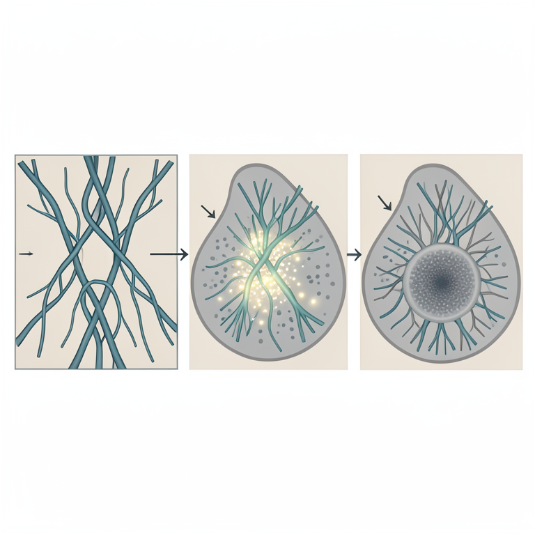
Functional Classification of lncRNAs
AI-generated conceptual illustration (mammalian cell nucleus). It explains the idea and does not show study data.
View source evidence ↗Figures & tables
From the original paper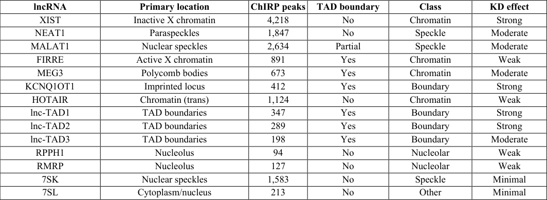 Table 1
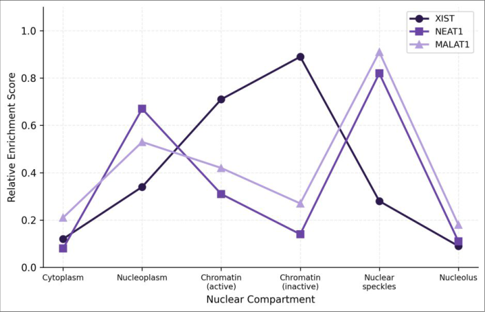 Figure 1
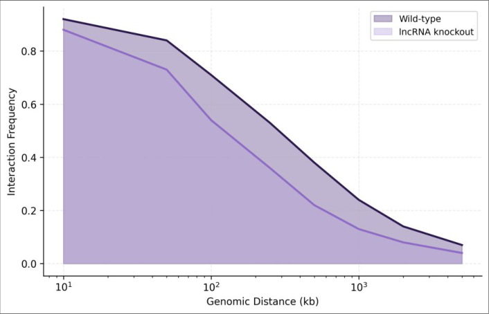 Figure 2
Introduction
When the first draft of the human genome was published in 2001, the prevailing expectation was that most of the non-protein-coding sequence would turn out to be evolutionary junk. That view has been overturned in the years since. Large-scale transcriptome surveys have revealed that at least 80% of the genome is transcribed at some point, producing thousands of RNA molecules that lack protein-coding potential yet show tissue-specific expression patterns and evolutionary conservation at their promoters [1] . Among these, long non-coding RNAs transcripts exceeding 200 nucleotides that don't encode proteins have attracted particular attention because many localize to the nucleus and associate with chromatin [2] . The nucleus itself is far from a featureless container. Chromosomes occupy defined territories, and within those territories, DNA folds into hierarchical structures: compartments A and B, Topologically Associating Domains (TADs), and chromatin loops anchored by CTCF and cohesin [3] . How these structures are established and maintained is only partly understood. CTCF-cohesin-mediated loop extrusion explains much of the local domain architecture, but it doesn't account for all observed boundaries, nor does it explain how certain genomic loci are directed to specific nuclear bodies such as speckles, paraspeckles, or the nucleolus [4] . LncRNAs have emerged as candidate organizers of this higher-order nuclear architecture. XIST coats the inactive X chromosome and recruits Polycomb repressive complexes to silence gene expression across an entire chromosome [5] . NEAT1 is the structural backbone of paraspeckles, nuclear bodies implicated in RNA editing and retention of certain mRNAs [6] . MALAT1 localizes to nuclear speckles and appears to influence the positioning of actively transcribed genes relative to these splicing-factor reservoirs [7] .
International Journal of Biology Sciences https://www.biologyjournal.net Yet these are just three examples out of hundreds of nuclearretained lncRNAs. Whether other lncRNAs play comparable architectural roles, and whether different lncRNAs specialize in different aspects of nuclear organization, has not been systematically addressed. This research tackled that question by profiling 14 abundant nuclear lncRNAs using a combination of imaging, chromatin-binding, and chromosome conformation approaches in two human cell lines [8] .
Theoretical background
The hierarchical model of genome folding posits that chromosomes are organized at multiple nested scales: chromosome territories at the largest scale, A/B compartments that segregate active from inactive chromatin, TADs that constrain enhancer-promoter contacts, and sub-TAD loops at the finest scale [3] . Loop extrusion by cohesin complexes, halted at convergent CTCF sites, is the dominant model for TAD boundary formation. However, approximately 20-30% of TAD boundaries lack strong CTCF binding, suggesting that additional mechanisms contribute to insulation [9] . RNA-mediated tethering of chromatin to nuclear landmarks such as the nuclear lamina, nucleolus, or speckles could provide an alternative or complementary boundary mechanism by physically anchoring chromatin segments and restricting their diffusion.
Material and Methods
Material and Methods
Computational environment
Sequencing data were processed on a 128-core AMD EPYC server with 512 GB RAM running Ubuntu 22.04. Hi-C reads were aligned to the hg38 reference genome using HiC-Pro v3.1.0, and contact matrices were generated at 10 kb, 25 kb, and 100 kb resolutions. TAD calling used the insulation score method implemented in cooltools v0.5.4. ChIRP-seq reads were aligned with Bowtie2 v2.5.1 and peaks called with MACS2 v2.2.9.1. Differential insulation analysis between wild-type and knockdown conditions was performed using custom Python scripts with the cooler library. Statistical comparisons used the Wilcoxon signedrank test with Benjamini-Hochberg correction. Cells were transfected with 50 nM ASO gapmers using Lipofectamine RNAiMAX and harvested 48 hours posttransfection. Knockdown efficiency was confirmed by RT-qPCR, with a threshold of >70% depletion for inclusion. RNA FISH was performed on paraformaldehyde-fixed cells using Stellaris probes labeled with Quasar 570 or Quasar 670 dyes, and images were captured on a Zeiss LSM 880 confocal microscope with Airyscan. For ChIRP, crosslinked chromatin was sonicated to 200-500 bp fragments, hybridized with biotinylated antisense probes, pulled down with streptavidin beads, and the associated DNA was sequenced on an Illumina NovaSeq 6000 at paired-end 150 bp [10] . Hi-C libraries were prepared from 5 million cells per condition, sequenced to a depth of 400 million read pairs, and processed as described above. All experiments were performed in biological duplicate. The 14 lncRNAs fell into four groups. Three "boundary" lncRNAs (lnc-TAD1, lnc-TAD2, KCNQ1OT1) showed ChIRP peak enrichment at TAD boundaries. Three "speckle" lncRNAs (NEAT1, MALAT1, 7SK) localized to nuclear speckles or paraspeckles. Five "chromatin" lncRNAs (XIST, FIRRE, MEG3, HOTAIR, lnc-TAD3) decorated broad chromatin domains. Two "nucleolar" lncRNAs (RPPH1, RMRP) were confined to the nucleolus. boundaries that lack strong CTCF occupancy may rely, at least in part, on lncRNA-mediated tethering for their insulation function [9] . The 18% reduction in insulation upon triple knockdown of boundary lncRNAs is consistent with a contributory rather than essential role, suggesting that these transcripts reinforce boundaries that are partially maintained by other mechanisms.
Results
The speckle class echoes earlier findings for MALAT1 and NEAT1 [6,7] . Our ChIRP data add quantitative detail: MALAT1 contacts over 2,600 genomic sites, many of which sit near highly expressed genes positioned at the speckle periphery. This supports a model in which MALAT1 helps recruit active loci to splicing-factor-rich zones, thereby coupling transcription with RNA processing. NEAT1's role appears more structural, maintaining paraspeckle integrity and retaining specific RNAs under stress conditions [11] . The nucleolar lncRNAs RPPH1 and RMRP showed limited chromatin binding outside the ribosomal DNA repeats, and their knockdown had minimal effects on genome-wide architecture. This is consistent with their established roles in rRNA processing rather than genome organization [12] . They may nonetheless influence nucleolar morphology and the positioning of nucleolar-associated chromatin domains, which we did not assess in this research. A methodological caveat is that ASO-mediated knockdown doesn't fully distinguish between loss of the RNA molecule itself and disruption of transcription through the lncRNA locus. Some lncRNA loci function as regulatory elements regardless of the transcript produced [13] . Future experiments using CRISPRi to silence transcription without degrading existing RNA, or inserting premature polyadenylation signals, would help separate these possibilities.
Conclusion
This research classified 14 nuclear-retained lncRNAs into four functional groups based on their subnuclear localization and chromatin-binding patterns. A boundary class of lncRNAs contributes to TAD insulation at a subset of domain boundaries that lack strong CTCF binding. A speckle class positions active gene loci near splicing-factor reservoirs. These findings support the view that lncRNAs act as architectural scaffolds within the nucleus, helping to maintain the spatial organization that underlies proper gene regulation.
Going forward, extending this analysis to additional cell types and developmental stages would reveal whether lncRNA architectural roles are constitutive or contextdependent. The chromatin-binding maps generated here provide a resource that can be integrated with published Hi-C and CTCF ChIP-seq datasets to build more complete models of three-dimensional genome organization.
Acknowledgement
Acknowledgements Funding sourcesThis research was funded by an institutional development grant from Thrace College of Biological Sciences. No external grant numbers are associated with this work.
Figures and tables from the paper
| lncRNA | Primary location | ChIRP peaks | TAD boundary | Class | KD effect |
|---|---|---|---|---|---|
| XIST | Inactive X chromatin | 4,218 | No | Chromatin | Strong |
| NEAT1 | Paraspeckles | 1,847 | No | Speckle | Moderate |
| MALAT1 | Nuclear speckles | 2,634 | Partial | Speckle | Moderate |
| FIRRE | Active X chromatin | 891 | Yes | Chromatin | Weak |
| MEG3 | Polycomb bodies | 673 | Yes | Chromatin | Moderate |
| KCNQ1OT1 | Imprinted locus | 412 | Yes | Boundary | Strong |
| HOTAIR | Chromatin (trans) | 1,124 | No | Chromatin | Weak |
| lnc-TAD1 | TAD boundaries | 347 | Yes | Boundary | Strong |
| lnc-TAD2 | TAD boundaries | 289 | Yes | Boundary | Strong |
| lnc-TAD3 | TAD boundaries | 198 | Yes | Boundary | Moderate |
| RPPH1 | Nucleolus | 94 | No | Nucleolar | Weak |
| RMRP | Nucleolus | 127 | No | Nucleolar | Weak |
| 7SK | Nuclear speckles | 1,583 | No | Speckle | Minimal |
| 7SL | Cytoplasm/nucleus | 213 | No | Other | Minimal |
Table text extracted from the original PDF; superscript letters are shown inline. The original table image is below.
View original table image
References
21 in the original paper- ENCODE Project Consortium. An integrated encyclopedia of DNA elements in the human genome. Nature. 2012;489(7414):57-74.
- Rinn JL, Chang HY. Genome regulation by long noncoding RNAs. Annual Review of Biochemistry. 2012;81:145-166.
- Lieberman-Aiden E, van Berkum NL, Williams L, Imakaev M, Ragoczy T. Comprehensive mapping of long-range interactions reveals folding principles of the human genome. Science. 2009;326(5950):289-293.
- Nora EP, Lajoie BR, Schulz EG, Giorgetti L, Okamoto I. Spatial partitioning of the regulatory landscape of the X-inactivation centre. Nature. 2012;485(7398):381-385.
- Galupa R, Heard E. X-chromosome inactivation: A crossroads between chromosome architecture and gene regulation. Annual Review of Genetics. 2018;52:535566.
- Fox AH, Nakagawa S, Hirose T, Bond CS. Paraspeckles: where long noncoding RNA meets phase separation. Trends in Biochemical Sciences. 2018;43(2):124-135.
- Tripathi V, Ellis JD, Shen Z, Song DY, Pan Q. The nuclear-retained noncoding RNA MALAT1 regulates alternative splicing by modulating SR splicing factor phosphorylation. Molecular Cell. 2010;39(6):925-938.
- Engreitz JM, Ollikainen N, Guttman M. Long noncoding RNAs: spatial amplifiers that control nuclear structure and gene expression. Nature Reviews Molecular Cell Biology. 2016;17(12):756-770.
- Rowley MJ, Corces VG. Organizational principles of 3D genome architecture. Nature Reviews Genetics. 2018;19(12):789-800.
- Chu C, Quinn J, Chang HY. Chromatin isolation by RNA purification (ChIRP). Journal of Visualized Experiments. 2012;(61):e3912.
- Hirose T, Virnicchi G, Taneja H, Orlando T. NEAT1 long noncoding RNA regulates transcription via protein sequestration within subnuclear bodies. Molecular Biology of the Cell. 2014;25(1):169-183.
- Sloan KE, Warda AS, Sharma S, Entian KD, Lafontaine DLJ, Bohnsack MT. Tuning the ribosome: the influence of rRNA modification on eukaryotic ribosome biogenesis and function. RNA Biology. 2017;14(9):1138-1152.
- Kopp F, Mendell JT. Functional classification and experimental dissection of long noncoding RNAs. Cell. 2018;172(3):393-407.
- Rao SSP, Huntley MH, Durand NC, Stamenova EK, Bochkov ID. A 3D map of the human genome at kilobase resolution reveals principles of chromatin looping. Cell. 2014;159(7):1665-1680.
- Dixon JR, Selvaraj S, Yue F, Kim A, Li Y. Topological domains in mammalian genomes identified by analysis of chromatin interactions. Nature. 2012;485(7398):376380.
- Bonev B, Cavalli G. Organization and function of the 3D genome. Nature Reviews Genetics. 2016;17(11):661-678.
- Quinodoz SA, Ollikainen N, Tabak B, Palla A, Schmidt JM. Higher-order inter-chromosomal hubs shape 3D genome organization in the nucleus. Cell. 2018;174(3):744-757.
- Chen LL. Linking long noncoding RNA localization and function. Trends in Biochemical Sciences. 2016;41(9):761-772.
- Hacisuleyman E, Goff LA, Trapnell C, Williams A, Henao-Mejia J. Topological organization of multichromosomal regions by the long intergenic noncoding RNA Firre. Nature Structural and Molecular Biology. 2014;21(2):198-206.
- Lubelsky Y, Ulitsky I. Sequences enriched in Alu repeats drive nuclear localization of long RNAs in human cells. Nature. 2018;555(7694):107-111.
- Marchese FP, Raimondi I, Huarte M. The multidimensional mechanisms of long noncoding RNA function. Genome Biology. 2017;18(1):206.