
Research article · 2026 · Full text
International Journal of Biology SciencesMucuna pruriens leaves extraction, antifungal activity, Minimum Inhibitory Concentration (MIC) and Minimum Fungicidal Concentration (MFC)
Volume 8(4) · Pages 120–126 · DOI: 10.33545/26649926.2026.v8.i4b.805 ↗
Research areasEssential Oils and Antimicrobial ActivityMedicinal Plant ResearchAntifungal resistance and susceptibility
Abstract
Author-writtenMucuna pruriens (L.) DC., commonly known as Velvet Bean, is a medicinally important legume widely used in traditional systems of medicine for its therapeutic and antimicrobial potential. The present study was conducted to evaluate the antifungal activity of M. pruriens leaf extracts along with the determination of Minimum Inhibitory Concentration (MIC) and Minimum Fungicidal Concentration (MFC) values. Leaf samples were collected, shade-dried, powdered, and extracted using the Soxhlet extraction method with solvents of increasing polarity petroleum ether, chloroform, acetone, ethanol, and methanol. The methanolic extract, found to be the most potent, was tested for antifungal activity against A. niger (MTCC-282) and T. reesei (MTCC-164) using the agar well diffusion method. The extract exhibited significant antifungal activity, with inhibition zones increasing proportionally to concentration (25, 50, and 100 mg/ml) compared to the standard antifungal drug Itraconazole. MIC and MFC were determined using broth dilution and sub-culturing techniques, respectively, revealing the minimum concentrations required for growth inhibition and fungicidal action. The results indicated that M. pruriens possesses strong antifungal potential, suggesting that its bioactive phytochemicals could serve as promising natural alternatives to synthetic antifungal agents. This study provides a scientific basis for the traditional use of M. pruriens and supports its further exploration in pharmaceutical and agricultural applications.
Summary
Source-checkedPlain-language reading of the full paper, prepared with AI and checked against the text. Each point links to the authors' own words.
Why it matters
- Mucuna pruriens, also known as Velvet Bean, is a legume valued in traditional medicine for its potential antimicrobial and therapeutic properties. View evidence ↗
- Medicinal plants produce secondary metabolites like flavonoids and alkaloids that are essential for both the plant's own defense and human health. View evidence ↗
- Methanol is considered an effective solvent for extracting polar bioactive compounds, such as phenolics and flavonoids, from plant materials. View evidence ↗
The question
- The study aimed to evaluate the antifungal activity of Mucuna pruriens leaf extracts and determine their Minimum Inhibitory Concentration (MIC) and Minimum Fungicidal Concentration (MFC). View evidence ↗
What they did
- Researchers used a Soxhlet apparatus to extract 15 g of powdered leaf material using five solvents of increasing polarity: petroleum ether, chloroform, acetone, ethanol, and methanol. View evidence ↗
- The antifungal assays were tested against two specific pathogenic fungi: Aspergillus niger (MTCC-282) and Trichoderma reesei (MTCC-164). View evidence ↗
- To determine the MIC, the methanolic extract was prepared in serial twofold dilutions ranging from 1.25 to 100 mg/ml in Potato Dextrose Broth. View evidence ↗
What they found
- Against A. niger, the methanolic extract showed dose-dependent activity, with the 100 mg/ml concentration producing the largest mean inhibition zone of 13.00 ± 1.50 mm. View evidence ↗
- The extract exhibited lower antifungal activity against T. reesei, with inhibition zones ranging from 6.83 ± 0.28 mm to 7.23 ± 0.25 mm at concentrations of 5 to 15 mg/ml. View evidence ↗
- The Minimum Inhibitory Concentration (MIC) for A. niger was determined to be 25%, where no turbidity was observed. View evidence ↗
- T. reesei was more resistant to the extract than A. niger, with an MIC value greater than 25%. View evidence ↗
- The Minimum Fungicidal Concentration (MFC) for A. niger was established at 25%, as growth was completely inhibited at this level but observed at 12.5%. View evidence ↗
- T. reesei showed visible growth even at a 25% concentration, indicating it is less susceptible to the fungicidal action of the extract. View evidence ↗
What it means
- The authors conclude that Mucuna pruriens is a potential natural source of antifungal agents, particularly effective against A. niger. View evidence ↗
- The study suggests that M. pruriens could be used to develop eco-friendly antifungal formulations. View evidence ↗
- The limited effect on T. reesei indicates a need for further purification and fractionation to isolate more potent bioactive compounds. View evidence ↗
- Future research is required to identify specific bioactive constituents through compound isolation, GC-MS profiling, and mode-of-action analysis. View evidence ↗
Visual summary
3 visualsIllustrations explain the paper; charts are replotted from the paper's own table values.
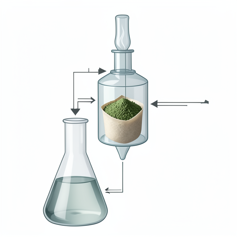
Soxhlet Extraction Process
AI-generated conceptual illustration (Mucuna pruriens leaves and a Soxhlet extraction apparatus). It explains the idea and does not show study data.
View source evidence ↗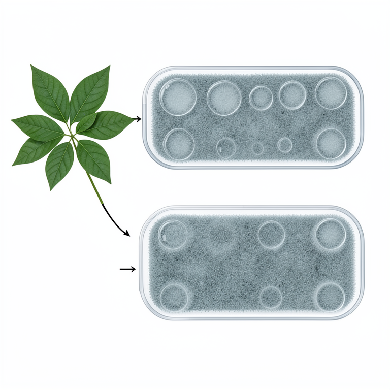
Dose-Dependent Inhibition
AI-generated conceptual illustration (Mucuna pruriens leaves and Aspergillus niger and Trichoderma reesei fungi). It explains the idea and does not show study data.
View source evidence ↗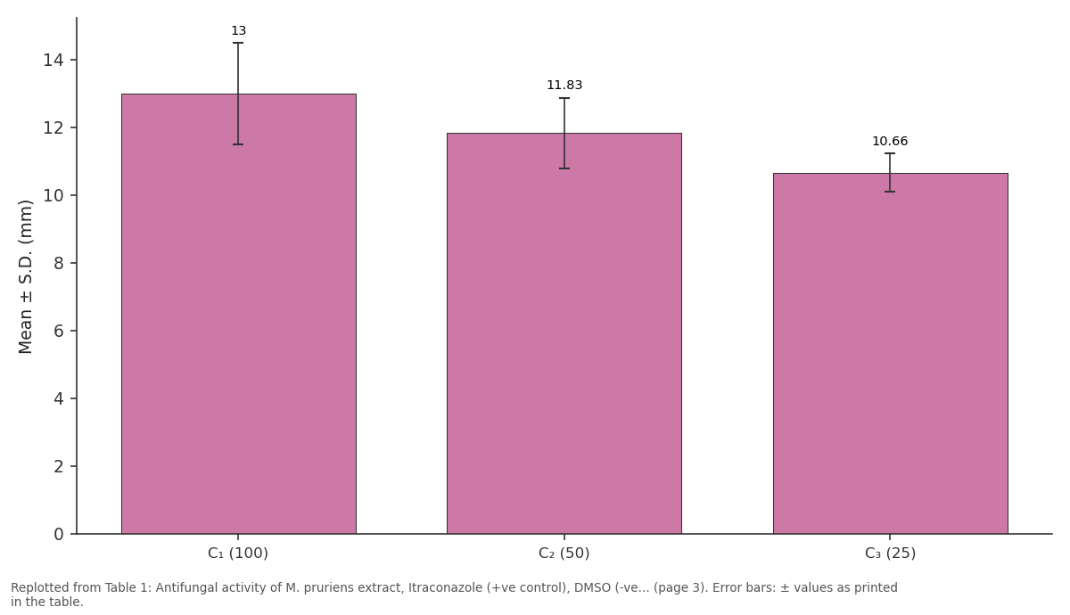
Mean ± S.D.: C₁ (100) vs C₂ (50) vs C₃ (25)
Replotted from Table 1 (page 3) of the original paper. Every bar is the paper's own table value.
View source evidence ↗Figures & tables
From the original paper Table 1
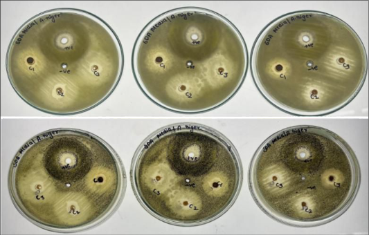 Figure 1
 Table 2
 Table 3
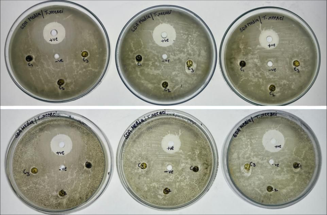 Figure 2
 Table 4
Introduction
The extraction of bioactive compounds from medicinal plants has become an essential step in identifying and characterizing natural products with therapeutic and antimicrobial potential. Medicinal plants synthesize a wide range of secondary metabolites such as alkaloids, flavonoids, phenolics, tannins, terpenoids, and saponins, which play important roles in plant defense and human health (Simsek & Whitney, 2024) [7] . These compounds can be efficiently recovered through solvent extraction techniques that utilize the polarity of solvents to selectively dissolve different classes of phytochemicals (Tiwari et al., 2011) [13] . Mucuna pruriens (L.) DC., commonly known as Velvet Bean, is a tropical leguminous plant belonging to the family Fabaceae, well known for its nutritional and pharmacological significance (Simmons, 2018) [9] . It has been used traditionally in Ayurvedic and folk medicine for the treatment of neurological disorders, infections, and metabolic diseases (Devhade et al., 2015) [4] . The extraction and phytochemical analysis of M. pruriens have revealed the presence of numerous bioactive constituents that contribute to its therapeutic potential (Ahmad & Singh, 2014; Bhusare et al., 2021) [1,2,22] . Plants are constantly exposed to abiotic and biotic stresses that trigger the accumulation of Reactive Oxygen Species (ROS) and secondary metabolites. These bioactive compounds, including flavonoids and phenolics, play crucial roles in stress tolerance and pathogen defense. Similar responses have been reported in Gmelina arborea under salinity stress, where increased antioxidant enzyme activity (catalase and peroxidase) contributed to oxidative stress mitigation (Ahmad, 2020) [3] .
International Journal of Biology Sciences https://www.biologyjournal.net
Recent studies have employed Soxhlet extraction using solvents of varying polarity-such as petroleum ether, chloroform, acetone, ethanol, and methanol to isolate bioactive metabolites from M. pruriens leaves and seeds (Narayanan et al., 2021) [5] . Among these, methanol has been found to be the most effective solvent for recovering polar compounds including phenolics, flavonoids, and fatty acids (Shelke et al., 2022) [6] . The extracts obtained were later characterized through Gas Chromatography-Mass Spectrometry (GC-MS), which identified several biologically active compounds such as catechol, 4vinylphenol, p-cymene-2,5-diol, dimethyl phthalate, and nhexadecanoic acid (Sruthi et al., 2023) [10] . These findings suggest that optimized extraction procedures play a crucial role in enhancing the yield and biological activity of plantderived compounds. In recent years, several studies from the Gopalganj region of Bihar have reported the potent biological activity of invasive alien species such as Ageratum conyzoides, Eupatorium adenophorum, and Mikania micrantha, which exhibited significant cytotoxic, antioxidant, and anti-inflammatory properties (Siddiquee & Ahmad, 2022) [8] . These findings indicate that naturally occurring phytochemicals, including phenolics, flavonoids, and terpenoids, play a major role in the antimicrobial defense mechanisms of plants. Such evidence further supports the investigation of native and medicinally important species like Mucuna pruriens (L.) DC., which belongs to the Fabaceae family and has a rich history of traditional medicinal use, for its potential antifungal efficacy. Besides its antimicrobial potential, Mucuna pruriens is also recognized for its high content of L-DOPA, a pharmacologically important amino acid precursor used in the treatment of Parkinson's disease (Srivastava et al., 2024) [11] . The conventional propagation of M. pruriens is limited by poor seed viability and germination rate. Therefore, in vitro propagation techniques such as callus culture and somatic embryogenesis have been explored as alternative methods for large-scale plant production and conservation, while also enabling the quantification of L-DOPA content in regenerated plants. Bihar's floristic diversity has undergone notable transformation due to the introduction and establishment of several exotic and alien plant species, particularly from tropical regions. Studies conducted in the Gopalganj district have documented around twenty such dicotyledonous species belonging to families like Amaranthaceae, Tiliaceae, Euphorbiaceae, and Apocynaceae, many of which possess medicinal and economic importance (Siddiquee & Ahmad, 2021) [12] . This highlights the phytochemical richness and adaptability of tropical flora in this region, supporting the need to explore indigenous species such as Mucuna pruriens (L.) DC. for their antimicrobial and pharmacological potential. In the present study, extraction of Mucuna pruriens leaf material was carried out using the Soxhlet extraction method to obtain crude solvent extracts for further evaluation of their antifungal activity, minimum inhibitory concentration (MIC), and Minimum Fungicidal Concentration (MFC). The extraction efficiency and the quality of phytochemical constituents directly influence the antifungal efficacy of the obtained extracts, emphasizing the importance of precise extraction techniques in pharmacological investigations.
Material and Methods
Material and Methods
Collection of plant material
Fresh and healthy leaves of Mucuna pruriens (L.) DC. were collected from the local market of Lucknow, India. The collected samples were thoroughly washed with clean tap water followed by distilled water to remove surface impurities and dust particles. The cleaned leaves were shade-dried at room temperature to preserve heat-sensitive bioactive compounds. The dried samples were then pulverized into fine powder using an electric grinder, sieved through standard mesh filters, and stored in airtight containers at 4 °C for further extraction and antifungal assays.
Preparation of plant material
The dried leaves and pods of Mucuna pruriens were processed separately. Seeds were carefully removed from the pods, while immature, discolored, or diseased leaves were discarded. The selected leaves were finely chopped and ground using a sterile grinder. The powdered samples were stored in sterile airtight containers and preserved at 4 °C until extraction. Grinding the leaves to a fine consistency increased the surface area for solvent penetration and improved extraction efficiency (Tiwari et al., 2011) [13] .
Preparation of plant extracts
The extraction of phytochemical constituents from Mucuna pruriens leaves was carried out using the Soxhlet extraction method as described by Narayanan et al. (2021) [5] with minor modifications. About 15 g of powdered leaf material was packed into a cellulose thimble and extracted successively with petroleum ether, chloroform, acetone, ethanol, and methanol (150 ml each) for 24 hours using a Soxhlet apparatus. The temperature of extraction was maintained at the respective boiling point of each solvent to ensure maximum recovery of bioactive metabolites. The obtained extracts were filtered through Whatman No. 1 filter paper and evaporated to dryness using a rotary evaporator. The concentrated extracts were then redissolved in a small volume (10 ml) of their respective solvents and stored in sterile, airtight vials at 4 °C for subsequent antifungal analysis. Among all extracts, the methanolic extract was primarily used for antifungal, MIC, and MFC studies owing to its higher extraction efficiency and phytochemical richness (Shelke et al., 2022; Sruthi et al., 2023) [6,10] .
Antifungal Activity Assay
The antifungal activity of the Mucuna pruriens leaf extracts was evaluated using the agar well diffusion method as described by Bauer et al. (1966) [14] with slight modifications. The assay was conducted against selected pathogenic fungi, namely A. niger (MTCC-282), and T. reesei (MTCC-164) which are known to cause significant human and plant infections. Pure fungal cultures were maintained on Potato Dextrose Agar (PDA) medium and incubated at 27 °C for 5-7 days to ensure active growth (Tiwari et al., 2011) [13] . A fungal spore suspension (1 × 10⁶ spores/ml) was prepared in sterile distilled water containing 0.1% Tween 80, and 0.1 ml of this suspension was uniformly spread over the surface of sterile PDA plates. Wells of 6 mm diameter were made using a sterile corn borer, and each well was loaded with 100 µL of different concentrations (25, 50, and 100 mg/ml) of the methanolic extract of M. pruriens. Dimethyl sulfoxide (DMSO) served as the negative control, while Itraconazole (10 µg/ml) was used as the positive control for comparative evaluation. The inoculated plates were incubated at 27 °C for 72 hours, and the antifungal activity was determined by measuring the zone of inhibition (in mm) around each well using a Vernier caliper. All tests were carried out in triplicate, and the results were expressed as mean ± Standard Deviation (SD). The antifungal efficacy of the M. pruriens extracts was compared with that of the standard antifungal agent to determine their relative inhibitory potential (Shelke et al., 2022; Sruthi et al., 2023) [6,10] .
Minimum Inhibitory Concentration (MIC)
The Minimum Inhibitory Concentration (MIC) of the Mucuna pruriens leaf extracts was determined using the broth dilution method as described by Andrews (2001) and modified by Wiegand et al. (2008) [17] . The assay was performed to establish the lowest concentration of the plant extract capable of completely inhibiting visible fungal growth. Serial twofold dilutions of the methanolic extract were prepared in Potato Dextrose Broth (PDB) to obtain concentrations ranging from 1.25 to 100 mg/ml. Each dilution (1 ml) was inoculated with 0.1 ml of standardized fungal suspension (1 × 10⁶ spores/ml) and incubated at 27 °C for 48-72 hours. A positive control containing Itraconazole (10 µg/ml) and a negative control containing only DMSO were maintained under identical conditions. After incubation, tubes were visually examined for turbidity to assess fungal growth. The MIC value was recorded as the lowest concentration of extract showing no visible growth compared to the control (Balouiri et al., 2016) [16] . All assays were performed in triplicate to ensure reproducibility and accuracy of results.
Minimum Fungicidal Concentration (MFC)
The Minimum Fungicidal Concentration (MFC) of the Mucuna pruriens leaf extract was determined according to the standard procedure described by Andrews (2001) [15] and Wiegand et al. (2008) [17] , following the MIC assay. After determining the MIC, aliquots (0.1 ml) from each tube that showed no visible growth were aseptically sub-cultured onto fresh Potato Dextrose Agar (PDA) plates. The inoculated plates were incubated at 27 °C for 48-72 hours, and the fungal growth was carefully observed. The MFC was defined as the lowest concentration of the extract that completely inhibited fungal growth on the agar surface, indicating a fungicidal effect (Balouiri et al., 2016) [16] . All tests were performed in triplicate to ensure reliability and reproducibility. The ratio of MFC to MIC was also used to distinguish between fungistatic and fungicidal activity, where an MFC/MIC ratio ≤4 indicated fungicidal potential of the extract (Cos et al., 2006) [18] .
Result and Discussion
Result and Discussion
Antifungal Activity
The methanolic extract of Mucuna pruriens showed concentration-dependent antifungal activity against A. niger and T. reesei, indicating the presence of potent bioactive compounds. The higher inhibition zones at increased concentrations suggest that phytochemicals such as flavonoids and phenolics may contribute to the antifungal effect. These findings highlight the potential of M. pruriens as a natural source of antifungal agents.
Mucuna pruriens extract against A. niger
The methanolic extract of Mucuna pruriens showed noticeable antifungal activity against A. niger (MTCC-282). As the extract concentration increased from 25 mg/ml to 100 mg/ml, the zone of inhibition also increased, suggesting that the antifungal activity of the extract is concentrationdependent. The comparative activity of the plant extract, standard antifungal drug (Itraconazole), and solvent control is summarized in Table 3.1.1.1. The antifungal potential of the Mucuna pruriens methanolic leaf extract was evaluated against A. niger using the agar well diffusion method. The extract exhibited measurable inhibitory zones, which increased proportionally with concentration, indicating a dose-dependent antifungal effect. The highest activity was observed at 100 mg/ml, producing a mean inhibition zone of 13.00 ± 1.50 mm, while lower concentrations (50 mg/ml and 25 mg/ml) produced inhibition zones of 11.83 ± 1.04 mm and 10.66 ± 0.57 mm, respectively. The standard antifungal agent, Itraconazole (300 ppm), produced a comparable inhibition zone of 12.83 ± 0.28 mm, whereas no inhibitory effect was observed in the negative control (DMSO). These findings confirm that the antifungal activity is attributed to the bioactive constituents of the M. pruriens extract rather than the solvent.
| Sample | Organism | Tube label | Concentration (%) | Absorbance/OD | Turbidity |
|---|---|---|---|---|---|
| M. pruriens extract | A. niger | C1 | 25 | 0.089 | NT |
| C2 | 12.5 | 0.257 | WT |
Table text extracted from the original PDF; superscript letters are shown inline. The original table image is below.
View original table image
Mucuna pruriens extract against T. reesei
The methanolic extract of Mucuna pruriens showed noticeable activity against T. reesei (MTCC-164). As the extract concentration increased from 25 mg/ml to 100 mg/ml, the zone of inhibition also increased, suggesting that the antifungal activity of the extract is concentration-dependent. The comparative activity of the plant extract, standard antifungal drug (Itraconazole), and solvent control is summarized in Table 3.1.2. The antifungal efficacy of the methanolic extract of Mucuna pruriens leaves was assessed against two fungal pathogens, A. niger and T. reesei, using the agar well diffusion method. The extract exhibited measurable inhibition zones that increased with concentration, indicating a dose-dependent antifungal effect. Against A. niger, the mean inhibition zones were 13.00 ± 1.50 mm, 11.83 ± 1.04 mm, and 10.66 ± 0.57 mm at concentrations of 100, 50, and 25 mg/ml, respectively. For T. reesei, the extract showed relatively lower activity, with inhibition zones of 7.23 ± 0.25 mm, 6.83 ± 0.28 mm, and 6.83 ± 0.28 mm at 15, 10, and 5 mg/ml concentrations, respectively. The standard antifungal drug, Itraconazole (300 ppm), produced inhibition zones of 12.83 ± 0.28 mm for A. niger and 19.00 ± 3.60 mm for T. reesei, whereas the negative control (DMSO) exhibited no inhibitory effect.
Minimum Inhibitory Concentration (MIC)
The Minimum Inhibitory Concentration (MIC) assay was conducted to determine the lowest concentration of Mucuna pruriens methanolic extract capable of inhibiting visible fungal growth. The antifungal potential of the extract was tested against two pathogenic fungi, A. niger and T. reesei, using the broth dilution method. Fungal growth was quantified spectrophotometrically by measuring the absorbance (OD, and visually assessed based on turbidity levels categorized as NT (No Turbidity), WT (Weak Turbidity), and HT (High Turbidity). The results of MIC determination are presented in the Table 3.2.1. In contrast, the extract showed limited inhibitory potential against T. reesei. Even at the highest concentration tested (25%), weak turbidity persisted, indicating incomplete growth inhibition. Therefore, the MIC value for T. reesei was considered greater than 25%, suggesting that this strain is more resistant to M. pruriens extract compared to A. niger.
Minimum Fungicidal Concentration (MFC)
The Minimum Fungicidal Concentration (MFC) of the methanolic extract of M. pruriens was evaluated against A. niger and T. reesei using the broth dilution method. Different concentrations of the extract were tested to determine the lowest concentration capable of completely inhibiting fungal growth. The observations of visible or no growth at each concentration are presented in Table 3.3.1. The results indicate that Mucuna pruriens methanolic extract completely inhibited A. niger growth at 25% concentration, whereas partial growth was observed at 12.5%, establishing 25% as the effective fungicidal concentration for this fungus. In contrast, T. reesei showed visible growth even at 25%, indicating that this fungus is less susceptible to the extract under the tested conditions. These findings suggest that the antifungal efficacy of M. pruriens is both concentration and species dependent, with higher concentrations required to inhibit more resistant fungi like T. reesei.
Discussion
The methanolic leaf extract of Mucuna pruriens demonstrated considerable antifungal activity, particularly against A. niger, while the activity against T. reesei was comparatively weaker. The observed concentration dependent increase in inhibition zones implicates that bioactive phytochemicals within the extract exert their effect more strongly at higher dosages. This trend is consistent with previous reports of plant extracts showing dose dependent antimicrobial effects (Vaou et al., 2021) [23] .
The efficiency of extraction solvents directly influences the recovery of phytoconstituents, as also reported for cannabinoids where methanol-chloroform mixtures enhanced extraction yield and analyte recovery. Similar solvent polarity effects could explain the high yield of M. pruriens extract obtained in the present study (Mandrioli et al., 2019) [24] . In our study, A. niger was completely inhibited (no visible turbidity) at 25% concentration, establishing the MIC at this level, and subculturing further confirmed it as the MFC (i.e., fungicidal action at the same concentration). In contrast, T. reesei maintained weak turbidity even at 25%, denoting partial inhibition only, and in subcultures, growth reappeared, indicating fungistatic or weak effect rather than fungicidal. These interspecific differences suggest that T. reesei either has stronger innate resistance mechanisms (e.g., robust cell wall, efflux pumps, enzymatic detoxification) or the bioactive compounds in M. pruriens do not penetrate or act effectively in that fungus. The bioactive components identified in Mucuna pruriens leaf extracts might act through mechanisms similar to those reported in Cannabis sativa and Salvia divinorum, where compounds such as terpenoids, phenolics, and saponins exhibit potent antimicrobial and anti-inflammatory effects (Priyanka & Ahmad 2023) [19] . Phytochemicals such as phenolics, flavonoids, terpenoids, alkaloids, and saponins are known to disrupt fungal cell walls, alter membrane permeability, inhibit enzymes, or generate oxidative stress (Khameneh et al., 2019) [20] . The superior activity of the methanolic extract is plausible because methanol efficiently extracts polar constituents (e.g. phenolics, flavonoids) that are often more bioactive (Theansungnoen et al., 2022) [21] . Additionally, Mucuna pruriens has been previously reported to show broad antimicrobial potential in other studies, supporting the legitimacy of our antifungal results (Salau et al., 2007) [25] . The antifungal effect of Mucuna pruriens observed in this study could be linked to the presence of similar bioactive compounds reported in Achyranthes aspera extracts, where antioxidant and antibacterial activities were attributed to phenolics and flavonoids. The FTIR spectra of Achyranthes aspera showed the presence of hydroxyl, carboxylic, and alkene functional groups that are also known to contribute to antimicrobial and antioxidant behavior, suggesting a comparable biochemical mechanism (Singh et al., 2020) [26] . The antifungal activity observed in the present study is consistent with the traditional medicinal use of Mucuna pruriens and other ethnomedicinal plants documented in the Gopalganj region, where local healers (Vaidyas) employ M. pruriens seeds to treat conditions such as gonorrhea, menstrual disorders, and nervous diseases. Such ethnopharmacological evidence supports the pharmacological potential of M. pruriens extracts as reported in previous studies (Ahmad & Singh, 2014) [1] . While the extract showed promising fungicidal action against A. niger, its limited effect on T. reesei suggests that further fractionation and purification may be needed to isolate more potent compounds or synergistic mixtures. Also, optimization of extraction (solvent system, time, temperature) could enhance yield and antifungal efficacy (Vaou et al., 2021) [23] . Future work might employ GC-MS or LC-MS to identify active constituents, and time-kill kinetics or microscopy to understand mode of action. Moreover, combining M. pruriens extracts with known antifungals might yield synergistic effects that reduce effective doses. The antifungal efficacy of Mucuna pruriens may be attributed to the presence of phenolic and esterbased compounds, which are also commonly detected in other Fabaceae members. For instance, GC-MS profiling of A. lebbeck, C. fistula, and D. regia identified bioactive molecules such as diisooctyl adipate and terephthalic acid derivatives known for their antimicrobial potential (Kumar et al., 2023) [27] . Such phytoconstituents may contribute to the inhibitory activity observed against A. niger and T. reesei in the current study. In summary, the study supports Mucuna pruriens as a potential natural antifungal source, with pronounced efficacy against A. niger under the tested conditions. The variable response across fungal species underscores the importance of species-level evaluation and the need for further mechanistic and phytochemical investigations.
Conclusion
The present study concludes that the methanolic leaf extract of Mucuna pruriens exhibits significant antifungal potential, particularly against A. niger, while showing comparatively weaker inhibition against T. reesei. The complete growth inhibition of A. niger at 25% concentration (MIC = 25%) and the absence of fungal regrowth at the same concentration (MFC = 25%) confirm its fungicidal nature. In contrast, T. reesei demonstrated partial inhibition even at the highest concentration tested, indicating higher resistance. The study supports that medicinal climbers like Mucuna pruriens and Quisqualis indica L. contain diverse bioactive metabolites, confirming the therapeutic potential of phytochemicals in combating oxidative and inflammatory stress (Baitha et al., 2024) [28] . The antifungal efficacy of M. pruriens is likely attributed to the synergistic action of its phytochemicals such as flavonoids, phenolics, alkaloids, and terpenoids which disrupt fungal cell wall integrity and inhibit essential metabolic enzymes (Khameneh et al., 2019; Theansungnoen et al., 2022) [20,21] . These findings align with previous reports highlighting the broad-spectrum antimicrobial properties of M. pruriens and its potential as a natural source of bioactive compounds (Salau et al., 2007; Vaou et al., 2021) [23,25] . Therefore, Mucuna pruriens may serve as a promising candidate for developing eco-friendly antifungal formulations. However, further studies involving compound isolation, GC-MS profiling, and mode-of-action analysis are required to identify the key bioactive constituents responsible for its antifungal activity and to validate its practical applicability in pharmaceutical and agricultural fields.
Figures and tables from the paper
| S. No. | Sample | Fungus | Concentration (mg/ml) | R₁ | R₂ | R₃ | Mean ± S.D. |
|---|---|---|---|---|---|---|---|
| 1 | M. pruriens extract | A. niger | C₁ (100) | 14.5 | 11.5 | 13 | 13.00 ± 1.50 |
| 2 | C₂ (50) | 13 | 11 | 11.5 | 11.83 ± 1.04 | ||
| 3 | C₃ (25) | 11 | 11 | 10 | 10.66 ± 0.57 | ||
| 4 | Positive Control (Itraconazole) | 300 ppm | 13 | 13 | 12.5 | 12.83 ± 0.28 | |
| 5 | Negative Control (DMSO) | - | Nil | Nil | Nil | Nil |
Table text extracted from the original PDF; superscript letters are shown inline. The original table image is below.
View original table image
| S.No. | Sample | Bacteria | Conc. (mg/ml) | Zone of Inhibition (mm) | — | — | — |
|---|---|---|---|---|---|---|---|
| R1 | R2 | R3 | Mean±S.D. | ||||
| 1 | M. pruriens extract | T. reesei | C1(15) | 7.5 | 7.2 | 7 | 7.23±0.25 |
| C2 (10) | 7 | 6.5 | 7 | 6.83±0.28 | |||
| C3 (5) | 7 | 7 | 6.5 | 6.83±0.28 | |||
| 2 | Pos Control (Itraconazole) | 300ppm | 23 | 16 | 18 | 19.00±3.60 | |
| 3 | Neg. Control (DMSO) | - | Nil | Nil | Nil | Nil |
Table text extracted from the original PDF; superscript letters are shown inline. The original table image is below.
View original table image
| Sample | Organism | Tested concentration (%) | Observation | Growth |
|---|---|---|---|---|
| M. pruriens | A. niger | 25 | No visible colonies | No Growth Observed |
| 12.5 | Visible colonies | Growth Observed | ||
| T. reesei | 25 | Visible colonies | Growth Observed | |
| 12.5 | Visible colonies | Growth Observed |
Table text extracted from the original PDF; superscript letters are shown inline. The original table image is below.
View original table image
References
28 in the original paper- Ahmad MS, Singh MK. Folk medicinal uses and pharmacological studies of few plants of Distt. Gopalganj. Indian Journal of Applied & Pure Bio. 2014;29(2):231-242.
- Bhusare BP, Ahire ML, John CK, Nikam TD. Uraria picta: a comprehensive review on evidence of utilization and strategies of conservation. Journal of Phytology. 2021;13:41-47.
- Ahmad MS. Micropropagation study on Gmelina arborea Roxb. under salinity stress. The Biobrio. 2020;7(1-2):495-502.
- Devhade JB, Kalwaghe SS, Devade MJ. Mucuna nivea DC., a member of Fabaceae: preliminary phytochemical analysis. The Pharma Innovation Journal. 2015;4:96-98.
- Narayanan M, Gopi A, Natarajan D, Kandasamy S, Saravanan M, El Askary A, et al. Retracted: hepato and nephroprotective activity of methanol extract of Hygrophila spinosa and its antibacterial potential against multidrug resistant Pandoraea sputorum. 2021.
- Shelke DB, Tayade S, Gawande P, Sonawane HB. GCMS analysis and antioxidant potential of Mucuna pruriens. Notulae Scientia Biologicae. 2022;14(1):11098.
- Simsek M, Whitney K. Examination of primary and secondary metabolites associated with a plant-based diet and their impact on human health. Foods. 2024;13(7):1020.
- Siddiquee A, Ahmad S. Cytotoxic and antimicrobial activity of invasive plant species of Gopalganj, Bihar (India). Advances in Zoology and Botany. 2022;10(1):11-17.
- Simmons AD. Parkinson's disease. In: Rakel D, editor. Integrative medicine. 4th ed. Elsevier; 2018. p.143-151.
- Sruthi D, Jagannathan A, Chandran AB, Rao HY, Jayabaskaran C. Chemical profiling of Mucuna pruriens (L.) DC. and its effect against oxidative stress in HEK 293T cells. South African Journal of Botany. 2023;159:85-97.
- Srivastava M, Siddiquee A, Baitha RR, Ahmad MS. Somatic embryogenesis and quantification of L-Dopa in Mucuna pruriens (L.) DC. Indian Journal of Applied & Pure Biology. 2024;39(2):1057-1064.
- Siddiquee A, Ahmad MS. Study of exotic plant species of dicot families and their economic importance. The Biobrio. 2021;8(3-4):676-687.
- Tiwari P, Kumar B, Kaur M, Kaur G, Kaur H. Phytochemical screening and extraction: a review. Internationale Pharmaceutica Sciencia. 2011;1:98-106.
- Bauer AW, Kirby WM, Sherris JC, Turck M. Antibiotic susceptibility testing by a standardized single disk method. American Journal of Clinical Pathology. 1966;45(4):493-496.
- Andrews JM. Determination of minimum inhibitory concentrations. Journal of Antimicrobial Chemotherapy. 2001;48(Suppl 1):5-16.
- Balouiri M, Sadiki M, Ibnsouda SK. Methods for in vitro evaluating antimicrobial activity: a review. Journal of Pharmaceutical Analysis. 2016;6(2):71-79.
- Wiegand I, Hilpert K, Hancock RE. Agar and broth dilution methods to determine the minimal inhibitory concentration (MIC). Nature Protocols. 2008;3(2):163175.
- Cos P, Vlietinck AJ, Vanden Berghe D, Maes L. Antiinfective potential of natural products. Journal of Ethnopharmacology. 2006;106(3):290-302.
- Priyanka K, Ahmad MS. Phytoconstituents characterization of extracts from seeds of hallucinogenic plants. Indian Journal of Applied & Pure Bio. 2020;35(2):1953333333204.
- Khameneh B, Iranshahy M, Soheili V, Fazly Bazzaz BS. Review on plant antimicrobials: a mechanistic viewpoint. Antimicrobial Resistance & Infection Control. 2019;8(1):118.
- Theansungnoen T, Nitthikan N, Wilai M, Chaiwut P, Kiattisin K, Intharuksa A. Phytochemical analysis and biological activities of Mucuna species. Cosmetics. 2022;9(1):14.
- Ahmad MS, Singh MK. Folk medicinal uses and pharmacological studies of plants of Distt. Gopalganj. Indian Journal of Applied & Pure Bio. 2014;29(2):231242.
- Vaou N, Stavropoulou E, Voidarou C, Tsigalou C, Bezirtzoglou E. Advances in medicinal plant antimicrobial activity: challenges and future perspectives. Microorganisms. 2021;9(10):2041.
- Mandrioli M, Tura M, Scotti S, Gallina Toschi T. Detection of cannabinoids in Cannabis sativa L. Molecules. 2019;24(11):2113.
- Salau AO, Odeleye OM. Antimicrobial activity of Mucuna pruriens on selected bacteria. African Journal of Biotechnology. 2007;6(18):1-5.
- Singh MK, Singh AK, Ahmad MS. Antimicrobial and antioxidant potential of Achyranthes aspera Linn. Indian Journal of Applied & Pure Bio. 2020;35(2):243254.
- Kumar A, Siddiquee A, Ahmad MS. Extraction and characterization of bioactive components of Albizia lebbeck (L.) Benth., Cassia fistula Linn., and Delonix regia (Hook.) Raf. Biospectra. 2023;18(2):337-344.
- Baitha RR, Siddiquee A, Srivastava M, Ahmad MS. Anti-inflammatory and phytochemical properties of Quisqualis indica L. Indian Journal of Applied & Pure Bio. 2024;39(2):1009-1028.