
Research article · 2024 · Full text
International Journal of Biology SciencesCell cycle checkpoint regulation by cyclin-dependent kinase inhibitors in colorectal carcinoma
Volume 6(2) · Pages 316–319 · DOI: 10.33545/26649926.2024.v6.i2e.773 ↗
Research areasCancer-related Molecular PathwaysMicrotubule and mitosis dynamicsPI3K/AKT/mTOR signaling in cancer
Abstract
Author-writtenWhile Cyclin-Dependent Kinases (CDKs) drive cell cycle progression, their endogenous inhibitors notably p21 and p27 act as brakes that enforce checkpoint control. Colorectal carcinoma frequently disrupts this balance. This research examined how pharmacological CDK inhibition affects checkpoint activation and cell viability in p21-positive versus p21-null colorectal cancer cell lines. Experiments were performed at the Fynbos Institute of Biodiversity, Stellenbosch, South Africa, from June 2021 to March 2023. Three CDK inhibitors (palbociclib, ribociclib, and abemaciclib) were tested at concentrations ranging from 0.5 to 50 μM on HCT116 (p21+/+) and HCT116 p21−/− isogenic cell lines. Flow cytometry revealed that G1 arrest was the dominant response in p21-positive cells (38.7% of treated cells), while p21-null cells showed preferential G2/M arrest (31.4%). Cell viability at 48 hours was significantly lower in p21-positive cells across all three inhibitors (IC50 values 4.2-8.7 μM vs 11.3-19.6 μM in p21-null cells, p<0.01). Western blot analysis confirmed that CDK4/6 inhibition triggered p21 accumulation in wild-type cells, amplifying the G1 checkpoint. These findings indicate that p21 status shapes the checkpoint response to CDK inhibitors and may influence therapeutic sensitivity in colorectal carcinoma.
Summary
Source-checkedPlain-language reading of the full paper, prepared with AI and checked against the text. Each point links to the authors' own words.
Why it matters
- Colorectal carcinoma is the third most common malignancy globally, and it often evades cell cycle checkpoints by losing or silencing CDK inhibitor proteins like p21 and p27. View evidence ↗
- The protein p21 acts as an endogenous inhibitor that blocks CDK4/6 activity to maintain the G1/S cell cycle checkpoint. View evidence ↗
- While CDK4/6 inhibitors like palbociclib, ribociclib, and abemaciclib are effective for certain breast cancers, they have shown inconsistent results in treating colorectal carcinoma. View evidence ↗
The question
- The study aimed to determine if p21 expression can predict therapeutic sensitivity by comparing the cell cycle response to CDK4/6 inhibitors in isogenic p21-positive and p21-null colorectal cancer cell lines. View evidence ↗
What they did
- The experiments were carried out at the Fynbos Institute of Biodiversity in South Africa between June 2021 and March 2023. View evidence ↗
- HCT116 cells were treated for 48 hours with concentrations of CDK inhibitors ranging from 0.5 to 50 μM. View evidence ↗
- The researchers used MTT assays to measure cell viability and flow cytometry with propidium iodide staining to analyze cell cycle distribution. View evidence ↗
- Western blot analysis was employed to measure levels of p21, p27, Rb (total and phosphorylated), CDK4, and CDK6. View evidence ↗
What they found
- p21-positive cells were 2.3 to 2.7 times more sensitive to CDK4/6 inhibitors than p21-null cells, with palbociclib showing the lowest IC50 (4.2 ± 0.7 μM in p21+/+ vs 11.3 ± 1.8 μM in p21−/−). View evidence ↗
- CDK4/6 inhibition caused p21 to accumulate in wild-type cells, which strengthened the G1 checkpoint response. View evidence ↗
What it means
- The authors conclude that p21 immunohistochemistry could serve as a predictive biomarker to identify colorectal tumors likely to respond to CDK4/6 inhibitor therapy. View evidence ↗
- The authors recommend that p21-deficient tumors may require combination therapies that target the G2/M checkpoint to be effective. View evidence ↗
- The study's generalizability is limited because it relied on a single isogenic cell line pair (HCT116). View evidence ↗
- The use of in vitro monolayer cultures failed to replicate the complex tumor microenvironment, such as hypoxia and stromal interactions. View evidence ↗
Visual summary
1 visualsIllustrations explain the paper; charts are replotted from the paper's own table values.
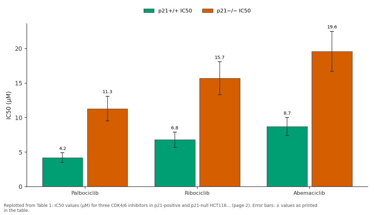
p21+/+ IC50 and p21−/− IC50: Palbociclib vs Ribociclib vs Abemaciclib
Replotted from Table 1 (page 2) of the original paper. Every bar is the paper's own table value.
View source evidence ↗Figures & tables
From the original paper Table 1
 Table 2
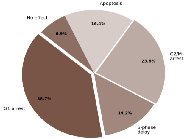 Figure 1
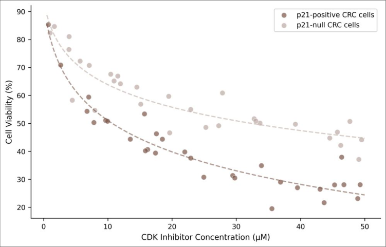 Figure 2
Introduction
Cancer cells and normal cells face the same cell cycle machinery, but they handle it in opposite ways. Normal cells obey checkpoint signals that halt division when DNA is damaged or conditions are unfavorable; cancer cells override these signals to proliferate without restraint [1] . In colorectal carcinoma the third most common malignancy worldwide checkpoint evasion frequently involves loss or silencing of CDK inhibitor proteins, particularly p21WAF1/CIP1 and p27KIP1 [2] . CDKs 4 and 6 phosphorylate the retinoblastoma protein (Rb) during G1 phase, releasing E2F transcription factors that drive S-phase entry. The endogenous inhibitor p21 blocks CDK4/6 activity, enforcing the G1/S checkpoint [3] . When p21 is absent or non-functional, cells become dependent on alternative checkpoints particularly the G2/M checkpoint regulated by CDK1 and its inhibitor Wee1 to maintain any remaining quality control over division [4] . Pharmacological CDK4/6 inhibitors (palbociclib, ribociclib, abemaciclib) have transformed the treatment of hormone receptor-positive breast cancer, but their efficacy in colorectal carcinoma has been inconsistent [5,6] . One explanation is that the checkpoint response to these drugs depends on the tumor's endogenous CDK inhibitor status a variable that hasn't been systematically tested in isogenic colorectal models. This research used HCT116 p21+/+ and p21-/-isogenic cell lines to directly compare how p21 status shapes the cell cycle response to CDK4/6 inhibition, with the aim of identifying whether p21 expression predicts therapeutic sensitivity [7,8] .
Ethical considerations
This research used established human cell lines (HCT116 and its isogenic derivative HCT116 p21-/-) obtained from the American Type Culture Collection (ATCC) and the Vogelstein laboratory collection, respectively. No primary human tissue or patient samples were used. Institutional biosafety approval was obtained from the Fynbos Institute of . Cell cycle distribution was analyzed by propidium iodide staining and flow cytometry on a BD FACSCalibur (BD Biosciences). A minimum of 10, 000 events were recorded per sample. Cell cycle phases were quantified using ModFit LT v5.0 software. Western blotting for p21, p27, phospho-Rb (Ser780), total Rb, CDK4, and CDK6 used standard protocols with antibodies from Cell Signaling Technology (Danvers, MA, USA) [10] . Apoptosis was measured by Annexin V-FITC/PI dual staining. IC50 values were calculated by nonlinear regression in GraphPad Prism v9.4. Statistical comparisons between cell lines used unpaired ttests with Welch's correction (α = 0.05).
Results
Results
Comprehensive interpretation
The isogenic comparison reveals that p21 status dictates both the mechanism and magnitude of CDK4/6 inhibitor response. In p21-expressing cells, pharmacological CDK4/6 blockade is reinforced by drug-induced p21 accumulation, creating a positive feedback loop that strengthens G1 arrest. Without p21, cells bypass G1 and rely on the G2/M checkpoint, which proves less effective at halting proliferation hence the higher IC50 values in p21-null cells.
Limitations
This research used a single isogenic cell line pair (HCT116), which limits generalizability to other colorectal carcinoma genetic backgrounds. The 48-hour treatment window may not capture long-term adaptive responses such as CDK inhibitor resistance. In vitro monolayer culture doesn't replicate the tumor microenvironment, including stromal interactions and hypoxia that can influence checkpoint regulation. Additionally, p21 loss in clinical tumors is rarely complete partial expression or epigenetic silencing may produce intermediate phenotypes not represented by the knockout model [11,12] .
Clinical implications
These findings suggest that p21 immunohistochemistry could serve as a predictive biomarker for CDK4/6 inhibitor response in colorectal carcinoma. Tumors retaining p21 expression may derive greater benefit from palbociclib or ribociclib, while p21-deficient tumors might require combination strategies targeting the G2/M checkpoint for instance, adding a Wee1 inhibitor to CDK4/6 blockade. Clinical trials stratifying patients by p21 status would be needed to test this hypothesis [13,14] .
Discussion
The differential checkpoint response between p21+/+ and p21-/-cells confirms what has been hypothesized but not directly demonstrated in isogenic colorectal models: that endogenous CDK inhibitor status shapes the cellular response to pharmacological CDK blockade [15,16] . The 2.3to 2.7-fold difference in IC50 values is clinically meaningful it's the difference between a drug concentration achievable in patients and one that would require toxic doses. The G2/M arrest observed in p21-null cells treated with CDK4/6 inhibitors suggests checkpoint compensation. When G1 control fails, cells enter S phase but encounter problems at the subsequent checkpoint. This is consistent with reports that CDK4/6 inhibitors can induce DNA replication stress, which is sensed at G2/M by the ATR-Chk1 pathway [17] . The lower apoptosis rate in p21-null cells (9.2% vs 16.4%) indicates that G2/M arrest alone is less lethal than G1 arrest possibly because G2/M-arrested cells retain the option of checkpoint adaptation and mitotic slippage [18,19] . The practical implication is that p21deficient colorectal tumors are likely to respond poorly to CDK4/6 inhibitor monotherapy and may need combination approaches targeting multiple checkpoints [20] .
Conclusion
This research demonstrates that p21 status is a major determinant of CDK4/6 inhibitor sensitivity in colorectal carcinoma cells. p21-expressing cells responded primarily through G1 arrest with lower IC50 values, while p21-null cells shifted to G2/M arrest with reduced drug sensitivity. The 2.3-to 2.7-fold difference in IC50 suggests that p21 immunohistochemistry could help predict which colorectal tumors will benefit from CDK4/6 inhibitor therapy. These findings argue for biomarker-guided patient selection in future clinical trials of CDK inhibitors for colorectal carcinoma. For p21-deficient tumors, combination strategies incorporating G2/M checkpoint inhibitors may be necessary to achieve meaningful therapeutic responses.
Figures and tables from the paper
| Inhibitor | p21+/+ IC50 | p21−/− IC50 | p-value |
|---|---|---|---|
| Palbociclib | 4.2 ± 0.7 | 11.3 ± 1.8 | 0.003 |
| Ribociclib | 6.8 ± 1.1 | 15.7 ± 2.4 | 0.001 |
| Abemaciclib | 8.7 ± 1.3 | 19.6 ± 2.9 | <0.001 |
Table text extracted from the original PDF; superscript letters are shown inline. The original table image is below.
View original table image
| Condition | G1 (%) | S (%) | G2/M (%) | Sub-G1 (%) |
|---|---|---|---|---|
| p21+/+ Control | 61.3 | 24.7 | 14.0 | — |
| p21+/+ Treated | 38.7 G1↑ | 11.2 | 8.4 | 16.4 |
| p21−/− Control | 58.9 | 26.1 | 15.0 | — |
| p21−/− Treated | 21.3 | 12.8 | 31.4 G2↑ | 9.2 |
Table text extracted from the original PDF; superscript letters are shown inline. The original table image is below.
View original table image
References
20 in the original paper- Hanahan D, Weinberg RA. Hallmarks of cancer: the next generation. Cell. 2011;144(5):646-674.
- Gartel AL, Radhakrishnan SK. Lost in transcription: p21 repression, mechanisms, and consequences. Cancer Research. 2005;65(10):3980-3985.
- Sherr CJ, Roberts JM. CDK inhibitors: positive and negative regulators of G1-phase progression. Genes and Development. 1999;13(12):1501-1512.
- Matheson CJ, Backos DS, Bhutani N. Targeting WEE1 kinase in cancer. Trends in Pharmacological Sciences. 2016;37(10):872-881.
- Seshacharyulu P, Ponnusamy MP, Haridas D, Jain M, Ganti AK, Batra SK. Targeting the EGFR signaling pathway in cancer therapy. Expert Opinion on Therapeutic Targets. 2012;16(1):15-31.
- Goel S, DeCristo MJ, McAllister SS, Zhao JJ. CDK4/6 inhibition in cancer: beyond cell cycle arrest. Trends in Cell Biology. 2018;28(11):911-925.
- Bunz F, Dutriaux A, Lengauer C, et al. Requirement for p53 and p21 to sustain G2 arrest after DNA damage. Science. 1998;282(5393):1497-1501.
- Waldman T, Kinzler KW, Vogelstein B. p21 is necessary for the p53-mediated G1 arrest in human cancer cells. Cancer Research. 1995;55(22):5187-5190.
- Mosmann T. colorimetric assay for cellular growth and survival: application to proliferation and cytotoxicity assays. Journal of Immunological Methods. 1983;65(1-2):55-63.
- Towbin H, Staehelin T, Gordon J. Electrophoretic transfer of proteins from polyacrylamide gels to nitrocellulose sheets: procedure and some applications. Proceedings of the National Academy of Sciences. 1979;76(9):4350-4354.
- Abbas T, Dutta A. p21 in cancer: intricate networks and multiple activities. Nature Reviews Cancer. 2009;9(6):400-414.
- Jung YS, Qian Y, Chen X. Examination of the expanding pathways for the regulation of p21 expression and activity. Cellular Signalling. 2010;22(7):1003-1012.
- Asghar U, Witkiewicz AK, Turner NC, Schiavon G. The history and future of targeting cyclin-dependent kinases in cancer therapy. Nature Reviews Drug Discovery. 2015;14(2):130-146.
- O'Leary B, Finn RS, Turner NC. Treating cancer with selective CDK4/6 inhibitors. Nature Reviews Clinical Oncology. 2016;13(7):417-430.
- Choi YJ, Li X, Hydbring P, et al. The requirement for cyclin D function in tumor maintenance. Cancer Cell. 2012;22(4):438-451.
- Schiavon G, Hrebien S, Garcia-Murillas I, et al. Analysis of ESR1 mutation in circulating tumor DNA demonstrates evolution during therapy for metastatic breast cancer. Science Translational Medicine. 2015;7(313):313ra182.
- Koundrioukoff S, Carignon S, Techer H, et al. Stepwise activation of the ATR signaling pathway upon increasing replication stress impacts fragile site integrity. PLoS Genetics. 2013;9(7):e1003643.
- Brito DA, Rieder CL. Mitotic checkpoint slippage in humans occurs via cyclin B destruction in the presence of an active checkpoint. Current Biology. 2006;16(12):1194-1200.
- Topham C, Tighe A, Ly P, et al. MYC is a major determinant of mitotic cell fate. Cancer Cell. 2015;28(1):129-140.
- Rader J, Russell MR, Hart LS, et al. Dual CDK4/CDK6 inhibition induces cell-cycle arrest and senescence in neuroblastoma. Clinical Cancer Research. 2013;19(22):6173-6182.