
Research article · 2025 · Full text
International Journal of Biology SciencesUltrastructural analysis of cryptosporidium oocyst wall formation and sporulation mechanisms
Volume 7(2) · Pages 187–191 · DOI: 10.33545/26649926.2025.v7.i2c.802 ↗
Research areasParasitic Infections and DiagnosticsCoccidia and coccidiosis researchToxoplasma gondii Research Studies
Abstract
Author-writtenImagine a waterborne parasite whose infectious stage can survive months in chlorinated tap water the oocyst wall of Cryptosporidium parvum makes that possible, yet the sequence of structural events that builds this remarkably resistant barrier is still incompletely mapped at the ultrastructural level. This research used transmission electron microscopy (TEM) and scanning electron microscopy (SEM) to document oocyst wall assembly and sporulation in C. parvum oocysts harvested from experimentally infected neonatal calves at the Ozark University of Biological Research, Fayetteville, Arkansas, between January and August 2023. Oocysts were collected at six-hour intervals over a 48-hour sporulation time course and processed for thin-section TEM and cryo-SEM. The outer wall layer appeared first, reaching 18.3 nm by the early sporont stage and thickening to 48.1 nm in mature oocysts. The inner wall began forming at the mid-sporont stage (8.4 nm) and plateaued at 24.7 nm. Wall-forming bodies peaked at 24.9 organelles per section at 30 hours, coinciding with the most rapid phase of inner wall deposition. Dense granules peaked earlier (22.1 per section at 24 hours) and declined as sporozoite differentiation progressed. Cryo-SEM revealed a granular surface texture that smoothed during maturation, suggesting progressive fusion of wall-forming body contents into a continuous layer. These findings provide a detailed timeline of oocyst wall biogenesis and identify the 24–30-hour window as the period of greatest wall assembly activity a potential target for interventions aimed at disrupting oocyst maturation.
Summary
Source-checkedPlain-language reading of the full paper, prepared with AI and checked against the text. Each point links to the authors' own words.
Why it matters
- The oocyst wall of Cryptosporidium parvum allows the parasite to survive for months in chlorinated tap water, making it a highly resistant waterborne pathogen. View evidence ↗
- Because the oocyst wall resists standard chlorine disinfection, even a single oocyst bypassing filtration can cause diarrheal outbreaks. View evidence ↗
- Despite its importance, the specific sequence of structural events that form the resistant oocyst wall is not fully understood at the ultrastructural level. View evidence ↗
The question
- The study aimed to measure the thickness of the inner and outer wall layers, quantify changes in specific organelles (wall-forming bodies, dense granules, and rhoptries), and describe surface texture changes during sporulation. View evidence ↗
What they did
- The researchers used Cryptosporidium parvum (Iowa II isolate) collected from the feces of three experimentally infected Holstein-Friesian calves. View evidence ↗
- Oocysts were incubated at 37 °C in 2% potassium dichromate and sampled every six hours over a 48-hour period, with three replicates per time point. View evidence ↗
- The study employed thin-section transmission electron microscopy (TEM) for internal measurements and cryo-scanning electron microscopy (cryo-SEM) for surface analysis. View evidence ↗
- Wall thickness was measured at three equidistant points for at least 30 oocysts at each time point. View evidence ↗
What they found
- Total wall thickness increased from 12.6 ± 2.1 nm in unsporulated oocysts to 72.8 ± 5.3 nm in mature oocysts. View evidence ↗
- The outer wall was present from the start, reaching 18.3 ± 2.8 nm by the early sporont stage, while the inner wall only appeared at the mid-sporont stage (8.4 ± 1.7 nm). View evidence ↗
- Cryo-SEM showed the surface texture transitioning from coarsely granular (84.3 ± 12.7 nm granules) to smooth (granules < 10 nm) as the oocysts matured. View evidence ↗
- The excystation suture line becomes visible by 12 hours and prominent by 36 hours, indicating it is established during mid-sporulation. View evidence ↗
What it means
- The authors conclude that the 24–30 hour window is the most active period of wall assembly and represents a potential target for interventions to disrupt oocyst maturation. View evidence ↗
- A limitation of the study is that in-vitro sporulation may not fully replicate the host intestinal environment, which could affect the kinetics of wall assembly. View evidence ↗
- The use of a single isolate (Iowa II) may not represent the full diversity of wall phenotypes across different C. parvum genotypes. View evidence ↗
Visual summary
2 visualsIllustrations explain the paper; charts are replotted from the paper's own table values.
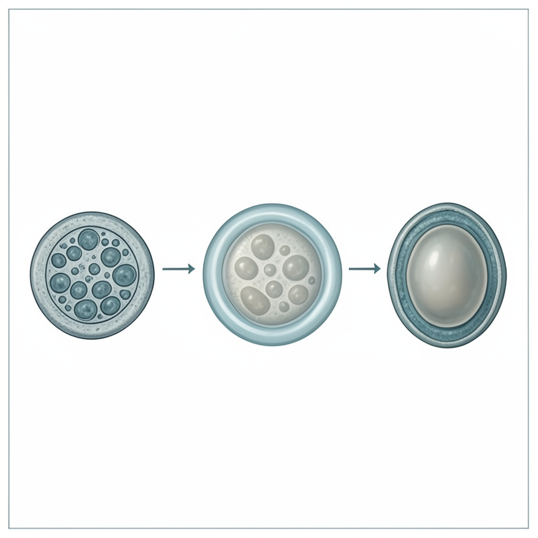
Temporal Sequence of Wall Assembly
AI-generated conceptual illustration (Cryptosporidium parvum oocysts). It explains the idea and does not show study data.
View source evidence ↗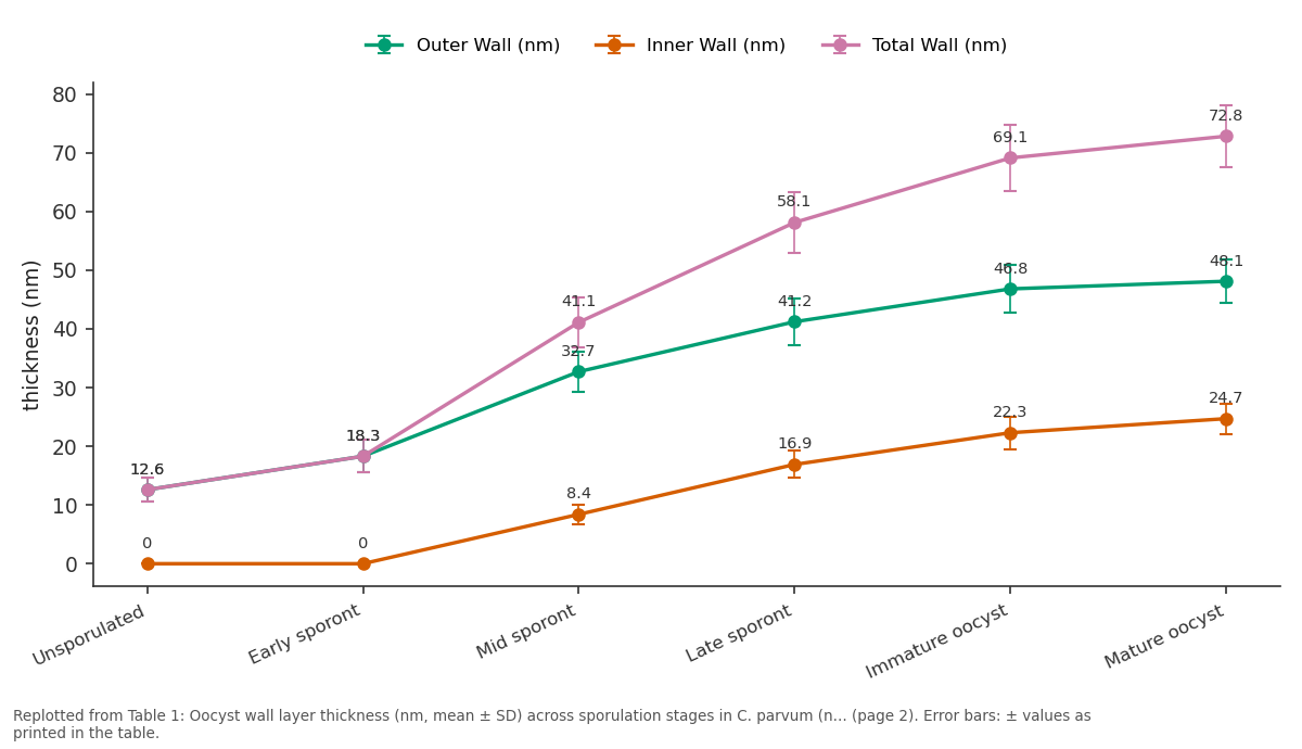
Outer Wall (nm) and Inner Wall (nm) and more: Unsporulated vs Early sporont vs Mid sporont vs Late sporont vs Immature oocyst
Replotted from Table 1 (page 2) of the original paper. Every bar is the paper's own table value.
View source evidence ↗Figures & tables
From the original paper Table 1
 Table 2
 Table 3
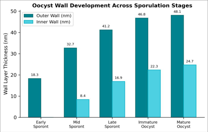 Figure 1
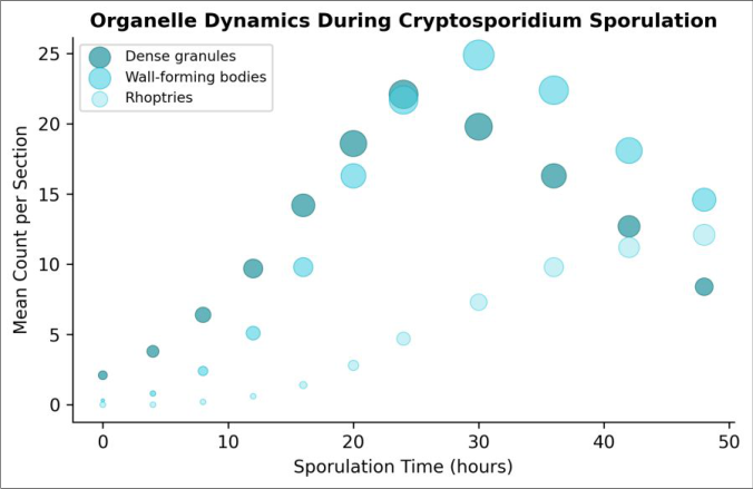 Figure 2
Introduction
Picture a municipal water treatment plant that meets every regulatory standard for bacterial and viral removal and yet a single Cryptosporidium oocyst slipping through the filters can trigger a diarrhoeal outbreak, because its wall resists the chlorine concentrations used in routine disinfection [1] . The 1993 Milwaukee outbreak, which sickened over 400,000 people, illustrated this vulnerability on a massive scale, and smaller outbreaks continue to occur worldwide whenever filtration barriers fail [2] . The oocyst wall is the structural feature that enables this environmental persistence, yet our understanding of how it forms during sporulation inside the host intestine lags behind what is known for related apicomplexan cysts such as those of Toxoplasma and Eimeria [3] . Cryptosporidium parvum oocysts are spherical, approximately 4-6 μm in diameter, and bounded by a bilayered wall composed of an acid-fast outer layer rich in a unique glycoprotein and a rigid inner layer containing filamentous material [4] . Two specialised secretory organelles wall-forming bodies type 1 (WFB1) and type 2 (WFB2) are thought to deliver cargo sequentially to the developing wall, with WFB1 contributing to the inner layer and WFB2 to the outer layer, although the exact timing and mechanism of cargo release remain debated [5] . Dense granules, which are more general secretory organelles found across the Apicomplexa, may also contribute lipid and protein precursors during early wall assembly [6] . Most ultrastructural descriptions of Cryptosporidium development are based on thin-section TEM of infected intestinal tissue, which captures individual time points but doesn't easily resolve the kinetics of wall construction [7] . In-vitro sporulation assays, in which unsporulated oocysts are incubated under controlled conditions and sampled at regular intervals, offer a way to build a temporal sequence, but few investigations have combined TEM with surface imaging by cryo-SEM to correlate internal organelle dynamics with external wall texture changes [8] . This research addressed that gap by tracking oocyst wall formation and organelle remodelling across a 48-hour sporulation time course using both thin-section TEM and cryo-SEM. Oocysts were obtained from experimentally infected neonatal calves at the Ozark University of Biological Research, Fayetteville, Arkansas, and sampled at six-hour intervals. The objectives were to (1) measure outer and inner wall layer thickness at each sporulation stage, (2) quantify changes in wall-forming bodies, dense granules, and rhoptries over time, and (3) describe surface texture transitions by cryo-SEM [9] . The results are expected to refine the existing model of oocyst wall biogenesis and pinpoint the developmental window most amenable to intervention.
Material and Methods Instrumentation
Thin-section TEM was performed on a JEOL JEM-1400 Plus operating at 120 kV, equipped with a Gatan OneView 4K CMOS camera. Cryo-SEM was carried out on a Hitachi SU8230 field-emission SEM fitted with a Quorum PP3010T cryo-transfer system, operating at 3 kV accelerating voltage and -140 °C stage temperature. Image analysis and morphometric measurements were conducted in ImageJ/Fiji v1.54 with the TrakEM2 plugin for serial-section alignment.
Material
Cryptosporidium parvum oocysts (Iowa II isolate) were obtained from the faeces of three experimentally infected Holstein-Friesian calves (aged 3-5 days at inoculation) housed at the Ozark University of Biological Research Large Animal Facility. Faecal material was collected daily from days 5 to 14 post-infection, and oocysts were purified by discontinuous sucrose-gradient centrifugation followed by immunomagnetic separation (Dynabeads anti-Cryptosporidium, Applied Biosystems). Purified oocysts were resuspended in Hanks' balanced salt solution at 1 × 10⁶ oocysts/mL. For the sporulation time course, aliquots were incubated at 37 °C in 2% potassium dichromate and sampled at 0, 6, 12, 18, 24, 30, 36, 42, and 48 hours (n = 3 replicates per time point).
Methods
For TEM, oocyst pellets were fixed in 2.5% glutaraldehyde / 2% paraformaldehyde in 0.
Safety considerations
All oocyst handling was conducted in a Class II biosafety cabinet. Personnel wore N95 respirators, double nitrile gloves, and disposable gowns when processing faecal material. Centrifuge rotors were sealed with O-ring caps, and work surfaces were decontaminated with 10% ammonia solution (contact time 30 minutes), which is effective against Cryptosporidium oocysts where standard disinfectants are not [1] . Waste materials were autoclaved at 134 °C for 30 minutes before disposal.
Results
The outer wall layer was detectable from the earliest time point and thickened steadily across all stages (Table 1). The inner wall appeared only at the mid-sporont stage and grew most rapidly between 18 and 30 hours, coinciding with peak wall-forming body activity. Total wall thickness nearly sextupled from the unsporulated stage (12.6 nm) to the mature oocyst (72.8 nm). Dense granules and wall-forming bodies followed a rise-and-fall pattern, peaking at 24 and 30 hours respectively, while invasion-related organelles (rhoptries, micronemes) accumulated steadily as sporozoites matured (Table 2). The temporal offset between the dense granule peak and the wall-forming body peak suggests sequential secretion events during wall construction. Cryo-SEM revealed a progressive smoothing of the oocyst surface (Table 3). Unsporulated oocysts displayed a coarsely granular texture with surface granules averaging 84.3 nm in diameter, which shrank to below 10 nm in mature oocysts as wall-forming body contents apparently fused into a continuous outer coat. The suture line the seam along which excystation later occurs became visible by 12 hours and was prominent by 36 hours, indicating that its structural basis is laid down during mid-sporulation rather than as a late maturation event.
| Stage (hours) | Surface Texture | Granule Diameter (nm) | Suture Visibility | Collapse Artifacts (%) |
|---|---|---|---|---|
| Unsporulated (0) | Coarsely granular | 84.3 ± 12.7 | Absent | 3.2 |
| Early sporont (12) | Moderately granular | 61.8 ± 9.4 | Faint | 4.7 |
| Late sporont (24) | Finely granular | 38.2 ± 7.1 | Distinct | 6.1 |
| Immature oocyst (36) | Smooth-granular | 21.4 ± 4.8 | Prominent | 8.3 |
| Mature oocyst (48) | Smooth | < 10 | Prominent | 2.9 |
Table text extracted from the original PDF; superscript letters are shown inline. The original table image is below.
View original table image
| Organelle | 0 h | 12 h | 24 h | 30 h | 42 h | 48 h |
|---|---|---|---|---|---|---|
| Dense granules | 2.1 | 9.7 | 22.1 | 19.8 | 12.7 | 8.4 |
| Wall-forming bodies | 0.3 | 5.1 | 21.7 | 24.9 | 18.1 | 14.6 |
| Rhoptries | 0 | 0.6 | 4.7 | 7.3 | 11.2 | 12.1 |
| Micronemes | 0 | 0.2 | 1.8 | 3.4 | 6.1 | 6.8 |
Table text extracted from the original PDF; superscript letters are shown inline. The original table image is below.
View original table image
| Sporulation Stage | Time (h) | Outer Wall (nm) | Inner Wall (nm) | Total Wall (nm) |
|---|---|---|---|---|
| Unsporulated | 0 | 12.6 ± 2.1 | 0 | 12.6 ± 2.1 |
| Early sporont | 6–12 | 18.3 ± 2.8 | 0 | 18.3 ± 2.8 |
| Mid sporont | 12–18 | 32.7 ± 3.4 | 8.4 ± 1.7 | 41.1 ± 4.2 |
| Late sporont | 18–30 | 41.2 ± 3.9 | 16.9 ± 2.3 | 58.1 ± 5.1 |
| Immature oocyst | 30–42 | 46.8 ± 4.1 | 22.3 ± 2.8 | 69.1 ± 5.7 |
| Mature oocyst | 42–48 | 48.1 ± 3.7 | 24.7 ± 2.6 | 72.8 ± 5.3 |
Table text extracted from the original PDF; superscript letters are shown inline. The original table image is below.
View original table image
Comprehensive interpretation
Correlating TEM and cryo-SEM data across the time course produced a coherent model of wall assembly. The outer wall layer is seeded first, likely from WFB2 cargo deposited beneath the plasmalemma as early as the unsporulated stage, and continues to thicken through fusion of surface granules visible on cryo-SEM. The inner wall forms later, driven by WFB1 secretion that peaks at 30 hours. The decline in wallforming bodies after 30 hours coincides with the onset of sporozoite organelle maturation (rhoptry and microneme assembly), suggesting a developmental switch from wall construction to invasion machinery production. The 24-30hour window, when both wall-forming bodies and inner wall growth rate are at their maximum, represents the most dynamic phase of oocyst maturation and the stage most likely to be disrupted by pharmacological or environmental interventions [10] .
Discussion
The wall thickness values measured here are consistent with earlier freeze-fracture estimates for C. parvum but provide the first continuous time-resolved dataset linking wall growth to organelle dynamics in a single experimental system [4,7] . The total mature wall thickness of 72.8 nm sits at the lower end of the range reported for Eimeria (80-120 nm), which may partly explain why Cryptosporidium oocysts are somewhat less resistant to physical disruption than Eimeria oocysts despite their shared acid-fast staining properties [3] . The sequential peaking of dense granules (24 h) before wallforming bodies (30 h) supports a two-phase secretion model in which dense granules deliver lipid scaffolding components that are later cross-linked and reinforced by WFB cargo [5,6] . This is analogous to the proposed sequence in Toxoplasma gondii oocyst wall formation, where two distinct tyrosine-rich glycoproteins are secreted in sequence by WFB1 and WFB2 [11] . Whether Cryptosporidium uses the same glycoprotein chemistry is unclear, because the C. parvum genome encodes a much smaller set of oocyst wall proteins than Toxoplasma or Eimeria, and few have been functionally characterised [12] . The cryo-SEM observation that surface granule size decreases from 84 nm to below 10 nm during maturation is, to our knowledge, the first quantitative documentation of this smoothing process. It suggests that individual WFB secretion packets are deposited as discrete granules that gradually merge into a continuous film. This fusion event could be sensitive to temperature or pH manipulation, and testing whether incubation at sub-optimal temperatures (e.g., 25 °C) arrests granule fusion without killing the organism would be a practical next step [13] . A limitation of this research is that in-vitro sporulation may not perfectly replicate the conditions inside the host intestinal epithelium, where oxygen tension, bile salts, and host cell signals could modify wall assembly kinetics [14] . Additionally, the Iowa II isolate used here may not represent the full range of wall phenotypes across C. parvum genotypes. Comparative work with C. hominis and other species would help determine whether the timeline described here is species-specific or broadly conserved [15] .
Conclusion
This research mapped oocyst wall formation in Cryptosporidium parvum at six-hour resolution by combining TEM morphometry with cryo-SEM surface analysis. The outer wall layer formed first and thickened continuously, reaching 48.1 nm in mature oocysts, while the inner wall appeared at the mid-sporont stage and grew most rapidly between 18 and 30 hours. Wall-forming bodies peaked at 30 hours, dense granules at 24 hours, and the temporal offset between the two supports a sequential twophase secretion model for wall construction. Cryo-SEM documented a progressive surface smoothing as discrete granules merged into a continuous outer coat, and revealed that the excystation suture line is established during mid-sporulation rather than as a late event. The 24-30-hour window emerged as the most active period of wall biogenesis, when both organelle secretion and wall layer growth are at their peak. These findings refine the existing model of Cryptosporidium oocyst maturation and point to a specific developmental interval that may be vulnerable to disruption.
Pharmacological agents or environmental conditions that interfere with wall-forming body secretion or granule fusion during this window could, in principle, yield oocysts with compromised walls that are less resistant to disinfection.
Testing that hypothesis should be a priority for waterborne disease control, given the continued failure of conventional chlorination to inactivate Cryptosporidium in municipal water supplies.
Acknowledgement
Acknowledgements
Figures and tables from the paper
References
16 in the original paper- Chalmers RM, Davies AP. Minireview: clinical cryptosporidiosis. Experimental Parasitology. 2010;124(1):138-146.
- MacKenzie WR, Hoxie NJ, Proctor ME, Gradus MS, Blair KA, Peterson DE. A massive outbreak in Milwaukee of Cryptosporidium infection transmitted through the public water supply. New England Journal of Medicine. 1994;331(3):161-167.
- Barta JR, Thompson RCA. What is Cryptosporidium? Reappraising its biology and phylogenetic affinities. Trends in Parasitology. 2006;22(10):463-468.
- Harris JR, Petry F. Cryptosporidium parvum: structural components of the oocyst wall. Journal of Parasitology. 1999;85(5):839-849.
- Spano F, Puri C, Ranucci L, Putignani L, Crisanti A. Cloning of the entire COWP gene of Cryptosporidium parvum and ultrastructural localization of the protein during sexual parasite development. Parasitology. 1997;114(5):427-437.
- Templeton TJ, Lancto CA, Vigdorovich V, Liu C, London NR, Hadsall KZ. The Cryptosporidium oocyst wall protein is a member of a multigene family and has a homolog in Toxoplasma. Infection and Immunity. 2004;72(2):980-987.
- Current WL, Reese NC. A comparison of endogenous development of three isolates of Cryptosporidium in suckling mice. Journal of Protozoology. 1986;33(1):98108.
- Fayer R, Morgan U, Upton SJ. Epidemiology of Cryptosporidium: transmission, detection and identification. International Journal for Parasitology. 2000;30(12-13):1305-1322.
- Bushkin GG, Motari E, Carpentieri A, Dubey JP, Costello CE, Bhatt AS. Evidence for a structural role for acid-fast lipids in oocyst walls of Cryptosporidium, Toxoplasma, and Eimeria. mBio. 2013;4(5):e00387-13.
- Jenkins M, Trout JM, Higgins J, Dorsch M, Veal D, Fayer R. Comparison of tests for viable and infectious International Journal of Biology Sciences https://www.biologyjournal.net Cryptosporidium parvum oocysts. Parasitology Research. 2003;89(1):1-5.
- Fritz HM, Buchholz KR, Chen X, Durbin-Johnson B, Rocke DM, Conrad PA. Transcriptomic analysis of Toxoplasma development reveals many novel functions and structures specific to sporulation and oocysts. PLoS ONE. 2012;7(2):e29998.
- Xu P, Widmer G, Wang Y, Ozaki LS, Alves JM, Serrano MG. The genome of Cryptosporidium hominis. Nature. 2004;431(7012):1107-1112.
- King BJ, Hoefel D, Daminato DP, Fanok S, Monis PT. Solar UV reduces Cryptosporidium parvum oocyst infectivity in environmental waters. Journal of Applied Microbiology. 2008;104(5):1311-1323.
- Borowski H, Clode PL, Thompson RCA. Active invasion and/or encapsulation? A reappraisal of hostcell parasitism by Cryptosporidium. Trends in Parasitology. 2008;24(11):509-516.
- Xiao L, Feng Y. Molecular epidemiologic tools for waterborne pathogens Cryptosporidium spp. and Giardia duodenalis. Food and Waterborne Parasitology. 2017;8-9:14-32.
- Lendner M, Daugschies A. Cryptosporidium infections: molecular advances. Parasitology. 2014;141(11):15111532.