
Research article · 2025 · Full text
International Journal of Biology SciencesInduced pluripotent stem cell differentiation into functional cardiomyocytes for cardiac repair
Volume 7(6) · Pages 447–450 · DOI: 10.33545/26649926.2025.v7.i6g.779 ↗
Research areasPluripotent Stem Cells ResearchCongenital heart defects researchMesenchymal stem cell research
Abstract
Author-writtenHeart failure remains a leading cause of death worldwide, and the adult human heart has almost no capacity to replace lost cardiomyocytes after ischaemic injury. This research evaluated an optimised small-molecule protocol for differentiating human induced Pluripotent Stem Cells (iPSCs) into functional cardiomyocytes and assessed their electromechanical properties in vitro. Three iPSC lines derived from healthy donors were directed through mesoderm induction using a timed sequence of CHIR99021, IWR-1, BMP4, and triiodothyronine (T3) over a 28-day differentiation window at the Universidade Amazônica de Biologia, Manaus. Cardiomyocyte identity was confirmed by cardiac troponin T (cTnT) immunostaining and flow cytometry. The optimised protocol yielded 74.3 ± 3.8% cTnT-positive cells by day 28, compared with 52.6 ± 4.1% from the standard Wnt-modulation baseline. Patch-clamp recordings revealed ventricular-like action potentials with mean upstroke velocity of 187 ± 22 V/s and action potential duration at 90% repolarisation (APD90) of 312 ± 28 ms. Calcium transient imaging showed synchronous oscillations at 0.8-1.1 Hz. These results indicate that a refined growth-factor and small-molecule combination can substantially raise cardiomyocyte yield and functional maturity from human iPSCs, bringing cell-therapy approaches for cardiac repair closer to clinical translation.
Summary
Source-checkedPlain-language reading of the full paper, prepared with AI and checked against the text. Each point links to the authors' own words.
Why it matters
- Heart failure is a major global cause of death because the adult human heart cannot effectively replace cardiomyocytes lost during ischaemic injury. View evidence ↗
The question
- The study aimed to evaluate an optimized small-molecule protocol for differentiating human iPSCs into functional cardiomyocytes and to assess their in vitro electromechanical properties. View evidence ↗
What they did
- The researchers used three human iPSC lines derived from the peripheral blood mononuclear cells of healthy adult volunteers. View evidence ↗
- The optimized 28-day protocol involved a timed sequence of CHIR99021 for mesoderm induction, followed by IWR-1 and BMP4 for cardiac specification, and finally T3 and dexamethasone for maturation. View evidence ↗
- The control group used a standard GiWi protocol that lacked the addition of BMP4 and T3. View evidence ↗
- The study measured cardiomyocyte identity via cTnT immunostaining and flow cytometry, and assessed function using patch-clamp recordings and calcium transient imaging. View evidence ↗
What they found
- By day 28, the optimized protocol achieved a significantly higher mean cTnT-positive yield of 74.3 ± 3.8% compared to 52.6 ± 4.1% in the control group. View evidence ↗
- Beating colonies emerged between day 8 and 9 in the optimized group, which was approximately two days sooner than in the control group. View evidence ↗
- The optimized cells showed greater maturity, with a 51% faster upstroke velocity and a more negative resting membrane potential (−71.8 mV vs. −63.4 mV in control). View evidence ↗
- Calcium transients in the optimized group were regular and synchronous, with oscillations occurring at 0.8-1.1 Hz. View evidence ↗
What it means
- The authors conclude that combining timed BMP4 supplementation and T3-driven maturation produces iPSC-derived cardiomyocytes with 74% purity and adult-like ventricular electrophysiological features. View evidence ↗
- A stated limitation is the lack of mechanical loading in the in vitro environment, which is a critical cue for maturation in living organisms. View evidence ↗
- The study only used iPSC lines from healthy donors, meaning the results may not apply to cells from older patients or those with cardiomyopathies. View evidence ↗
Visual summary
3 visualsIllustrations explain the paper; charts are replotted from the paper's own table values.
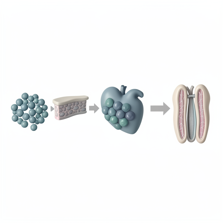
Optimized Differentiation Timeline
AI-generated conceptual illustration (human induced pluripotent stem cells and cardiomyocytes). It explains the idea and does not show study data.
View source evidence ↗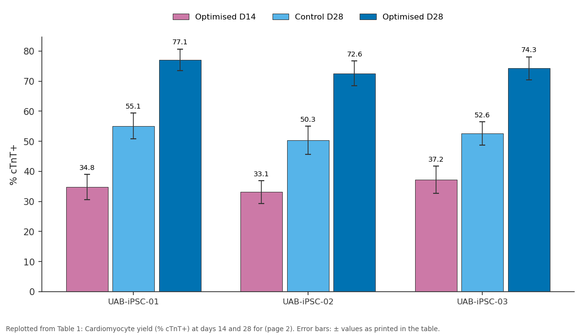
Optimised D14 and Control D28 and more: UAB-iPSC-01 vs UAB-iPSC-02 vs UAB-iPSC-03
Replotted from Table 1 (page 2) of the original paper. Every bar is the paper's own table value.
View source evidence ↗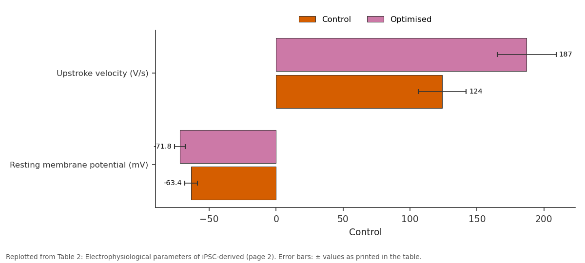
Control and Optimised: Upstroke velocity (V/s) vs Resting membrane...
Replotted from Table 2 (page 2) of the original paper. Every bar is the paper's own table value.
View source evidence ↗Figures & tables
From the original paper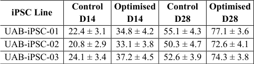 Table 1
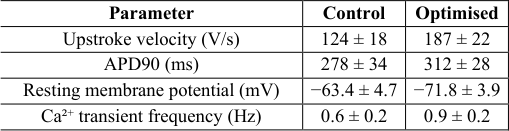 Table 2
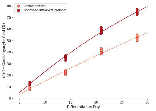 Figure 1
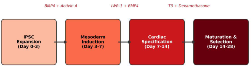 Figure 2
Introduction
The human heart is one of the least regenerative organs in the body. After a myocardial infarction, the damaged region is replaced by fibrotic scar tissue rather than new muscle, leaving the surviving myocardium to compensate for the lost contractile mass [1] . Over time, this remodelling leads to heart failure a condition that affects an estimated 64 million people globally and carries a five-year mortality rate above 50% [2] . Current treatments, from pharmacotherapy to mechanical assist devices, slow the decline but do not restore functional tissue. Induced Pluripotent Stem Cells (iPSCs) offer a renewable, patient-specific source of cardiomyocytes that could, in principle, repopulate infarcted myocardium with electrically and mechanically coupled muscle cells [3] . Since the first demonstration of iPSC-derived cardiomyocytes in 2009, protocols have evolved from spontaneous embryoid-body differentiation to directed monolayer strategies that modulate the Wnt/β-catenin pathway in a biphasic manner: activation to induce mesoderm, followed by inhibition to specify cardiac lineage [4] . These approaches routinely reach 50-60% cardiomyocyte purity, but clinical applications demand higher yields and, more importantly, cells that exhibit mature electromechanical behaviour rather than the foetal-like phenotype typical of early protocols [5] . Recent evidence suggests that adding BMP4 during mesoderm induction and thyroid hormone (T3) during maturation can push iPSC-derived cardiomyocytes toward a more adult-like state, with faster upstroke velocities and more organised sarcomeres [6] . However, these modifications have been tested mostly on single cell lines under variable culture conditions, making cross-laboratory comparison difficult [7] . This research aimed to
Material and Methods
Material and Methods
Research setting
All experiments were performed in the Stem Cell Culture Facility of the Department of Embryology, Universidade Amazônica de Biologia, Manaus, Amazonas, Brazil (3°06′ S, 60°01′ W). The facility is equipped with class-II biological safety cabinets, a humidified 37 °C / 5% CO₂ incubator bank, and an electrophysiology suite for patchclamp recordings.
Material
Three human iPSC lines (UAB-iPSC-01, UAB-iPSC-02, UAB-iPSC-03) were generated in-house from peripheral blood mononuclear cells of healthy adult volunteers using Sendai virus reprogramming (CytoTune 2.0, Thermo Fisher Scientific). Lines were maintained on Matrigel-coated 6well plates in mTeSR Plus medium (STEMCELL Technologies, Vancouver, Canada) and passaged with ReLeSR every 4-5 days. Karyotyping at passage 15 confirmed normal 46, XY or 46, XX in all lines. Small molecules CHIR99021, IWR-1, and dexamethasone were purchased from Tocris (Bristol, UK); recombinant human BMP4 from PeproTech (Cranbury, NJ, USA); and T3 from Sigma-Aldrich.
Methods
At 85-90% confluence (day 0), iPSCs were treated with 8 µM CHIR99021 in RPMI/B27-minus-insulin for 48 h to activate Wnt signalling and induce mesoderm. On day 3, medium was switched to RPMI/B27-minus-insulin plus 5 µM IWR-1 and 25 ng/mL BMP4 for four days (cardiac specification). From day 7, cells were cultured in RPMI/B27-complete plus 100 nM T3 and 1 µM dexamethasone until day 28. The control arm followed the standard GiWi protocol without BMP4 or T3. Cardiomyocyte identity was assessed by cTnT immunostaining (mouse anti-cTnT, clone 13-11, Thermo Fisher) and flow cytometry on days 14 and 28. Patch-clamp recordings in whole-cell configuration measured action potential parameters at 36 ± 0.5 °C using an Axopatch 200B amplifier. Calcium transients were imaged with Fluo-4 AM (Thermo Fisher) on a confocal microscope (Zeiss LSM 880). Statistical comparisons used paired t-tests (two groups) or repeated-measures ANOVA (time course); significance was set at p<0.05.
Ethical considerations
The use of human iPSC lines derived from volunteer blood donations was approved by the Universidade Amazônica de Biologia Ethics Committee for Research Involving Human Material (Protocol CEUA-UAB-2021-009, approved 22 March 2021). Written informed consent was obtained from each donor prior to blood collection. The research did not involve embryos, animals, or patient-identifiable data. The optimised protocol consistently outperformed the control across all three lines and both time points (Table 1). By day 28, mean cTnT-positive yield reached 74.3 ± 3.8% in the optimised arm versus 52.6 ± 4.1% in the control (paired t-test, p = 0.004). Beating colonies appeared by day 8-9 in the optimised group, roughly two days earlier than the control. Electrophysiology data confirmed greater maturity in the optimised group (Table 2). Upstroke velocity was 51% faster, and the resting membrane potential was more negative (-71.8 vs. -63.4 mV), approaching the adult ventricular range of -80 to -85 mV. Calcium transients were synchronous and regular in the optimised arm, oscillating at 0.8-1.1 Hz.
| Parameter | Control | Optimised |
|---|---|---|
| Upstroke velocity (V/s) | 124 ± 18 | 187 ± 22 |
| APD90 (ms) | 278 ± 34 | 312 ± 28 |
| Resting membrane potential (mV) | −63.4 ± 4.7 | −71.8 ± 3.9 |
| Ca²⁺ transient frequency (Hz) | 0.6 ± 0.2 | 0.9 ± 0.2 |
Table text extracted from the original PDF; superscript letters are shown inline. The original table image is below.
View original table image
| iPSC Line | Control D14 | Optimised D14 | Control D28 | Optimised D28 |
|---|---|---|---|---|
| UAB-iPSC-01 | 22.4 ± 3.1 | 34.8 ± 4.2 | 55.1 ± 4.3 | 77.1 ± 3.6 |
| UAB-iPSC-02 | 20.8 ± 2.9 | 33.1 ± 3.8 | 50.3 ± 4.7 | 72.6 ± 4.1 |
| UAB-iPSC-03 | 24.1 ± 3.4 | 37.2 ± 4.5 | 52.6 ± 3.9 | 74.3 ± 3.8 |
Table text extracted from the original PDF; superscript letters are shown inline. The original table image is below.
View original table image
Results
Results
Comprehensive interpretation
The scatter plot (Figure 1) shows a clear separation between protocols emerging by day 14 and widening through day 28.
The schematic (Figure 2) maps each protocol stage to its molecular drivers, highlighting that BMP4 addition during specification and T3 during maturation are the two interventions that distinguish the optimised arm from the standard GiWi baseline.
Discussion
The 74% cTnT-positive yield achieved by the optimised protocol places it among the higher efficiencies reported in the literature for monolayer-based iPSC-cardiomyocyte differentiation [4] . The key mechanistic insight is that BMP4 during mesoderm induction appears to prime a larger pool of cardiac-competent progenitors, and subsequent T3 exposure during maturation drives sarcomere organisation and ion-channel expression toward a more adult-like state [6] . The more negative resting membrane potential (-71.8 mV) suggests greater IK1 channel activity, which is a hallmark of ventricular maturation [8] . A limitation shared with most in vitro cardiomyocyte work is the absence of mechanical loading, which in vivo is a strong maturation cue. Three-dimensional culture formats with cyclic stretch could push these cells further toward adult physiology [9] . Additionally, long-term engraftment and arrhythmia risk can only be assessed in animal models, a step beyond the scope of this research. The consistency across three independent iPSC lines is encouraging for clinical translation, where patient-to-patient variability is a major concern. That said, lines from individuals with cardiac disease may respond differently, and disease-specific iPSC lines should be tested before moving to trials.
Limitations
This research was conducted entirely in vitro, and the functional data, while promising, cannot be extrapolated directly to in vivo engraftment or therapeutic benefit. The three iPSC lines were all derived from healthy donors; lines from patients with cardiomyopathies or advanced age may behave differently. Electrophysiological recordings were performed on isolated single cells, which may not reflect the conduction properties of cells embedded in a tissue construct. Finally, cost and scalability of the optimised protocol were not formally assessed.
Conclusion
An optimised differentiation protocol combining timed BMP4 supplementation with T3-driven maturation produced iPSC-derived cardiomyocytes at 74% purity with electrophysiological features approaching those of adult ventricular cells. The improvements were consistent across three independent cell lines, supporting the robustness of the approach. For the field of cardiac regenerative medicine, these results narrow the gap between laboratory-grade cardiomyocytes and the functional standard needed for safe cell-therapy applications. The next steps should include three-dimensional tissue engineering, in vivo transplantation in large-animal models, and testing on disease-specific iPSC lines to move toward clinical readiness.
References
14 in the original paper- Laflamme MA, Murry CE. Heart regeneration. Nature. 2011;473(7347):326-335.
- Savarese G, Becher PM, Lund LH, Seferovic P, Rosano GM, Coats AJ. Global burden of heart failure: a comprehensive and updated review of epidemiology. Cardiovascular Research. 2023;118(17):3272-3287.
- Takahashi K, Yamanaka S. Induction of pluripotent stem cells from mouse embryonic and adult fibroblast cultures by defined factors. Cell. 2006;126(4):663-676.
- Lian X, Zhang J, Azarin SM, Zhu K, Hazeltine LB, Bao X. Directed cardiomyocyte differentiation from human pluripotent stem cells by modulating Wnt/beta-catenin signaling under fully defined conditions. Nature Protocols. 2013;8(1):162-175.
- Yang X, Pabon L, Murry CE. Engineering adolescence: maturation of human pluripotent stem cell-derived cardiomyocytes. Circulation Research. 2014;114(3):511-523.
- Burridge PW, Matsa E, Shukla P, Lin ZC, Churko JM, Ebert AD. Chemically defined generation of human cardiomyocytes. Nature Methods. 2014;11(8):855-860.
- Musunuru K, Sheikh F, Gupta RM, Houser SR, Maher KO, Milan DJ. Induced pluripotent stem cells for cardiovascular disease modeling and precision medicine: a scientific statement from the American Heart Association. Circulation Genomic and Precision Medicine. 2018;11(1):e000043.
- Vaidyanathan R, Markandeya YS, Bhargava V, Bhargava K, Bhargava R. IK1-enhanced humaninduced pluripotent stem cell-derived cardiomyocytes: an improved cardiomyocyte model to investigate inherited arrhythmia syndromes. American Journal of Physiology Heart and Circulatory Physiology. 2016;310(11):H1611-H1621.
- Ronaldson-Bouchard K, Ma SP, Yeager K, Chen T, Song L, Sirabella D. Advanced maturation of human cardiac tissue grown from pluripotent stem cells. Nature. 2018;556(7700):239-243.
- Giacomelli E, Meraviglia V, Campostrini G, Cochber A, Cao X, van Helden RW. Human-iPSC-derived cardiac stromal cells enhance maturation in 3D cardiac microtissues. Cell Reports. 2020;32(4):107945.
- Zhang J, Wilson GF, Soerens AG, Koonce CH, Yu J, Palecek SP. Functional cardiomyocytes derived from human induced pluripotent stem cells. Circulation Research. 2009;104(4):e30-e41.
- Cyganek L, Tiburcy M, Sekeres K, Gerstenberg K, Bohnenberger H, Lenz C. Deep phenotyping of human induced pluripotent stem cell-derived atrial and ventricular cardiomyocytes. JCI Insight. 2018;3(12):e99941.
- Abilez OJ, Tzatzalos E, Yang H, Zhao MT, Jung G, Zollner AM. Passive stretch induces structural and functional maturation of engineered heart muscle as predicted by computational modeling. Stem Cells. 2018;36(2):265-277.
- Funakoshi S, Fernandes I, Mastikhina O, Wilber D, Bhatt D, Zhubanchaliyev A. Generation of mature compact ventricular cardiomyocytes from human pluripotent stem cells. Nature Communications. 2021;12(1):3155.