
Research article · 2026 · Full text
International Journal of Biology SciencesA review on utilization of infrared thermography in cattle production
Volume 8(4) · Pages 101–108 · DOI: 10.33545/26649926.2026.v8.i4b.799 ↗
Research areasInfrared Thermography in MedicineEffects of Environmental Stressors on LivestockThermography and Photoacoustic Techniques
Abstract
Author-writtenThermal imaging is ‘real-time’ as it permits very fast scanning of the objects. Infrared thermography (IRT) is a noninvasive technology that provides real-time thermal data. Heat detected from the body is measured by IRT and displayed as a temperature distribution image. Each pixel in radiometric thermal image represents a temperature measurement that can be monitored, recorded and analyzed. Its uses in the livestock sector, principally in bovine include early identification of inflammatory condition as in case of mastitis, lameness and bovine respiratory disease virus; determination of the appropriateness of milking equipment, assessment of animal welfare concerns such as tail docking. This review mainly focuses on the veterinary aspects of the thermal imaging and its uses in the veterinary field.
Summary
Source-checkedPlain-language reading of the full paper, prepared with AI and checked against the text. Each point links to the authors' own words.
Why it matters
- Infrared thermography (IRT) is a non-invasive, safe, and accurate diagnostic tool used in veterinary medicine. View evidence ↗
- Traditional invasive methods for assessing animal health, such as rectal temperature and blood collection, often require significant time and laboratory resources. View evidence ↗
- In cattle, IRT is used for early detection of inflammatory conditions like mastitis and lameness, as well as assessing animal welfare and milking equipment. View evidence ↗
The question
- The aim of this review is to examine the veterinary aspects and applications of thermal imaging in the veterinary field. View evidence ↗
What they did
- IRT works by converting infrared images into radiometric images where each pixel represents a specific temperature measurement. View evidence ↗
- To ensure reliable skin temperature measurements, hair must be removed in areas of high density because it acts as a heat insulator. View evidence ↗
What they found
- Under normal physiological conditions, the surface temperature of the scrotum is 5-6°C lower than that of the abdomen. View evidence ↗
- The ocular surface temperature in cows is typically approximately 2°C lower than the rectal temperature. View evidence ↗
- In buffalo bulls, the maximum temperature of the orbital area shows a high correlation (0.58, P<0.0001) with rectal temperature. View evidence ↗
- Udder skin surface temperature correlates positively with California Mastitis Test scores, making IRT a viable tool for detecting subclinical mastitis. View evidence ↗
- Hooves with lesions exhibit higher surface temperatures compared to healthy hooves. View evidence ↗
- IRT imaging of the muzzle and vulval surfaces shows potential for the detection of estrus in cattle. View evidence ↗
- External parasite loads, such as ticks, can be detected using IRT due to the temperature difference between the parasite and the animal's skin. View evidence ↗
What it means
- The authors conclude that IRT is an effective tool for evaluating the pathophysiological state of various animal systems, including breeding soundness in males and general health in dairy cattle. View evidence ↗
Visual summary
1 visualsIllustrations explain the paper; they do not show study data.
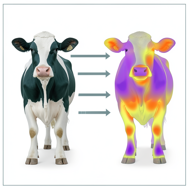
Applications of IRT in Cattle
AI-generated conceptual illustration (Holstein Friesian cows). It explains the idea and does not show study data.
View source evidence ↗Figures & tables
From the original paper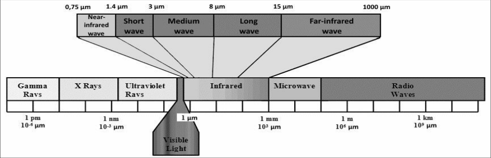 Figure 1
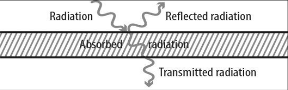 Figure 2
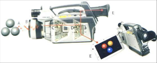 Figure 3
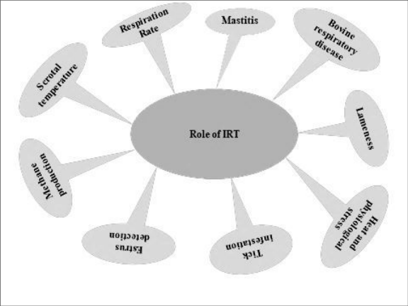 Figure 4
 Table 1
Introduction
Infrared thermography; is an example of a visualization technique that can be used in many different areas of science. Infrared thermography (IRT) is a safe, simple, non-contact, noninvasive disease diagnostics technique with high accuracy and effectiveness in the field of veterinary medicine. The infrared thermographic method has found abundant applications largely to industrial and civil engineering (Tran et al., 2023) [51] , military and police patrolling (Kristo, 2016) [26] , different rescue operation as well as biology (Harrap et al., 2018) [19] , ecology (Still et al., 2019) [48] and human medicine (Kumar et al., 2022) [27] . For the assessment of patho-physiological conditions various invasive methods like rectal temperature measurement, blood collection, respiration, heart and pulse rate measurement are commonly used. These invasive methods require adequate laboratory diagnostic resources and significant time also for reliable result. But Infrared thermography technique can be used very efficaciously in prediction and detection of disease, in research, and in other applications in the field of veterinary within due time (McManus et al., 2022) [35][36] . In veterinary practice infrared thermography can be used by following different ways:
• Diagnostic tool: IRT becomes a physiological imaging method. The difference of 1 °C between two anatomically symmetric regions indicates inflammation. • Physical examination: The possibilities of physical examination thermography can be used to determine doubtful areas where the heat is increased or decreased; • Routine monitoring programs: Animals are monitored on a routine basis weekly. It can be used to detect subclinical problems, as clinical signs occur few days after thermographic changes.
Discovery and background of Infrared thermography
In 1800 British astronomer Sir William Herschel discovered Infrared radiation (IR); he conducted an experimentation evaluating the difference in temperature between the colors in the visible spectrum and found that an invisible radiation in the spectrum lower in energy than the red light. This radiation is termed as infrared radiation; this infrared word comes from the Latin word infra that means below and the English word red. These radiations are that part of the electromagnetic spectrum that is closely adjacent to the red light of approximately. 760 nm on the long-wave side of the visible spectrum. The infrared spectrum is usually divided into near, mid, and far infrared (Sangwan and Balhara, 2022) [44] . Near-
International Journal of Biology Sciences https://www.biologyjournal.net Infrared (NIR) begins just beyond the red portion of the spectrum at 0.7 micrometers to about 3 micrometers. The mid-infrared, consisting of wavelengths from around 3-15 micrometers, contains the region of the spectrum also known as thermal infrared (Figure 1). On the basis of fundamental physics, every object at any given absolute temperature above 0 Kelvin (-273.15˚C) emits radiation (Balhara and Sangwan, 2022) [5] .
The maximum radiant power that can be emitted by any object depends only on the temperature of the object. Therefore, this emitted radiation is called thermal radiation. We experience infrared radiation every day.
The heat that we sunlight, a fire or a radiator is all infrared. The warmer the object, the more infrared radiation it emits. Infrared radiation has its origin from the alteration in the movement of electrons (Sangwan and Balhara, 2022) [44] . Radiation Wave type is, Transverse Wave and has its Speed of 3 ×10⁸ m/s also exhibits the property of refraction.
Absorption and Reflection characteristics of Infrared
Radiation is that they can be absorbed, reflected or transmitted depending on the nature of the surface that it strikes (Figure 2). Incident radiations are all the radiation that strikes an object from its surroundings, of the total radiation incident on a target, a certain proportion will be absorbed (Wα), reflected (Wρ) and transmitted (Wτ).
Hence: Wα + Wρ + Wτ = WINCID = 100% The coefficients α, ρ, and τ describe the object's incident energy absorption (α), reflection (ρ), and transmission (τ).
Each coefficient can have a value from zero to one, depending on how well an object absorbs, reflects, or transmits incident radiation. For example, if ρ = 0, τ = 0, and α = 1, then there is no reflected or transmitted radiation, and 100% of incident radiation is absorbed. This is called a perfect blackbody. In the real world there are no objects that are perfect absorbers, reflectors, or transmitters, although some may come very close to one of these properties. Nonetheless, the concept of a perfect blackbody is very important in the science of thermography, because it is the foundation for relating IR radiation to an object's temperature. Fundamentally, a perfect blackbody is a perfect absorber and emitter of radiant energy. This concept is stated mathematical as Kirchhoff's Law. The radiative properties of a body are denoted by the symbol , the emittance or emissivity of the body. Kirchhoff's law states that = , and since both values vary with the radiation wavelength, the formula can take the form
where denotes the wavelength. The total radiation law can thus take the mathematical form 1 = + + , which for an opaque body ( = 0) can be simplified to 1 = + or = 1 - (i.e., reflection = 1emissivity). Since a perfect blackbody is a perfect absorber, = 0 and = 1.
The radiative properties of a perfect blackbody can also be described mathematically by Planck's Law. Since this has a complex mathematical formula, and is a function of temperature and radiation wavelength, a blackbody's radiative properties are usually shown as a series of curves. These curves show the radiation per wavelength unit and area unit, called the spectral radiant emittance of the blackbody. The higher the temperature, the more intense the emitted radiation. However, each emittance curve has a distinct maximum value at a certain wavelength. This maximum can be calculated from Wien's displacement law,
where T is the absolute temperature of the blackbody, measured in Kelvin (K), and max is the wavelength at the maximum intensity. Using blackbody emittance curves, one can find that an object at 30°C has a maximum near 10μm, whereas an object at 1000 °C has a radiant intensity with a maximum of near 2.3μm. The latter has a maximum spectral radiant emittance about 1400 times higher than a blackbody at 30°C, with a considerable portion of the radiation in the visible spectrum. From Planck's law, the total radiated energy from a blackbody can be calculated. This is expressed by a formula known as the Stefan-Boltzmann law,
where σ is the Stefan-Boltzmann's constant (5.67 × 10 -8 W/m 2 K 4 ), A is the area of surface.
As a rule of thumb, the effective body surface is 1m 2 , and radiates about 0.5kW-a substantial heat loss. The equations described in this section provide important relationships between emitted radiation and temperature of a perfect blackbody. Since most objects of interest to thermographers are not perfect blackbodies, there needs to be some way for an IR camera to graph the temperature of a "normal" object.
For real targets, we must always consider that radiation from two sources exit the object, first is radiation emitted from the object itself (W) and reflected (Wρ) from the surroundings as expressed by the given formula:
Here , is emissivity Emissivity is the ratio of the radiation emitted by a real body, compared to the radiation emitted by a Blackbody, both at the same temperature, and at the same wavelength.
= W RB /W BB
where W RB is the real body radiation and W BB is the black body radiation.
Hence infra-red thermography may be defined as the science of acquisition and analysis of thermal information from noncontact thermal imaging devices. Infrared thermography is the art of transforming an infrared image into a radiometric one, which allows temperature values to be read from the image. So every pixel in the radiometric image is in fact a temperature measurement. In order to do this, complex algorithms are incorporated into the thermal imaging camera.
Infrared thermal camera
Thermal cameras capture infrared energy (heat) and use that energy to create images through digital or analog video outputs. The camera is made up of a lens, a thermal sensor, processing electronics, and a mechanical housing. Infrared energy (A) coming from an object is focused by the optics (B) onto an infrared detector (C). The detector sends the information to sensor electronics (D) for image processing.
The electronics translate the data coming from the detector into an image (E) that can be viewed in the viewfinder or on a standard video monitor or LCD screen (Balhara, 2022) (Fig. 3). It is easy to operate the infrared camera and has very less chances of injury to animal and human (Westermann et al., 2013) [52] . Infrared thermography was initially developed for army and engineering purposes. The first electronic infrared camera was developed by the Germany army and used during the Second World War on tanks for night vision.
Role of IRT in measurement of skin surface temperature in bovines
Thermographic evaluation of temperature distribution within the examined skin surface is useful in quick, realtime non-invasive judgment about the variation in temperature. Thermal cameras capture infrared energy (heat) and use that energy to create images through digital or analog video out puts. Hair is effective insulator of heat; therefore, it is necessary to remove it to obtain reliable measurement of skin temperature in case the animal has high hair density or involves organs with high density of hair. IRT play significant role in the bovine diagnostics sector by means of measurement of skin surface temperature (Figure 4). IRT is supportive in early detection of mastitis (Machado et al., 2021) [32] , determination of the correctness of machine milking (Alejandro et al., 2014) [2] , bovine respiratory viral disease (Lowe et al., 2019) [31] , lameness (Alsaod et al., 2015); evaluation of productivity, including of heat and methane production (Jabbi et al., 2022) and feed efficiency (Lage et al., 2020) [28] , assessment of scrotal temperature for fertility traits ( [11,24] . Preceding studies had shown that the IRT has been used as a tool in the evaluation of breeding soundness in male animals (Menegassi et al., 2018) [37] . It has high sensitivity in determining temperature differences across the skin surface including the scrotal surface temperature (Belay et al., 2016) [7] . A thermal image encompassing infrared emissions could be used to assess scrotal thermoregulation in bull (De Ruediger et al., 2016) [11] . The surface temperature of the scrotum is highly correlated with deep testicular temperature (Kastelic, 2014) [24] and the infrared temperature thermograms provide accurate information about testicular thermoregulation ( [22,11,37] . It has proven that under normal conditions, scrotal surface temperature is 5-6°C lower than abdominal temperature (Menegassi et. al., 2018; Junior et. al., 2015) [37] and there is a positive top-to-bottom gradient on scrotum -top warmer than the bottom (Kastelic and Rizzoto, 2021) [25] and it could be because the scrotum is vascularized from the top to the bottom.
Core body temperature:
The core body temperature is related to the body's internal temperature and it may evaluate from the internal organs, the thorax, the abdomen, central nervous system. Normally mammals have an internal body temperature between 36.5 ˚C to 39.5˚ C and this temperature is comparatively constant. The core body temperature is regulated mainly by reflexes in the body, which is an impulsive motor pattern that the spinal cord or the extended marrow triggers as a respond to a specific sensory stimulus. Core body temperature is an accepted indicator of normal health or illness in animals. Homeothermic animals, under standard physiological conditions, are capable to continue a constant body temperature independently from adjacent environmental temperature. The measurements of internal body temperature play an important role in clinical examination of animal. Various methods are used for assessing core body temperature, by approaching through the rectum (Farrar et al., 2020) [12] , rumen (Lees et al., 2019) [29] , abdominal cavity (Giro et al., 2019) [16] and tympanic membrane of the ear (Schmid et al., 2021) [46] . The use of rectal thermometer is a common method and more standardized practice used by veterinarians and farmers for the evaluation of internal body temperature, although this method has some limitations (Kang et al., 2022) [23] . Usually the rectal temperature obtained by inserting rectal thermometer can be subjective to the presence of fecal material, it can cause stress to animal and may be harmful also and it can be difficult in restless animals (Hall, 2017; Opersteny et al., 2017) [18,40] . Areas of the eye, specifically around the inner corner or the region of lacrimal gland, have large capillary network that respond to alterations in flow of blood resulting in change in temperature (Lokesh Babu et al., 2018) [30] possibly because the proximity of eyes to the thermosensitive site of hypothalamic, which decreases lag time in response. Cows have been reported to have approximately 2 o C lower reading of ocular surface temperature as compared to rectal temperature (Lokesh Babu et al., 2018) [30] . McManus et al. (2022) [35][36] and Hoffmann et al. (2013) [20] reported that the surface temperature of the orbital region can be used to measure the rectal temperature in bovine as an indicator of core body temperature. Likewise, Barros et al. (2016) [6] conclude that the maximum temperature of the orbital area revealed highest correlation (0.58, P<0.0001) to the rectal temperature in buffalo bulls.
Heat and physiological stress: body temperature and rate of respiration are the main physiological parameters which are mainly used for the assessment of heat tolerance and adaptability in animals (Costa et al., 2015) [10] . Nevertheless, proper restraining is required for the evaluation of these parameters and which can causes handling stress. Thus, infrared thermography; a noninvasive and remote sampling tool can be used for the detection of animal stress. Stressful environmental condition correlates with change in body temperature that leads to change in blood flow and this change in blood flow assessed by infrared thermography.
Therefore the temperature at the particular region like eye, ear, dewlap, flank, rump, udder and feet may be used for the prediction of physiological parameter and stress in bovines. (Gabbi et al., 2022, Idris et al., 2021) [14,21] .
Mastitis: is a disease of dairy cow having persistent inflammatory condition in udder parenchyma that is responsible for huge economic losses for the dairy farmers.
It is necessary to detect mastitis in early stage because it responsible for decrease in milk production, milk quality and increase in labor cost, medicine cost. Among various diagnostic test for mastitis like external examination of udder, pH and color milk, somatic cell count, California mastitis test, microbial culture test, infrared thermography has got a potential application (Sathiyabarathi et al., 2016) [45] . Machado et al., 2021) [32] studied daily variation in the udder surface temperature of Holstein-Friesian dairy cows during different stage of lactation by infrared thermography for the detection of mastitis. They conclude that infrared thermography shows potential in its application as an early detection method for mastitis. Machado et al. (2021) [32] conducted an experiment using infrared thermography to evaluate subclinical mastitis and compare the results with California Mastitis Test and Somatic cell count in Brown Swiss dairy cows. The authors found that the udder skin surface temperature was positively correlated with the California Mastitis Test score so they concluded that the infrared thermography can be used as a quick and noninvasive tool for the detection of subclinical mastitis by measuring udder skin surface temperature.
Bovine respiratory disease (BRD):
is the most common disease affecting bovine in all over the world and has significant economic impact in dairy farming. It is usually caused by variety of viral (Adenovirus, Bovine Viral Diarrhea Virus) and bacterial (Pasteurella multocida, Mycoplasma bovis) agents. Lowe et al. (2019) [31] conducted a study to explore the use of infrared thermography in early diagnosis of bovine respiratory disease in weaned calves. They revealed that infrared thermography scans of the eye area in calves were effective as an early indicator of bovine respiratory disease. During bacterial infection there is increase in body temperature and this increased body temperature is evaluated by infrared thermal camera. The observation that radiated heat increment can serve as an early indicator of disease conditions is perhaps logical. Lameness in bovines is one of the most serious welfare issues but it is over-looked in dairy industry. According to Schulz et al. (2016) [47] duration of laying bouts and locomotion scoring may be used diagnosis of lameness in dairy cows. Traditional methods for the diagnosis of lameness are change in locomotion of animal and lifting the foot in a claw trimming chute. Basically, these are time consuming approaches so infrared thermography is used as non-invasive, non-contact diagnostic tool (Ramirez-GarciaLuna et al., 2022) [42] . The inflammation or injury accompanied with limb lesions have change in blood flow that leads to change in skin surface temperature and the thermographs can be used to recognize these thermal abnormalities in animals (Alsaaod et al., 2015) [3] . The healthy hoof has lower surface temperature than the hoof with a lesion (Bobic et al., 2017) [8] . The coronary band and the skin surrounding this region has been used for the measurement of foot surface temperature because; (a) the coronary band has very much blood supply so any foot lesion or inflammation may be recognized through raised coronary band temperature, (b) during examination thermograph of coronary band and surrounding skin can be obtained with minimum stress and without physical contact of animal. In 2018, Gianesella et al. 2018 [15] conducted an experiment to investigate the potential role of using infrared thermography for detection of hoof lesions. The thermographs were analysed before and after claw trimming with respect to the temperature of skin and coronary band, and there was difference in temperature between skin and coronary band of the hind limbs. The authors accomplished that the infrared thermography may be used for the detection and prevention of hoof lesions. Wood et al. (2015) [53] conducted an experiment whether infrared thermography could be used to detect feet with digital dermatitis. They revealed that there is increase in feet temperature related to feet having pathological lesions and suggested that thermography could be used as a monitoring tool for limb and hoof lesion. As a summary these findings may be tabulated as follow: [8] Methane production: Emission of methane from livestock is through enteric fermentation and manure management. Total 8% of metabolic energy is lost through methane production in ruminant animals. Hence, by improving the efficiency of feed utilization by ruminant animals makes livestock industry more economic and environmentally sustainable. Calculation of residual feed intake is an important parameter for the assessment of feed efficiency but it is costly and laborious to measure residual feed intake (Fitzsimons et al., 2014) [13] . Therefore, livestock industry requires an alternate method for the assessment of feed efficiency. Infrared thermography can be effectively used for evaluating methane and heat production (Gabbi et al., 2022) [14] . The authors conducted a trail with four lactating Holstein dairy cattle, the gas was measured by open-circuit indirect calorimetry system and ThermaCamTM SC2000; FLIR Systems, infrared camera was used for thermography. The images were taken from fore feet, left side of rump, left and right flank area. In ruminant heat production and energy loss is corelated with methane production during microbial fermentative process (Morgavi et al., 2023) [38] . The skin surface temperature depends on the body's internal circulatory activities and the tissue metabolic rate. So, the differences in the skin surface temperature captured by infrared thermography can be hypothetically used to measure the heat production and methane emission in ruminants (Guadagnin et al., 2023) [17] .
Estrus detection: incompetent detection of estrus in bovine causes lessening in reproductive efficiency and milk production where artificial insemination is practice (Reith and Hoy 2018) [43] . Several methods can be used for the detection of estrous viz. mounting activity, visual observation, variations in body temperature, alteration in vaginal mucus membrane. For measuring vaginal or rectal temperature and using implant within reproductive organs animals need to be restrain and this may cause animal stress that may change in body temperature. So, infrared thermography is an alternate and safe diagnostic technique with minimal discomfort (Racewicz et al., 2018) [41] . Talukder et al. (2014) [49] reported that infrared thermography has potential in estrus detection when image was taken from vulval and muzzle surface.
Tick infestation: ticks are most significant ectoparasites of livestock that are accountable for large amount of economic losses either blood sucking parasite or a vector of toxin and pathogens. Heavy load of tick infestation leads to anemia, reduced body weight, and poor quality of hides (Naseem et al., 2023) [39] . Usually, there are temperature variations between the surface of the animal and the surface of the parasite. Thus, this temperature variation is used to detect external parasite load in animals using infrared thermal camera (Cortivo et al., 2016) [9] .
Conclusion
Infrared thermography (IRT) is a new emerging, noninvasive and lowcost technique of thermal profile visualization. Each living and non-living object on earth produce radiation in the infrared part of the light spectrum and the intensity of these radiation depends on the temperature of its surface. IRT of animals comprises the measurement of the body surface temperature. In warmblooded animals, the temperature values obtained in IRT depend on the impact of animal's thermoregulatory response because the change in body temperature depends on change in blood flow. In addition to the volume of blood perfusion, skin temperature depends on the intensity of tissue metabolism, the type and color of the hair coat, and the thickness of adipose tissue. Thermographic technique has found abundant advantages not only in military and industries, but also in human and veterinary medicine, predominantly for diagnostic purposes. The thermographic approach can be effectively used to evaluate the pathophysiological condition of different systems of animals. The infrared thermography has been used as a tool in the evaluation of breeding soundness in male animals and assessment of health conditions in dairy cattle.
Figures and tables from the paper
| Type of animals | Type of infrared camera | Type of foot lesion observed | Area of animal | Author |
|---|---|---|---|---|
| Dairy cows | FLIR Inframetric | Healthy hooves | Hooves | Gianesella et al., 2018 [15] |
| Cattle | TIR1 imager | Foot-and-mouth disease | Feet and Eye | Lokesh Babu et al., 2018 [30] |
| Dairy cows | Ti25 Thermal Imager | Digital dermatitis | Foot | Alsaaod et al., 2015 [3] |
| Dairy cows | FLIR and 7 | Sole ulcer, inter- digital hyperplasia, dermatitis digitalis | Hooves | Bobic et al., 2017 [8] |
Table text extracted from the original PDF; superscript letters are shown inline. The original table image is below.
View original table image
References
54 in the original paper- Ahirwar MK, Kataktalware MA, Ramesha KP, Pushpadass HA, Jeyakumar S, et al. Influence of season, age and management on scrotal thermal profile in Murrah bulls using scrotal infrared digital thermography. International Journal of Biometeorology. 2017;61(12):2119-2125. DOI:10.1007/s00484-0171415-0.
- Alejandro M, Romero G, Sabater JM, Díaz JR. Infrared thermography as a tool to determine teat tissue changes caused by machine milking in Murciano-Granadina goats. Livestock Science. 2014;160:178-185. DOI:10.1016/j.livsci.2013.11.029.
- Alsaaod M, Schaefer AL, Büscher W, Steiner A. The role of infrared thermography as a non-invasive tool for the detection of lameness in cattle. Sensors. 2015;15(6):14513-14525. DOI:10.3390/s150614513.
- Balhara AK. Infrared measurement techniques. In: Infrared thermal imaging in animal research, health and welfare. ICAR-CIRB; 2022. p. 21-31.
- Balhara S, Sangwan S. Basic thermal science. In: Infrared thermal imaging in animal research, health and welfare. ICAR-CIRB; 2022. p. 6-11.
- Barros DV, Silva LK, Kahwage PR, Lourenço Júnior JD, Sousa JS, et al. Assessment of surface temperatures of buffalo bulls (Bubalus bubalis) raised under tropical conditions using infrared thermography. Arquivo Brasileiro de Medicina Veterinaria e Zootecnia. 2016;68:422-430. DOI:10.1590/1678-4162-8327.
- Belay RE, Huang GO, Shen JK, Ko EY. Diagnosis of clinical and subclinical varicocele: how has it evolved? Asian Journal of Andrology. 2016;18(2):182. DOI:10.4103/1008-682X.169991.
- Bobic T, Mijić P, Gregić M, Bagarić A, Gantner V. Early detection of the hoof diseases in Holstein cows using thermovision camera. Agriculturae Conspectus Scientificus. 2017;82(2):197-200.
- Cortivo PD, Dias E, Barcellos JO, Peripolli V, Costa JB Jr, et al. Use of thermographic images to detect external parasite load in cattle. Computers and Electronics in Agriculture. 2016;127:413-417. DOI:10.1016/j.compag.2016.07.002.
- Costa AN, Feitosa JV, Montezuma PA, de Souza PT, de Araújo AA. Rectal temperatures, respiratory rates, production, and reproduction performances of crossbred Girolando cows under heat stress in northeastern Brazil. International Journal of Biometeorology. 2015;59:1647-1653. DOI:10.1007/s00484-015-0971-4.
- De Ruediger FR, Chacur MG, Alves FC, Oba E, de Amorim Ramos A. Digital infrared thermography of the scrotum, semen quality, serum testosterone levels in Nellore bulls (Bos taurus indicus) and their correlation with climatic factors. Semina: Ciencias Agrarias. 2016;37(1):221-232. DOI:10.5433/16790359.2016v37n1p221.
- Farrar KL, Field AE, Norris SL, Jacobsen KO. Comparison of rectal and infrared thermometry temperatures in anesthetized swine (Sus scrofa). Journal of the American Association for Laboratory Animal Science. 2020;59(2):221-225. DOI:10.30802/AALASJAALAS-19-000119.
- Fitzsimons C, Kenny DA, McGee M. Visceral organ weights, digestion and carcass characteristics of beef bulls differing in residual feed intake offered a high concentrate diet. Animal. 2014;8(6):949-959. DOI:10.1017/S1751731114000652.
- Gabbi AM, Kolling GJ, Fischer V, Pereira LGR, Tomich TR. Use of infrared thermography to estimate enteric methane production in dairy heifers. International Journal of Biology Sciences https://www.biologyjournal.net Quantitative Infrared Thermography Journal. 2022;19(3):187-195. DOI:10.1080/17686733.2021.1882075.
- Gianesella M, Arfuso F, Fiore E, Giambelluca S, Giudice E, et al. Infrared thermography as a rapid and non-invasive diagnostic tool to detect inflammatory foot diseases in dairy cows. Polish Journal of Veterinary Sciences. 2018;21(2):298-305. DOI:10.24425/122597.
- Giro A, de Campos Bernardi AC, Junior WB, Lemes AP, Botta D, et al. Application of microchip and infrared thermography for monitoring body temperature of beef cattle kept on pasture. Journal of Thermal Biology. 2019;84:121-128. DOI:10.1016/j.jtherbio.2019.06.009.
- Guadagnin AR, Matiello JP, Ribeiro RS, Pereira LG, Machado FS, et al. Assessment of heat production and methane emission using infrared thermography in lactating Holstein and Gyrolando-F1 cows. Journal of Thermal Biology. 2023;115:103628. DOI:10.1016/j.jtherbio.2023.103628.
- Hall EJ, Carter AJ. Comparison of rectal and tympanic membrane temperature in healthy exercising dogs. Comparative Exercise Physiology. 2017;13(1):37-44. DOI:10.3920/CEP160034.
- Harrap MJ, Hempel de Ibarra N, Whitney HM, Rands SA. Reporting of thermography parameters in biology: a systematic review of thermal imaging literature. Royal Society Open Science. 2018;5(12). DOI:10.1098/rsos.181281.
- Hoffmann G, Schmidt M, Ammon C, Rose-Meierhöfer S, Burfeind O. Monitoring the body temperature of cows and calves using video recordings from an infrared thermography camera. Veterinary Research Communications. 2013;37:91-99. DOI:10.1007/s11259-012-9549-3.
- Idris M, Uddin J, Sullivan M, McNeill DM, Phillips CJ. Non-invasive physiological indicators of heat stress in cattle. Animals. 2021;11(1):71. DOI:10.3390/ani11010071.
- Indriastuti R, Ulum MF, Arifiantini RI, Memili E, Purwantara B. Relationship among body weight, scrotal circumference and sperm quantity of Bali bulls in Baturiti Artificial Insemination Center. IOP Conference Series: Earth and Environmental Science. 2020;478:012004. DOI:10.1088/17551315/478/1/012004.
- Kang H, Sole-Guitart A, Mellor VA, Gaughan JB, Zsoldos RR. The use of percutaneous thermal sensing microchips for non-invasive measurement of body temperature in foals during summer seasons in a subtropical region. Animal. 2022;16(9):100620. DOI:10.1016/j.animal.2022.100620.
- Kastelic JP. Understanding and evaluating bovine testes. Theriogenology. 2014;81(1):18-23. DOI:10.1016/j.theriogenology.2013.09.001.
- Kastelic JP, Rizzoto G. Thermoregulation of the testes. In: Bovine reproduction. 2021. p. 40-46. DOI:10.1002/9781119602484.ch4.
- Kristo M. Review of methods for the surveillance and access control using the thermal imaging system. Review of Innovation and Competitiveness: Journal of Economic and Social Measurement. 2016;2(4):71-91.
- Kumar P, Gaurav A, Rajnish RK, Sharma S, Kumar V, et al. Applications of thermal imaging with infrared thermography in orthopaedics. Journal of Clinical Orthopaedics and Trauma. 2022;24. DOI:10.1016/j.jcot.2021.101722.
- Lage CF, Coelho SG, Diniz Neto HC, Malacco VM, Rodrigues JP, et al. Relationship between feed efficiency indexes and thermography, blood, and ruminal parameters in pre-weaning dairy heifers. PLoS One. 2020;15(7). DOI:10.1371/journal.pone.0236118.
- Lees AM, Sejian V, Lees JC, Sullivan ML, Lisle AT, et al. Evaluating rumen temperature as an estimate of core body temperature in Angus feedlot cattle during summer. International Journal of Biometeorology. 2019;63(7):939-947. DOI:10.1007/s00484-019-017060.
- Lokesh Babu DS, Jeyakumar S, Vasant PJ, Sathiyabarathi M, Manimaran A, et al. Monitoring foot surface temperature using infrared thermal imaging for assessment of hoof health status in cattle: a review. Journal of Thermal Biology. 2018;78:10-21. DOI:10.1016/j.jtherbio.2018.08.021.
- Lowe G, Sutherland M, Waas J, Schaefer A, Cox N, et al. Infrared thermography-A non-invasive method of measuring respiration rate in calves. Animals. 2019;9(8):535. DOI:10.3390/ani9080535.
- Machado NA, Da Costa LB, Barbosa-Filho JA, De Oliveira KP, De Sampaio LC, et al. Using infrared thermography to detect subclinical mastitis in dairy cows in compost barn systems. Journal of Thermal Biology. 2021;97:102881. DOI:10.1016/j.jtherbio.2021.102881.
- Marquez HP, Ambrose DJ, Schaefer AL, Cook NJ, Bench CJ. Infrared thermography and behavioral biometrics associated with estrus indicators and ovulation in estrus-synchronized dairy cows housed in tie stalls. Journal of Dairy Science. 2019;102(5):44274440. DOI:10.3168/jds.2018-15221.
- Marquez HP, Ambrose DJ, Schaefer AL, Cook NJ, Bench CJ. Evaluation of infrared thermography combined with behavioral biometrics for estrus detection in naturally cycling dairy cows. Animal. 2021;15(7):100205. DOI:10.1016/j.animal.2021.100205.
- McManus R, Boden LA, Weir W, Viora L, Barker R. Thermography for disease detection in livestock: a scoping review. Frontiers in Veterinary Science. 2022;9:965622. DOI:10.3389/fvets.2022.965622.
- McManus R, Boden LA, Weir W, Viora L, Barker R, Kim Y, et al. Thermography for disease detection in livestock: a scoping review. Frontiers in Veterinary Science. 2022;9:965622. DOI:10.3389/fvets.2022.965622.
- Menegassi SR, Pereira GR, Dias EA, Rocha MK, Carvalho HR. Infrared thermography as a noninvasive method to assess scrotal insulation on sperm production in beef bulls. Andrologia. 2018;50(3):e12904. DOI:10.1111/and.12904.
- Morgavi DP, Cantalapiedra-Hijar G, Eugène M, Martin C, Nozière P, et al. Reducing enteric methane emissions improves energy metabolism in livestock: is the tenet right? Animal. 2023:100830. DOI:10.1016/j.animal.2023.100830.
- Naseem MN, Raza A, Kamran M, Allavena R, Constantinoiu C. The development of cutaneous lesions in tropically adapted beef cattle is associated with hypersensitive immune response to buffalo fly antigens. Animals. 2023;13(12):2011. DOI:10.3390/ani13122011.
- Opersteny E, Anderson H, Bates J, Davenport K, Husby J, et al. Precision, sensitivity and patient preference of non-invasive thermometers in a pediatric surgical acute care setting. Journal of Pediatric Nursing. 2017;35:3641.
- Racewicz P, Sobek J, Majewski M, Różańska-Zawieja J. The use of thermal imaging measurements in dairy cow herds. Animal Science and Genetics. 2018;14(1):55-69.
- Ramirez-GarciaLuna JL, Bartlett R, Arriaga-Caballero JE, Fraser RD, Saiko G. Infrared thermography in wound care, surgery, and sports medicine: a review. Frontiers in Physiology. 2022;13:838528. DOI:10.3389/fphys.2022.838528.
- Reith S, Hoy S. Behavioral signs of estrus and the potential of fully automated systems for detection of estrus in dairy cattle. Animal. 2018;12(2):398-407. DOI:10.1017/S1751731117001975.
- Sangwan S, Balhara S. The infrared thermal light. In: Infrared thermal imaging in animal research, health and welfare. ICAR-CIRB; 2022. p. 1-5.
- Sathiyabarathi M, Jeyakumar S, Manimaran A, Pushpadass HA, Sivaram M, et al. Investigation of body and udder skin surface temperature differentials as an early indicator of mastitis in Holstein Friesian crossbred cows using digital infrared thermography technique. Veterinary World. 2016;9(12):1386-1391. DOI:10.14202/vetworld.2016.1386-1391.
- Schmid SM, Büscher W, Steinhoff-Wagner J. Suitability of different thermometers for measuring body core and skin temperatures in suckling piglets. Animals. 2021;11(4):1004. DOI:10.3390/ani11041004.
- Schulz T, Gundelach Y, Feldmann M, Hoedemaker M. Early detection and treatment of lame cows. Tierärztliche Praxis Ausgabe G: Großtiere/Nutztiere. 2016;44(1):5-11. DOI:10.15653/TPG-150534.
- Still C, Powell R, Aubrecht D, Kim Y, Helliker B, et al. Thermal imaging in plant and ecosystem ecology: applications and challenges. Ecosphere. 2019;10(6). DOI:10.1002/ecs2.2768.
- Talukder S, Kerrisk KL, Ingenhoff L, Thomson PC, Garcia SC, et al. Infrared technology for estrus detection and as a predictor of time of ovulation in dairy cows in a pasture-based system. Theriogenology. 2014;81(7):925-935. DOI:10.1016/j.theriogenology.2014.01.009.
- Teixeira VA, Coelho SG, Tomich TR, Rodrigues JP, Campos MM, et al. Reproductive characteristics of bulls from two breed compositions and their correlations with infrared thermography. Journal of Thermal Biology. 2019;85:102407. DOI:10.1016/j.jtherbio.2019.102407.
- Tran QH, Dang QM, Pham XT, Truong TC, Nguyen TX, et al. Passive infrared thermography technique for concrete structures health investigation: case studies. Asian Journal of Civil Engineering. 2023:1-9. DOI:10.1007/s42107-023-00571-y.
- Westermann S, Buchner HH, Schramel JP, Tichy A, Stanek C. Effects of infrared camera angle and distance on measurement and reproducibility of thermographically determined temperatures of the distolateral aspects of the forelimbs in horses. Journal of the American Veterinary Medical Association. 2013;242(3):388-395. DOI:10.2460/javma.242.3.388.
- Wood S, Lin Y, Knowles TG, Main DJ. Infrared thermometry for lesion monitoring in cattle lameness. Veterinary Record. 2015;176(12):308. DOI:10.1136/vr.102571.
- Yadav SK, Singh P, Kumar P, Singh SV, Singh A, et al. Scrotal infrared thermography and testicular biometry: indicator of semen quality in Murrah buffalo bulls. Animal Reproduction Science. 2019;209:106145. DOI:10.1016/j.anireprosci.2019.106145.