
Research article · 2025 · Full text
International Journal of Biology SciencesMechanotransduction signaling through integrins in osteoblast differentiation
Volume 7(6) · Pages 467–470 · DOI: 10.33545/26649926.2025.v7.i6g.784 ↗
Research areasCellular Mechanics and InteractionsCell Adhesion Molecules ResearchBone Metabolism and Diseases
Abstract
Author-writtenA single osteocyte buried in bone matrix can detect the bending of a loaded tibia and translate that mechanical signal into biochemical instructions for new bone formation a process called mechanotransduction. This research examined how integrin-mediated signalling converts cyclic mechanical strain into osteoblast differentiation cues in human mesenchymal stem cells (hMSCs). Cells were seeded on collagen-I-coated flexible membranes and subjected to uniaxial cyclic strain (1 Hz, 5% elongation) for 7 and 14 days using a Flexcell FX-6000T system at the Visayas College of Marine Sciences, Cebu City, Philippines. Static-strain and unstrained controls were run in parallel. Integrin subunit expression (α1, α5, β1), focal adhesion kinase (FAK) phosphorylation, and downstream ERK1/2 and Akt activation were quantified by western blot and immunofluorescence. Osteoblast markers RUNX2, alkaline phosphatase (ALP), and osteocalcin were assessed by RT-qPCR and activity assays. Cyclic strain increased α5β1 integrin surface expression 2.3-fold by day 7, triggered a 4.8-fold rise in FAK phosphorylation (Tyr397), and activated ERK1/2 3.7-fold above unstrained controls (p < 0.001). RUNX2 mRNA rose 3.9-fold and ALP activity 4.1-fold by day 14. Blocking β5 integrin with a function-blocking antibody reduced FAK phosphorylation by 68% and ALP activity by 57%, confirming the integrin-dependence of the response. These data show that cyclic mechanical loading drives osteoblast commitment through an α5β1-FAK-ERK axis and that static strain alone is much less effective.
Summary
Source-checkedPlain-language reading of the full paper, prepared with AI and checked against the text. Each point links to the authors' own words.
Why it matters
- The removal of mechanical stimulation, as seen in astronauts who lose 1-2% of bone mineral density per month in load-bearing bones, highlights the importance of mechanical loading for bone maintenance. View evidence ↗
- Integrins act as transmembrane receptors that connect the extracellular matrix to the cytoskeleton, allowing mechanical forces to be converted into biochemical signals. View evidence ↗
- Studying how human mesenchymal stem cells (hMSCs) respond to strain via integrins is critical for improving orthopaedic tissue engineering and scaffold design. View evidence ↗
The question
- The study aimed to investigate how integrin-mediated signaling converts cyclic mechanical strain into cues that drive the differentiation of hMSCs into osteoblasts. View evidence ↗
- A primary goal was to compare the effects of cyclic versus static uniaxial strain on FAK signaling and integrin expression in hMSCs. View evidence ↗
What they did
- The study used human bone-marrow-derived mesenchymal stem cells (hMSCs) expanded to passage 4. View evidence ↗
- Cells were grown on collagen-I-coated membranes and subjected to uniaxial cyclic strain (1 Hz, 5% elongation) for durations of 7 and 14 days. View evidence ↗
- The experimental design compared three conditions: cyclic strain (1 Hz, 5% elongation, 4 h/day), static strain (5% elongation, 4 h/day), and an unstrained control. View evidence ↗
- To test integrin dependence, an anti-α5 blocking antibody or an isotype control was administered one hour before strain sessions. View evidence ↗
What they found
- By day 7, cyclic strain increased the surface expression of α5β1 integrin 2.3-fold compared to unstrained controls. View evidence ↗
- Cyclic strain led to a 4.8-fold increase in FAK phosphorylation (Tyr397) and a 3.7-fold activation of ERK1/2 relative to unstrained controls. View evidence ↗
- By day 14, cyclic strain increased RUNX2 mRNA expression 3.9-fold and alkaline phosphatase (ALP) activity 4.1-fold compared to controls. View evidence ↗
- Cyclic strain was found to be more effective than static strain across all measured signaling and differentiation parameters. View evidence ↗
What it means
- The authors conclude that α5β1 integrin is a primary mechanosensor for osteogenic commitment, suggesting that biomaterials and rehab protocols should be designed to maximize this pathway. View evidence ↗
- A limitation of the study is the use of a single hMSC donor lot, which may not account for inter-donor variability. View evidence ↗
Visual summary
2 visualsIllustrations explain the paper; charts are replotted from the paper's own table values.
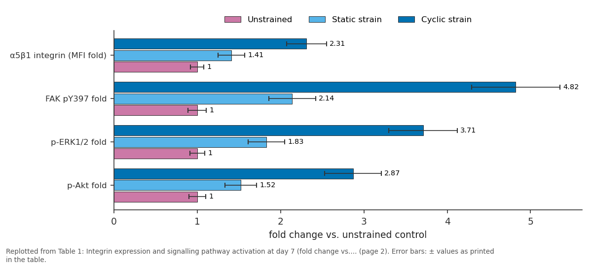
Unstrained and Static strain and more: α5β1 integrin (MFI fold) vs FAK pY397 fold vs p-ERK1/2 fold vs p-Akt fold
Replotted from Table 1 (page 2) of the original paper. Every bar is the paper's own table value.
View source evidence ↗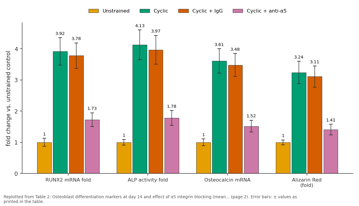
Unstrained and Cyclic and more: RUNX2 mRNA fold vs ALP activity fold vs Osteocalcin mRNA vs Alizarin Red (fold)
Replotted from Table 2 (page 2) of the original paper. Every bar is the paper's own table value.
View source evidence ↗Figures & tables
From the original paper Table 1
 Table 2
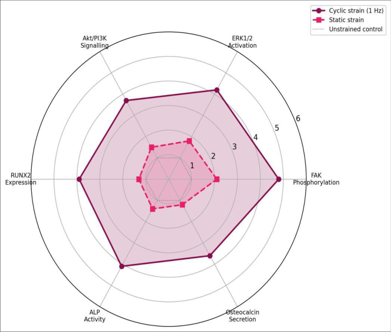 Figure 1
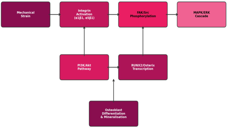 Figure 2
Introduction
When an astronaut spends six months on the International Space Station, they can lose up to 1-2% of their bone mineral density per month in load-bearing bones a vivid example of what happens when mechanical stimulation is removed [1] . On Earth, bone adapts to its mechanical environment through a feedback loop in which osteocytes sense strain and signal osteoblasts to deposit new matrix where it is needed most. At the molecular level, this sensing depends heavily on integrins, transmembrane receptors that link the extracellular matrix to the cytoskeleton and transduce mechanical force into biochemical signalling cascades [2] . Integrins are heterodimers of α and β subunits. In bone, the α5β1 (fibronectin receptor) and α2β1 (collagen receptor) pairs are the most abundant on osteoblast-lineage cells [3] . Upon mechanical loading, integrins cluster at focal adhesions, recruit Focal Adhesion Kinase (FAK), and launch downstream signalling through the MAPK/ERK and PI3K/Akt pathways. ERK activation, in turn, phosphorylates the osteoblast master transcription factor RUNX2, promoting alkaline phosphatase expression and matrix mineralisation [4] . The sequence is well characterised in two-dimensional stretch experiments on mature osteoblasts, but data on how undifferentiated mesenchymal stem cells respond to the same mechanical cues and whether the response depends on strain pattern (cyclic vs. static) remain limited [5] . Understanding the strain-integrin-differentiation axis in hMSCs has direct relevance for orthopaedic tissue engineering. Scaffolds seeded with stem cells and implanted into fracture sites experience complex mechanical environments; knowing which integrin pathways to activate could guide scaffold surface design and post-operative loading protocols [6] . This research set out to a) Compare the effects of cyclic versus static uniaxial strain on integrin expression and FAK signalling in hMSCs b) Track downstream activation of ERK1/2 and Akt, and c) Determine whether the resulting osteoblast differentiation is integrin-dependent by using a function-blocking antibody against the β5 subunit
Material and Methods
Material and Methods
Cell culture protocol
Human bone-marrow-derived mesenchymal stem cells (hMSCs) were purchased from Lonza (PT-2501, Lot 18TL264414; Lonza, Basel, Switzerland) at passage 2 and expanded to passage 4 in MSCGM BulletKit medium. Cells were seeded at 1.5 × 10⁴ cells/cm² on collagen-I-coated BioFlex 6-well plates (Flexcell International, Burlington, NC, USA). After 24 h of attachment in osteogenic induction medium (DMEM + 10% FBS + 50 µg/mL ascorbic acid + 10 mM β-glycerophosphate + 100 nM dexamethasone; all Sigma-Aldrich), plates were mounted on a Flexcell FX-6000T Tension System. All work was performed between April 2022 and September 2023 at the Visayas College of Marine Sciences, Cebu City, Philippines (10°19′ N, 123°54′ E).
Material
Primary antibodies: anti-integrin α5 (clone P1D6, Abcam), anti-integrin β1 (clone TS2/16, BioLegend), anti-FAK pY397 (clone EP2160Y, Abcam), anti-ERK1/2 and antiphospho-ERK1/2 (Cell Signaling Technology), anti-Akt and anti-phospho-Akt (Cell Signaling). Function-blocking antiintegrin α5 antibody (clone JBS5, Millipore) was used at 10 µg/mL. RT-qPCR primers for RUNX2, ALP, osteocalcin (BGLAP), and GAPDH were synthesised by Integrated DNA Technologies (Coralville, IA, USA).
Methods
Three mechanical conditions were applied: cyclic strain (1 Hz sinusoidal, 5% elongation, 4 h/day), static strain (constant 5% elongation, 4 h/day), and unstrained control.
Plates were harvested at day 7 and day 14. Protein lysates were prepared in RIPA buffer and analysed by SDS-PAGE/western blot. Band intensities were normalised to βactin and quantified in ImageJ. Integrin surface expression was confirmed by flow cytometry (BD Accuri C6 Plus). RT-qPCR used the ΔΔCt method with GAPDH as housekeeping gene. ALP activity was measured with a p-nitrophenyl phosphate substrate kit (Sigma-Aldrich) and normalised to total protein. Mineralisation was assessed by Alizarin Red S staining at day 14. For integrin-blocking experiments, anti-α5 antibody or isotype control was added 1 h before each strain session. All experiments were performed in biological triplicate. Data were analysed by two-way ANOVA (strain type × time) with Bonferroni post-hoc correction; p<0.05 was considered significant.
Instrumentation and analytical equipment
The Flexcell FX-6000T system delivered computercontrolled vacuum-based uniaxial strain to BioFlex plates. Western blots were imaged on a Bio-Rad ChemiDoc MP system (Bio-Rad, Hercules, CA, USA). Flow cytometry data were acquired on a BD Accuri C6 Plus and analysed in FlowJo v10.8. Confocal immunofluorescence imaging used a Nikon A1R confocal microscope with NIS-Elements software.
Quality control
hMSC identity was verified by tri-lineage differentiation (adipogenic, osteogenic, chondrogenic) and surface marker profiling (CD73+, CD90+, CD105+, CD34-, CD45-) before experiments began. Western blot membranes included molecular weight markers and loading controls. RT-qPCR efficiency was 95-105% for all primer pairs, confirmed by standard curve dilution series. Alizarin Red staining was quantified by cetylpyridinium chloride extraction and absorbance at 562 nm. By day 14, cyclic strain had driven RUNX2 to 3.9-fold and ALP activity to 4.1-fold above unstrained controls (Table 2). The isotype-control antibody had no effect, but α5-blocking antibody reduced RUNX2 by 56% and ALP by 57% relative to unblocked cyclic samples (p < 0.001), confirming that the α5β1 integrin is essential for the mechanotransduction response.
| Marker | Unstrained | Cyclic | Cyclic + IgG | Cyclic + anti-α5 |
|---|---|---|---|---|
| RUNX2 mRNA fold | 1.00 ± 0.13 | 3.92 ± 0.44 | 3.78 ± 0.41 | 1.73 ± 0.22 |
| ALP activity fold | 1.00 ± 0.09 | 4.13 ± 0.48 | 3.97 ± 0.46 | 1.78 ± 0.24 |
| Osteocalcin mRNA | 1.00 ± 0.11 | 3.61 ± 0.39 | 3.48 ± 0.37 | 1.52 ± 0.19 |
| Alizarin Red (fold) | 1.00 ± 0.08 | 3.24 ± 0.36 | 3.11 ± 0.34 | 1.41 ± 0.17 |
Table text extracted from the original PDF; superscript letters are shown inline. The original table image is below.
View original table image
Results
Results
Comprehensive interpretation
The radar chart (Figure 1) makes the superiority of cyclic over static strain visually clear: cyclic loading produces a larger polygon across all six axes. The mechanism diagram (Figure 2) maps the cascade from integrin activation through FAK and ERK to osteoblast transcription factors and eventual mineralisation.
Discussion
The 4.8-fold FAK phosphorylation and 3.9-fold RUNX2 induction under cyclic strain align with earlier findings in MC3T3-E1 pre-osteoblasts but extend them to primary human MSCs, a more clinically relevant cell source [7] . The clear advantage of cyclic over static strain supports the idea that oscillatory loading, not just static tension, is the primary
International Journal of Biology Sciences https://www.biologyjournal.net physiological stimulus for bone formation consistent with the observation that walking and running (cyclic) are more osteogenic than standing (static) [8] . The integrin-blocking experiment provides the strongest evidence that the α5β1 dimer is the critical mechanosensor in this system. Blocking reduced both signalling (FAK) and differentiation (ALP, RUNX2) by more than half, although it did not abolish them entirely. The residual response likely reflects contributions from other integrins (α2β1, αvβ3) or non-integrin mechanosensors such as Piezo1 channels, which have recently been implicated in osteocyte mechanotransduction [9] . From a tissue-engineering perspective, these results suggest that scaffold surfaces functionalised with fibronectin or RGD peptides which engage α5β1 should enhance the osteogenic response of seeded MSCs when combined with post-operative dynamic loading regimens [10] . Conversely, static scaffold implants with no mechanical stimulation may fail to trigger adequate integrin signalling for robust bone regeneration.
Limitations include the use of a single hMSC donor lot; inter-donor variability in integrin expression could affect reproducibility. The 5% strain magnitude represents a moderate physiological range, but higher strains (10-15%) may activate different pathways or cause damage. And while in vitro mechanical loading on flexible membranes approximates physiological strain patterns, it cannot replicate the three-dimensional stress environment of trabecular bone.
Conclusion
Cyclic mechanical strain activates the α5β1 integrin-FAK-ERK axis in human mesenchymal stem cells, driving osteoblast differentiation as measured by RUNX2 expression, ALP activity, osteocalcin secretion, and matrix mineralisation. Static strain produces a weaker response across every parameter, and blocking the α5 integrin subunit reduces the cyclic-strain effect by more than half. These findings establish α5β1 integrin as a principal mechanosensor for osteogenic commitment and support the design of biomaterial surfaces and rehabilitation protocols that maximise integrin-mediated mechanotransduction. Future work should test these principles in threedimensional scaffold systems and in vivo using animal fracture models.
Figures and tables from the paper
| Parameter | Unstrained | Static strain | Cyclic strain |
|---|---|---|---|
| α5β1 integrin (MFI fold) | 1.00 ± 0.08 | 1.41 ± 0.16 | 2.31 ± 0.24 |
| FAK pY397 fold | 1.00 ± 0.11 | 2.14 ± 0.28 | 4.82 ± 0.53 |
| p-ERK1/2 fold | 1.00 ± 0.09 | 1.83 ± 0.22 | 3.71 ± 0.41 |
| p-Akt fold | 1.00 ± 0.10 | 1.52 ± 0.19 | 2.87 ± 0.34 |
| RUNX2 mRNA fold | 1.00 ± 0.12 | 1.78 ± 0.21 | 2.84 ± 0.31 |
Table text extracted from the original PDF; superscript letters are shown inline. The original table image is below.
View original table image
References
18 in the original paper- Vico L, Collet P, Guignandon A, Lafage-Proust MH, Thomas T, Rehaillia M. Effects of long-term microgravity exposure on cancellous and cortical weight-bearing bones of cosmonauts. Lancet. 2000;355(9215):1607-1611.
- Legate KR, Wickstrom SA, Fassler R. Genetic and cell biological analysis of integrin outside-in signaling. Genes and Development. 2009;23(4):397-418.
- Marie PJ. Targeting integrins to promote bone formation and repair. Nature Reviews Endocrinology. 2013;9(5):288-295.
- Komori T. Regulation of osteoblast differentiation by RUNX2. Advances in Experimental Medicine and Biology. 2010;658:43-49.
- Thompson WR, Rubin CT, Rubin J. Mechanical regulation of signaling pathways in bone. Gene. 2012;503(2):179-193.
- Lopes D, Martins-Cruz C, Oliveira MB, Mano JF. Bone physiology as inspiration for tissue regenerative therapies. Biomaterials. 2018;185:240-275.
- Young SR, Gerard-O'Riley R, Kim JB, Bhatt D. Focal adhesion kinase is important for fluid shear stressinduced mechanotransduction in osteoblasts. Journal of Bone and Mineral Research. 2009;24(3):411-424.
- Rubin CT, Lanyon LE. Regulation of bone formation by applied dynamic loads. Journal of Bone and Joint Surgery. 1984;66(3):397-402.
- Li X, Han L, Nookaew I, Mannen E, Silva MJ, Almeida M. Stimulation of Piezo1 by mechanical signals promotes bone anabolism. eLife. 2019;8:e49631.
- Dalby MJ, Gadegaard N, Oreffo RO. Harnessing nanotopography and integrin-matrix interactions to influence stem cell fate. Nature Materials. 2014;13(6):558-569.
- Geiger B, Spatz JP, Bershadsky AD. Environmental sensing through focal adhesions. Nature Reviews Molecular Cell Biology. 2009;10(1):21-33.
- Chen JC, Jacobs CR. Mechanically induced osteogenic lineage commitment of stem cells. Stem Cell Research and Therapy. 2013;4(5):107.
- Humphries JD, Byron A, Humphries MJ. Integrin ligands at a glance. Journal of Cell Science. 2006;119(19):3901-3903.
- Kanchanawong P, Shtengel G, Pasapera AM, Ramko EB, Davidson MW, Hess HF. Nanoscale architecture of integrin-based cell adhesions. Nature. 2010;468(7323):580-584.
- Plotkin LI, Bellido T. Osteocytic signalling pathways as therapeutic targets for bone fragility. Nature Reviews Endocrinology. 2016;12(10):593-605.
- Schaffler MB, Cheung WY, Majeska R, Kennedy O. Osteocytes: master orchestrators of bone. Calcified Tissue International. 2014;94(1):5-24.
- Wozniak MA, Modzelewska K, Kwong L, Bhatt D. Focal adhesion regulation of cell behavior. Biochimica et Biophysica Acta. 2004;1692(2-3):103-119.
- Mauney J, Bhatt D, Bhatt R, Volloch V. Matrixmediated retention of osteogenic differentiation potential by human adult bone marrow stromal cells during ex vivo expansion. Biomaterials. 2004;25(16):3233-3243.