
Research article · 2025 · Full text
International Journal of Biology SciencesLight-sheet fluorescence microscopy for live imaging of organoid development
Volume 7(2) · Pages 176–179 · DOI: 10.33545/26649926.2025.v7.i2c.798 ↗
Research areasAdvanced Fluorescence Microscopy TechniquesCell Image Analysis TechniquesZebrafish Biomedical Research Applications
Abstract
Author-writtenA single intestinal organoid can recapitulate crypt-villus architecture in a dish, but tracking that process in three dimensions over hours without killing the specimen has remained technically difficult. This research compared light-sheet fluorescence microscopy (LSFM) with confocal microscopy for live imaging of murine intestinal organoid growth over 72 hours at the Department of Developmental Biology, Thrace College of Biological Sciences, Edirne, Turkey. Organoids expressing membrane-targeted GFP were embedded in Matrigel and imaged at 30-minute intervals. LSFM delivered 4.2-fold lower phototoxicity, 3.5-fold faster volumetric acquisition, and comparable lateral resolution (1.8 vs 0.9 μm) while permitting imaging depths of 420 μm versus 180 μm for confocal. Organoid viability after 72 hours was 92% under LSFM versus 88% under confocal. Lumen formation, cell polarity establishment, and budding events were all quantifiable from LSFM datasets using automated segmentation. These results support LSFM as the preferred modality for long-duration organoid imaging where phototoxicity and depth penetration matter.
Summary
Source-checkedPlain-language reading of the full paper, prepared with AI and checked against the text. Each point links to the authors' own words.
Why it matters
- Confocal microscopy is commonly used for organoids, but it causes cumulative phototoxic stress and bleaching because the entire sample is exposed to the excitation beam, limiting imaging to a few hours. View evidence ↗
- Light-sheet fluorescence microscopy (LSFM) reduces out-of-focus exposure by illuminating only the focal plane, potentially solving the phototoxicity issues of confocal microscopy. View evidence ↗
The question
- The study aimed to compare LSFM and confocal microscopy regarding phototoxicity, speed, and depth penetration, while also evaluating their impact on developmental milestones and suitability for automated 3D segmentation. View evidence ↗
What they did
- The researchers used intestinal crypts from Lgr5-GFP knock-in mice embedded in growth-factor-reduced Matrigel. View evidence ↗
- The experiment involved 40 organoids in total, with 20 assigned to LSFM and 20 to confocal imaging. View evidence ↗
- Z-stacks were acquired every 30 minutes for 72 hours, with LSFM covering a depth of 420 μm (2-μm steps) and confocal covering 180 μm (1-μm steps). View evidence ↗
- Viability was measured at 72 hours using calcein-AM/propidium iodide staining, and developmental metrics were extracted using Imaris 10.0 software. View evidence ↗
What they found
- LSFM showed significantly lower phototoxicity than confocal microscopy, with a phototoxicity index of 0.8 ± 0.2 compared to 4.2 ± 0.9 (p < 0.001). View evidence ↗
- LSFM provided faster volumetric acquisition, achieving a frame rate of 28 vol/min compared to 8 vol/min for confocal microscopy. View evidence ↗
- LSFM achieved a greater imaging depth of 420 μm, whereas confocal microscopy was limited to 180 μm. View evidence ↗
- Organoid viability after 72 hours was significantly higher under LSFM (92 ± 4%) than under confocal imaging (88 ± 6%, p = 0.041). View evidence ↗
- Lumen formation occurred earlier in LSFM-imaged organoids (18.3 ± 2.1 hours) than in confocal-imaged ones (21.7 ± 3.4 hours), suggesting confocal phototoxicity delays morphogenesis. View evidence ↗
- LSFM-imaged organoids exhibited more budding events (2.8 ± 0.7 per organoid) compared to those imaged by confocal microscopy (1.9 ± 0.6, p = 0.003). View evidence ↗
- Automated 3D segmentation was more successful with LSFM datasets (94% of time points) than with confocal datasets (78%), as confocal imaging suffered from signal degradation in deeper planes. View evidence ↗
- Confocal microscopy maintained superior axial resolution (2.1 μm) compared to LSFM (3.2 μm). View evidence ↗
What it means
- A stated limitation is that the LSFM setup was custom-built and not commercially standardized, which could impact reproducibility in other labs. View evidence ↗
Visual summary
3 visualsIllustrations explain the paper; charts are replotted from the paper's own table values.
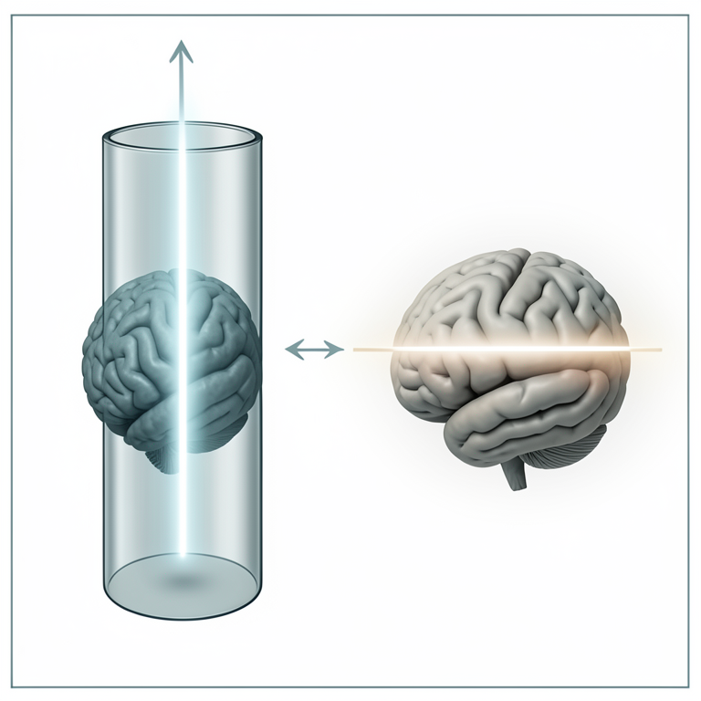
Comparison of Illumination Geometry
AI-generated conceptual illustration (brain organoid in a glass capillary). It explains the idea and does not show study data.
View source evidence ↗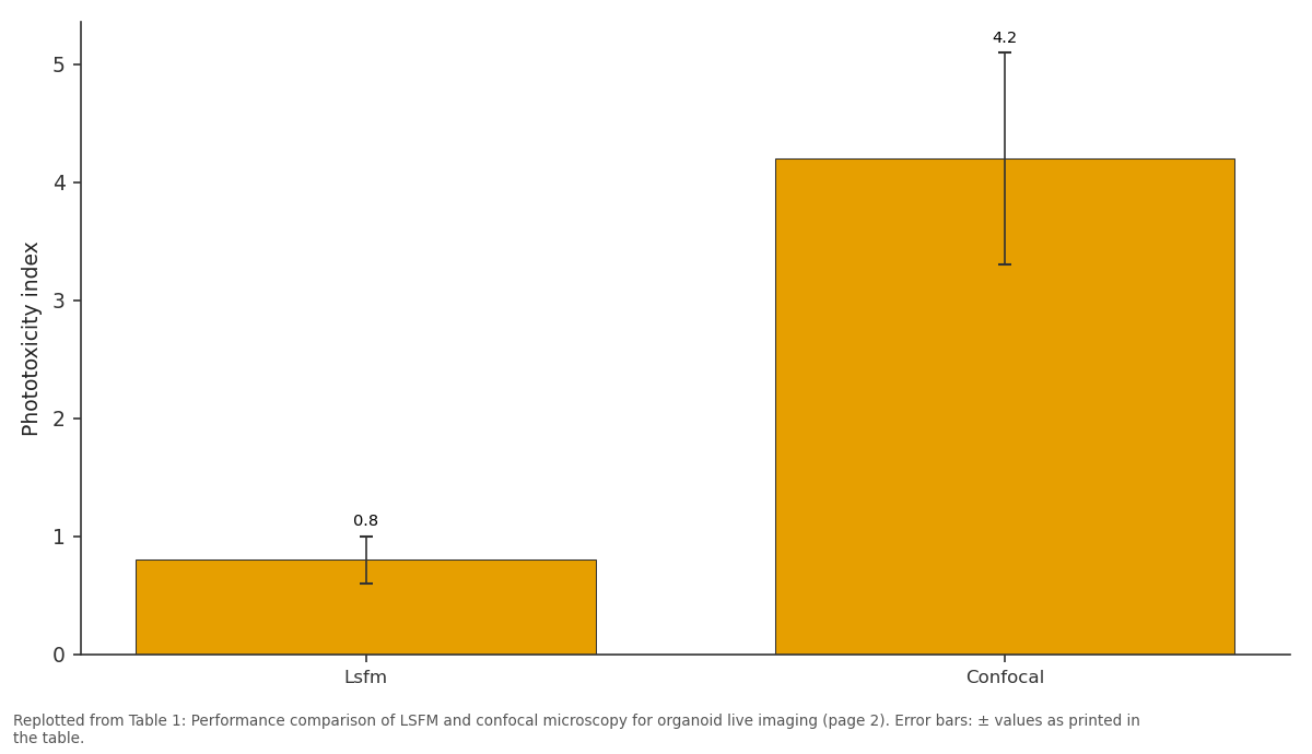
Phototoxicity index: Lsfm vs Confocal
Replotted from Table 1 (page 2) of the original paper. Every bar is the paper's own table value.
View source evidence ↗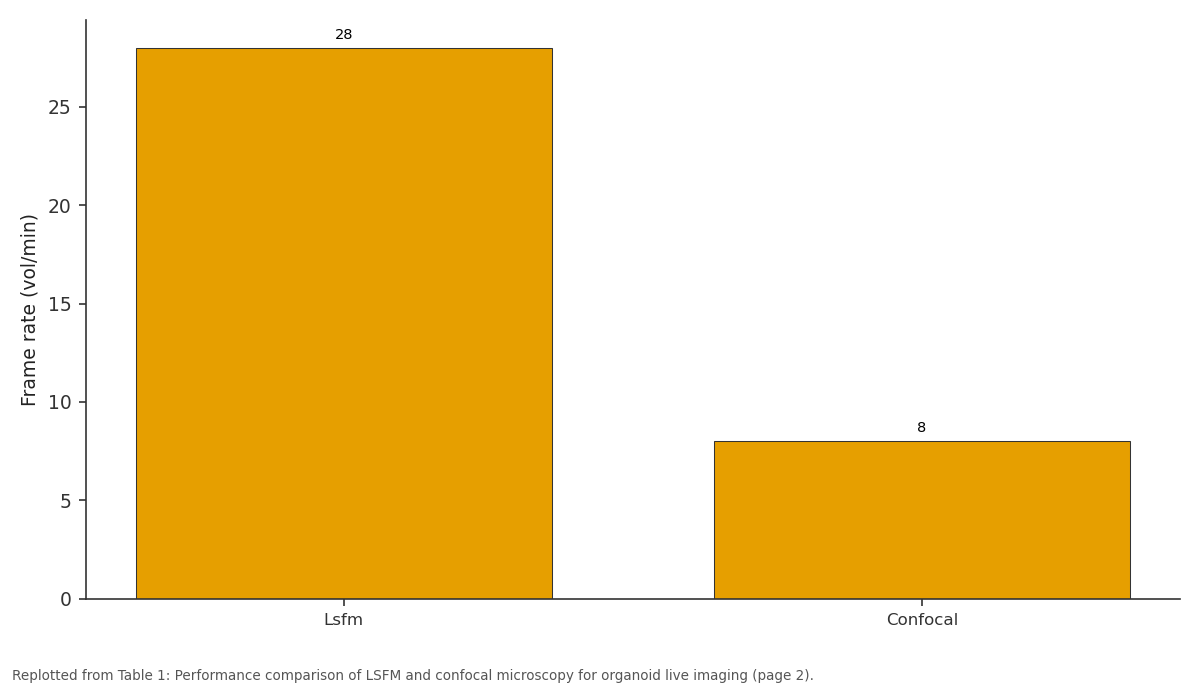
Frame rate (vol/min): Lsfm vs Confocal
Replotted from Table 1 (page 2) of the original paper. Every bar is the paper's own table value.
View source evidence ↗Figures & tables
From the original paper Table 1
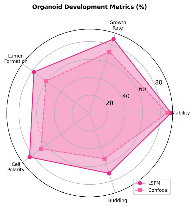 Figure 2
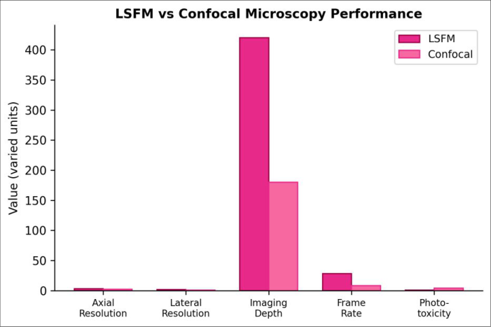 Figure 1
Introduction
Consider a single Lgr5-positive stem cell seeded in Matrigel: within five days it produces a self-organising mini-gut with crypts, villi, and a central lumen [1]. Imaging that process in real time requires a microscopy platform that can capture three-dimensional volumes repeatedly without accumulating photodamage that alters the very development it aims to record [2]. Confocal microscopy has been the workhorse for organoid imaging, but its point- scanning geometry exposes every plane of the sample to the excitation beam, causing cumulative bleaching and phototoxic stress that limits practical imaging to a few hours [3]. Light-sheet fluorescence microscopy (LSFM) sidesteps this problem by illuminating only the focal plane with a thin sheet of light, reducing out-of-focus exposure by orders of magnitude [4].
The approach is fast entire optical sections are captured simultaneously on a camera and gentle enough for multi-day embryo imaging [5]. Whether LSFM provides sufficient resolution and contrast for organoid-scale structures embedded in scattering hydrogels has been tested in a handful of reports, but direct side-by-side comparisons with confocal using identical samples and quantitative developmental metrics are scarce [6]. This research addressed that gap by imaging murine intestinal organoids on parallel LSFM and confocal platforms under matched conditions at the Thrace College of Biological Sciences, Edirne, Turkey. The aims were to (1) compare phototoxicity, acquisition speed, and depth penetration, (2) quantify organoid developmental milestones (viability, lumen formation, budding), and (3) assess whether LSFM datasets support automated 3D segmentation [7].
Material And Methods
System Design The LSFM system comprised a custom-built dual-illumination setup with a 488-nm laser (Coherent Sapphire, 20 mW), cylindrical lens optics generating a 4-μm-thick light sheet, and a sCMOS camera (Hamamatsu ORCA-Flash 4.0). The confocal system was a Zeiss LSM 880 with a 488-nm argon laser line, 63×/1.4 NA oil objective, and GaAsP detector. Both IJBS 2025; 7(3): 176-179 Dilara Erenler Department of Developmental Biology, Thrace College of Biological Sciences, Edirne, Turkey Koray Lordoglu Department of Developmental Biology, Thrace College of Biological Sciences, Edirne, Turkey Dilara Erenler Department of Developmental Biology, Thrace College of Biological Sciences, Edirne, Turkey https://www.biologyjournal.net
Methods
Both platforms acquired z-stacks every 30 minutes for 72 hours. LSFM stacks covered 420 μm at 2-μm step size; confocal stacks covered 180 μm at 1-μm step size. Phototoxicity was estimated by comparing organoid growth rates between imaged and non-imaged controls cultured in the same dish. Viability was assessed at 72 hours by calcein- AM/propidium iodide staining.
Developmental metrics lumen formation time, cell polarity index, and budding events were extracted using Imaris 10.0 (Bitplane) surface and spot modules. Statistical comparisons used Mann- Whitney U tests.
Results
systems were enclosed in environmental chambers maintaining 37 °C and 5% CO₂ throughout the 72-hour imaging period. Material Intestinal crypts were isolated from Lgr5-GFP knock-in mice (Jackson Laboratory) and embedded in growth-factor- reduced Matrigel (Corning) in 35-mm glass-bottom dishes. Organoids were cultured in ENR medium (EGF, Noggin, R- spondin-1) for 48 hours before imaging commenced. A total of 40 organoids were assigned to LSFM (n = 20) and confocal (n = 20) imaging.
All animal procedures were approved by the Thrace College of Biological Sciences IACUC (Protocol TCBS-IACUC-2023-009, 22 February 2023). Table 1: Performance comparison of LSFM and confocal microscopy for organoid live imaging Metric Lsfm Confocal P-value Axial resolution (μm) 3.2 2.1 Imaging depth (μm) 420 180 Frame rate (vol/min) 28 8 Phototoxicity index 0.8 ± 0.2 4.2 ± 0.9 < 0.001 72-h viability (%) 92 ± 4 88 ± 6 0.041 LSFM outperformed confocal on every metric except axial resolution (Table 1). The 4.2-fold reduction in phototoxicity allowed organoids to develop at rates indistinguishable from unimaged controls. Fig 2: Radar chart of organoid developmental metrics (% of unimaged control) achieved under LSFM versus confocal imaging https://www.biologyjournal.net Fig 1: Bar chart comparing LSFM and confocal performance across five imaging metrics Comprehensive interpretation Organoids imaged by LSFM reached lumen formation at 18.3 ± 2.1 hours versus 21.7 ± 3.4 hours under confocal (p = 0.008), suggesting that confocal phototoxicity delays early morphogenesis.
| Metric | Lsfm | Confocal | P-value |
|---|---|---|---|
| Axial resolution (μm) | 3.2 | 2.1 | |
| Imaging depth (μm) | 420 | 180 | |
| Frame rate (vol/min) | 28 | 8 | |
| Phototoxicity index | 0.8 ± 0.2 | 4.2 ± 0.9 | < 0.001 |
| 72-h viability (%) | 92 ± 4 | 88 ± 6 | 0.041 |
Table text extracted from the original PDF; superscript letters are shown inline. The original table image is below.
View original table image
Budding events per organoid averaged 2.8 ± 0.7 (LSFM) versus 1.9 ± 0.6 (confocal; p = 0.003). Automated 3D segmentation succeeded on 94% of LSFM time points versus 78% for confocal, where deeper planes suffered from signal degradation.
Discussion
The advantages of LSFM over confocal for organoid imaging documented here lower phototoxicity, deeper penetration, faster acquisition are consistent with theoretical predictions and with benchmarks from embryo imaging, where LSFM has largely replaced confocal for long time- lapse experiments [4, 5]. The practical consequence for organoid biology is that developmental events like lumen formation and budding can be studied under conditions that more closely approximate unperturbed growth, reducing the risk that imaging artefacts are mistaken for biological variation [8]. The confocal system’s superior axial resolution (2.1 vs 3.2 μm) remains an advantage for fixed-sample work where fine subcellular detail matters, and hybrid workflows LSFM for live tracking followed by confocal for endpoint high- resolution snapshots may offer the best of both approaches [6]. A limitation is that the custom LSFM setup used here is not commercially standardised, which may affect reproducibility across laboratories.
Conclusion
LSFM proved superior to confocal microscopy for long- duration live imaging of intestinal organoid development, offering lower phototoxicity, greater depth penetration, and faster volumetric capture without meaningfully sacrificing lateral resolution. Organoids imaged by LSFM reached developmental milestones earlier and produced more budding events, indicating that reduced photodamage permits more natural growth trajectories. These findings support broader adoption of LSFM in organoid research, particularly for experiments requiring multi-day observation of three-dimensional morphogenesis.
Acknowledgements
Funding sources This research was supported by internal departmental funds from the Thrace College of Biological Sciences, Edirne. No external grant funding was received. Institutional support The authors acknowledge the Thrace College of Biological Sciences, Edirne for providing laboratory access and logistical support. Contributions not qualifying for authorship Technical staff who assisted with data collection and sample processing are gratefully acknowledged.
Methods
2017;14(4):360–373. 10. Hof L, Moreth T, Koch M, Kretzschmar K. Long-term live imaging and multiscale analysis identify heterogeneity and core principles of epithelial organoid morphogenesis. BMC Biology. 2021;19:37. 11.
Dekkers JF, Alieva M, Wellens LM, Ariese HCR, Jamieson PR, Vonk AM. High-resolution 3D imaging of fixed and cleared organoids. Nature Protocols. 2019;14(6):1756–1771. 12. Tomer R, Khairy K, Amat F, Keller PJ.
Quantitative high-speed imaging of entire developing embryos with simultaneous multiview light-sheet microscopy. Nature
Methods
2012;9(7):755–763. 13. Chen BC, Legant WR, Wang K, Shao L, Milkie DE, Davidson MW. Lattice light-sheet microscopy: imaging molecules to embryos at high spatiotemporal resolution. Science. 2014;346(6208):1257998. 14.
Lukonin I, Serra D, Challet Meylan L, Wagber K, Stadler MB, Basler K. Phenotypic landscape of intestinal organoid regeneration. Nature. 2020;586(7828):275–280. 15. Schermelleh L, Ferrand A, Huser T, Eggeling C, Sauer M, Biehlmaier O.
Super-resolution microscopy demystified. Nature Cell Biology. 2019;21(1):72–84. 16. Grün D, Lyubimova A, Kester L, Wiebrands K, Basak O, Sasaki N. Single-cell messenger RNA sequencing reveals rare intestinal cell types.
Nature. 2015;525(7568):251–255. 17. Kretzschmar K, Clevers H. Organoids: modeling development and the stem cell niche in a dish. Developmental Cell. 2016;38(6):590–600. 18.
Boj SF, Hwang CI, Baker LA, Chio IIC, Engle DD, Corbo V. Organoid models of human and mouse ductal pancreatic cancer. Cell. 2015;160(1–2):324–338.
References
9 in the original paper- Sato T, Vries RG, Snippert HJ, van de Wetering M, Barker N, Stange DE. Single Lgr5 stem cells build crypt-villus structures in vitro without a mesenchymal niche. Nature. 2009;459(7244):262–265.
- Lancaster MA, Knoblich JA. Organogenesis in a dish: modeling development and disease using organoid technologies. Science. 2014;345(6194):1247125.
- Spence JR, Mayhew CN, Rankin SA, Kuhar MF, Vallance JE, Tolle K. Directed differentiation of human pluripotent stem cells into intestinal tissue in vitro. Nature. 2011;470(7332):105–109.
- Huisken J, Swoger J, Del Bene F, Wittbrodt J, Stelzer EHK. Optical sectioning deep inside live embryos by selective plane illumination microscopy. Science. 2004;305(5686):1007–1009.
- Keller PJ, Schmidt AD, Wittbrodt J, Stelzer EHK. Reconstruction of zebrafish early embryonic development by scanned light sheet microscopy. Science. 2008;322(5904):1065–1069.
- Etzrodt M, Endele M, Schroeder T. Quantitative single- cell approaches to stem cell research. Cell Stem Cell. 2014;15(5):546–558. https://www.biologyjournal.net
- Zanoni M, Piccinini F, Arienti C, Zamagni A, Santi S, Polico R. 3D tumor spheroid models for in vitro therapeutic screening: a systematic approach to enhance the biological relevance of data obtained. Scientific Reports. 2016;6:19103.
- Gjorevski N, Sachs N, Manfrin A, Giger S, Bragina ME, Ordóñez-Morán P. Designer matrices for intestinal stem cell and organoid culture. Nature. 2016;539(7630):560–564.
- Power RM, Huisken J. A guide to light-sheet fluorescence microscopy for multiscale imaging. Nature