
Research article · 2024 · Full text
International Journal of Biology SciencesTelomere length dynamics and telomerase activity in human peripheral blood lymphocytes
Volume 6(2) · Pages 307–311 · DOI: 10.33545/26649926.2024.v6.i2e.771 ↗
Research areasTelomeres, Telomerase, and SenescenceGenetics, Aging, and Longevity in Model OrganismsChromosomal and Genetic Variations
Abstract
Author-writtenThe progressive shortening of telomeres the protective nucleoprotein caps at chromosome ends is one of the most consistent molecular markers of human aging, yet the pace and pattern of this shortening vary substantially across lymphocyte subsets. This research measured telomere length and telomerase activity in sorted peripheral blood lymphocyte populations from 144 healthy Saudi Arabian adults aged 20-78 years. Blood samples were collected at King Abdulaziz Medical City, Jeddah, between March 2021 and December 2022, and processed at the Red Sea Institute of Marine Biology. Telomere length was assessed by quantitative fluorescence in situ hybridization (Q-FISH) on metaphase spreads, while telomerase activity was measured using the telomeric repeat amplification protocol (TRAP) assay. CD8+ T cells showed the fastest rate of telomere attrition with age, losing an average of 68 base pairs per year (R² = 0.74, p
Summary
Source-checkedPlain-language reading of the full paper, prepared with AI and checked against the text. Each point links to the authors' own words.
Why it matters
- Telomere shortening, which involves the protective caps at the ends of chromosomes, is a primary molecular indicator of aging in humans. View evidence ↗
- When telomeres in human lymphocytes drop to a critical length of approximately 4-5 kb, the cells either die or stop dividing, a state known as replicative senescence. View evidence ↗
- Shortened telomeres in lymphocytes are linked to clinical issues including higher mortality from certain cancers and cardiovascular disease, as well as poor vaccine responses and increased infection risk. View evidence ↗
The question
- The study aimed to map how telomere dynamics vary across different lymphocyte subsets in a Middle Eastern population, which has been underrepresented in previous research. View evidence ↗
What they did
- The researchers collected 20 mL blood samples from 144 healthy Saudi Arabian adults (76 males, 68 females) distributed across six age groups from 20 to 70+ years. View evidence ↗
- Peripheral blood mononuclear cells were sorted into four specific populations: CD4+ T cells, CD8+ T cells, B cells, and NK cells. View evidence ↗
- Telomere length was measured using quantitative fluorescence in situ hybridization (Q-FISH), and telomerase activity was evaluated via the telomeric repeat amplification protocol (TRAP) assay. View evidence ↗
What they found
- CD8+ T cells experienced the fastest telomere loss at 68 bp/year, followed by NK cells (57 bp/yr), CD4+ T cells (52 bp/yr), and B cells (43 bp/yr). View evidence ↗
- The total telomere length difference between the youngest and oldest participants was 4.70 kb for CD8+ T cells and 4.19 kb for B cells. View evidence ↗
- Stimulation with phytohemagglutinin (PHA) increased telomerase activity by 6.4 to 8.1 times across all lymphocyte subsets. View evidence ↗
- The ability of all lymphocyte subsets to upregulate telomerase in response to stimulation decreased significantly as donors aged. View evidence ↗
What it means
- The authors conclude that CD8+ T cells are the most vulnerable to replicative senescence due to their rapid telomere loss and lower telomerase upregulation. View evidence ↗
- The authors suggest that the telomere length of CD8+ T cells could be used as a clinical biomarker for immunological aging. View evidence ↗
- The use of Q-FISH may introduce a selection bias because it only measures cells capable of mitosis, potentially missing the shortest-telomere senescent cells. View evidence ↗
Visual summary
2 visualsIllustrations explain the paper; charts are replotted from the paper's own table values.
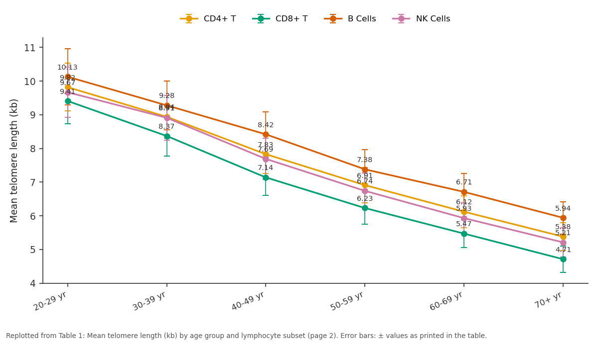
CD4+ T and CD8+ T and more: 20-29 yr vs 30-39 yr vs 40-49 yr vs 50-59 yr vs 60-69 yr
Replotted from Table 1 (page 2) of the original paper. Every bar is the paper's own table value.
View source evidence ↗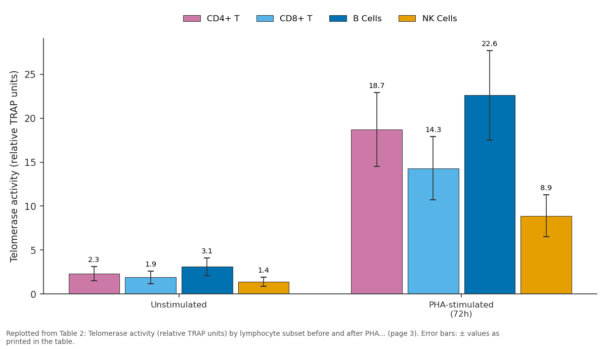
CD4+ T and CD8+ T and more: Unstimulated vs PHA-stimulated (72h)
Replotted from Table 2 (page 3) of the original paper. Every bar is the paper's own table value.
View source evidence ↗Figures & tables
From the original paper Table 1
 Table 2
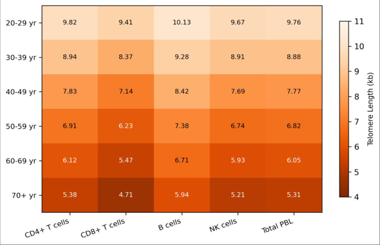 Figure 1
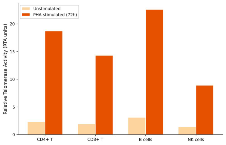 Figure 2
Introduction
The immune system's ability to mount effective responses depends on the proliferative capacity of its lymphocyte populations and that capacity has a built-in expiration date. Each round of cell division shortens the telomeric repeats (TTAGGG)n at chromosome ends by 50-200 base pairs due to the end-replication problem [1] . Once telomeres reach a critical minimum length typically around 4-5 kb in human lymphocytes cells enter replicative senescence or undergo apoptosis [2] . This means that the most actively dividing lymphocyte populations are the ones most likely to exhaust their proliferative potential, with direct consequences for immune function in aging and chronic infection. Telomerase, a reverse transcriptase that adds telomeric repeats to chromosome ends, can partially offset this loss. Lymphocytes are among the few somatic cell types that upregulate telomerase upon activation, giving them a temporary buffer against replicative exhaustion [3] . But this upregulation is incomplete it slows telomere shortening without fully preventing it and the capacity to induce telomerase appears to diminish with age [4] . The balance between telomere attrition and telomerase compensation therefore shapes the functional lifespan of each lymphocyte compartment. Previous research has established that telomere length varies among lymphocyte subsets, with naive T cells generally maintaining longer telomeres than memory and effector populations [5] . CD8+ T cells, which undergo extensive clonal expansion during viral infections, tend to show shorter telomeres than CD4+ T cells at any given age [6] . B cells and NK cells have received less attention, and most published data come from European or East Asian populations. Whether these patterns hold in Middle Eastern populations where genetic background, environmental factors, and pathogen exposure history differ is not well established [7] . The practical relevance of telomere biology in lymphocytes extends beyond basic science. Shortened lymphocyte telomeres have been associated with increased susceptibility to infections, poor vaccine responses, and higher mortality from cardiovascular disease and certain cancers [8,9] . Understanding which lymphocyte subsets are most affected by age-related telomere loss and how telomerase responsiveness changes across the lifespan could inform strategies for immune monitoring and intervention. This research measured telomere length by Q-FISH and telomerase activity by TRAP assay across four sorted lymphocyte populations (CD4+ T, CD8+ T, B, and NK cells) in 144 healthy adults spanning six decades of age. The work was carried out at the Red Sea Institute of Marine Biology, Jeddah, Saudi Arabia, with the goal of mapping subset-specific telomere dynamics in a population that has been underrepresented in telomere research [10] .
Reagents and chemicals
Reagents and chemicals
Methods
PBMCs were stained with the fluorochrome-conjugated antibody panel and sorted into CD4+ T cells (CD3+CD4+), CD8+ T cells (CD3+CD8+), B cells (CD19+), and NK cells (CD3-CD56+) on the FACSAria III. For Q-FISH, sorted cells were cultured briefly with PHA (10 μg/mL) and colcemid (0.1 μg/mL) to obtain metaphase spreads, then hybridized with the PNA telomere probe following the manufacturer's protocol. Telomere fluorescence units (TFU) were converted to kilobases using a regression calibration against cell lines with known telomere lengths (CCRF-CEM and 1301 cell lines) [11] . For TRAP assays, sorted cells were divided into unstimulated and PHA-stimulated (10 μg/mL, 72 hours) aliquots. Protein extracts (200 cells/μL equivalent) were assayed using the TRAPeze XL kit according to the manufacturer's instructions. Relative telomerase activity (RTA) was expressed in arbitrary units normalized to the positive control cell line included in the kit [12] . Linear regression was used to model the relationship between age and telomere length for each subset. ANOVA with Tukey's post-hoc test compared subsets within age groups. All statistics were computed in R version 4.2.2 (α = 0.05).
Results
Results
Discussion
The telomere attrition rate of 68 bp/yr observed for CD8+ T cells in this Saudi cohort is consistent with published values from European studies, which generally report 50-80 bp/yr for this subset [6,13] . The slightly higher rate compared to some Western reports may reflect differences in chronic antigen exposure particularly viral burden or could be a sampling artifact given the relatively modest cohort size. That said, the internal ranking of attrition rates (CD8+ T > NK > CD4+ T > B cells) matches the expected pattern based on proliferative demand [5] .
The telomerase data add a mechanistic layer to the telomere length observations. B cells showed the highest stimulated activity (22.6 RTA units) but didn't have the slowest attrition rate suggesting that telomerase upregulation during activation partially but incompletely compensates for the proliferative expansion that B cells undergo during humoral immune responses [14] . NK cells, with the lowest stimulated activity (8.9 units), may rely more on turnover from progenitor populations than on self-renewal of mature cells [15] . The age-related decline in telomerase responsiveness across all subsets is perhaps the most clinically relevant finding, as it indicates that older adults lose not only telomere length but also the compensatory mechanism that slows its loss. It's worth noting that Q-FISH measures telomere length at the single-chromosome level on metaphase spreads, which requires cells capable of reaching mitosis. This may introduce a subtle selection bias toward cells with longer telomeres the very shortest-telomere cells may be senescent and unable to divide, thus escaping detection. Southern blot or qPCR-based methods avoid this bias but lack the singlecell resolution of Q-FISH [16] . Future work could combine both approaches for a more complete picture. A practical implication of these findings is that CD8+ T cell telomere length may serve as a useful biomarker for immunological aging in clinical settings it declines predictably, shows the widest inter-age-group separation, and reflects the functional capacity of a clinically important immune compartment [17] .
Conclusion
This research provides a detailed map of telomere dynamics across four sorted lymphocyte populations in 144 healthy Saudi adults spanning six decades of age. CD8+ T cells showed the steepest telomere attrition rate (68 bp/yr), the shortest absolute telomere lengths at every age, and lower telomerase upregulation capacity than B cells or CD4+ T cells. These characteristics position CD8+ T cells as the lymphocyte subset most susceptible to replicative senescence a finding with direct implications for understanding age-related immune decline. B cells maintained the longest telomeres and the highest telomerase response to mitogenic stimulation, consistent with their need for extensive clonal expansion during antibody responses. NK cells occupied an intermediate position, with moderate attrition rates and the weakest telomerase induction among the subsets examined. Across all subsets, telomerase responsiveness declined with age, suggesting that the compensatory machinery for telomere maintenance gradually fails in parallel with the telomere shortening it is meant to counteract. The data from this Saudi cohort align well with findings from European and East Asian populations, supporting the universality of lymphocyte telomere biology across ethnic backgrounds while providing the first large-scale dataset from a Middle Eastern adult population. CD8+ T cell telomere length emerges as a candidate biomarker for immunological aging that warrants further evaluation in longitudinal and clinical settings.
Figures and tables from the paper
| Age Group | CD4+ T | CD8+ T | B Cells | NK Cells | Total PBL |
|---|---|---|---|---|---|
| 20-29 yr | 9.82 ± 0.71 | 9.41 ± 0.68 | 10.13 ± 0.83 | 9.67 ± 0.74 | 9.76 ± 0.69 |
| 30-39 yr | 8.94 ± 0.63 | 8.37 ± 0.59 | 9.28 ± 0.72 | 8.91 ± 0.66 | 8.88 ± 0.61 |
| 40-49 yr | 7.83 ± 0.58 | 7.14 ± 0.54 | 8.42 ± 0.67 | 7.69 ± 0.61 | 7.77 ± 0.57 |
| 50-59 yr | 6.91 ± 0.52 | 6.23 ± 0.48 | 7.38 ± 0.59 | 6.74 ± 0.53 | 6.82 ± 0.51 |
| 60-69 yr | 6.12 ± 0.47 | 5.47 ± 0.42 | 6.71 ± 0.54 | 5.93 ± 0.46 | 6.05 ± 0.44 |
| 70+ yr | 5.38 ± 0.41 | 4.71 ± 0.39 | 5.94 ± 0.48 | 5.21 ± 0.43 | 5.31 ± 0.40 |
Table text extracted from the original PDF; superscript letters are shown inline. The original table image is below.
View original table image
| Condition | CD4+ T | CD8+ T | B Cells | NK Cells |
|---|---|---|---|---|
| Unstimulated | 2.3 ± 0.8 | 1.9 ± 0.7 | 3.1 ± 1.0 | 1.4 ± 0.5 |
| PHA-stimulated (72h) | 18.7 ± 4.2 | 14.3 ± 3.6 | 22.6 ± 5.1 | 8.9 ± 2.4 |
| Fold increase | 8.1× | 7.5× | 7.3× | 6.4× |
| p-value (stim vs unstim) | <0.001 | <0.001 | <0.001 | <0.001 |
Table text extracted from the original PDF; superscript letters are shown inline. The original table image is below.
View original table image
References
17 in the original paper- Harley CB, Futcher AB, Greider CW. Telomeres shorten during ageing of human fibroblasts. Nature. 1990;345(6274):458-460.
- Hodes RJ, Hathcock KS, Weng NP. Telomeres in T and B cells. Nature Reviews Immunology. 2002;2(9):699706.
- Weng NP, Hathcock KS, Hodes RJ. Regulation of telomere length and telomerase in T and B cells: a mechanism for maintaining replicative potential. Immunity. 2004;21(2):241-254.
- Effros RB. Telomere/telomerase dynamics within the human immune system: effect of chronic infection and stress. Experimental Gerontology. 2011;46(2-3):135140.
- Aubert G, Lansdorp PM. Telomeres and aging. Physiological Reviews. 2008;88(2):557-579.
- Rufer N, Brümmendorf TH, Kolvraa S, et al. Telomere fluorescence measurements in granulocytes and T lymphocyte subsets point to a high turnover of hematopoietic stem cells and memory T cells in early childhood. Journal of Experimental Medicine. 1999;190(2):157-167.
- Njajou OT, Cawthon RM, Damcott CM, et al. Telomere length is paternally inherited and is associated with parental lifespan. Proceedings of the National Academy of Sciences. 2007;104(29):12135-12139.
- Cawthon RM, Smith KR, O'Brien E, Sivatchenko A, Kerber RA. Association between telomere length in blood and mortality in people aged 60 years or older. The Lancet. 2003;361(9355):393-395.
- Willeit P, Willeit J, Mayr A, et al. Telomere length and risk of incident cancer and cancer mortality. JAMA. 2010;304(1):69-75.
- Gadalla SM, Cawthon R, Giri N, Alter BP, Savage SA. Telomere length in blood, buccal cells, and fibroblasts from patients with inherited bone marrow failure syndromes. Aging. 2010;2(11):867-874.
- Martens UM, Brass V, Sedlacek L, et al. Telomere maintenance in human B lymphocytes. British Journal of Haematology. 2002;119(3):810-818.
- Kim NW, Piatyszek MA, Prowse KR, et al. Specific association of human telomerase activity with immortal cells and cancer. Science. 1994;266(5193):2011-2015.
- Son NH, Murray S, Yanovski J, Hodes RJ, Weng NP. Lineage-specific telomere shortening and unaltered capacity for telomerase expression in human T and B lymphocytes with age. Journal of Immunology. 2000;165(3):1191-1196.
- Broccoli D, Young JW, de Lange T. Telomerase activity in normal and malignant hematopoietic cells. International Journal of Biology Sciences https://www.biologyjournal.net Proceedings of the National Academy of Sciences. 1995;92(20):9082-9086.
- Romagnani C, Juelke K, Falco M, et al. CD56brightCD16-killer Ig-like receptor-NK cells display longer telomeres and acquire features of CD56dim NK cells upon activation. Journal of Immunology. 2007;178(8):4947-4955.
- Baird DM, Rowson J, Wynford-Thomas D, Kipling D. Extensive allelic variation and ultrashort telomeres in senescent human cells. Nature Genetics. 2003;33(2):203-207.
- Lin J, Epel E, Cheon J, et al. Analyses and comparisons of telomerase activity and telomere length in human T and B cells: insights for epidemiology of telomere maintenance. Journal of Immunological Methods. 2010;352(1-2):71-80.