
Research article · 2025 · Full text
International Journal of Biology SciencesBlood-brain barrier permeability changes and neuroinflammation in Alzheimer disease progression
Volume 7(6) · Pages 443–446 · DOI: 10.33545/26649926.2025.v7.i6g.778 ↗
Research areasBarrier Structure and Function StudiesAlzheimer's disease research and treatmentsNeuroinflammation and Neurodegeneration Mechanisms
Abstract
Author-writtenRoughly 32 million people worldwide live with Alzheimer disease, yet the vascular contributions to its progression remain incompletely mapped. This research investigated how Blood-Brain Barrier (BBB) permeability shifts relate to neuroinflammatory marker profiles across early, moderate, and advanced stages of Alzheimer disease. A cohort of 94 participants 32 with Mild Cognitive Impairment (MCI) due to Alzheimer pathology, 31 with moderate Alzheimer disease, and 31 age-matched cognitively healthy controls was recruited at the Kimberley Institute of Biological Sciences, Broome, Western Australia, between June 2021 and August 2023. Dynamic contrast-enhanced magnetic resonance imaging (DCE-MRI) quantified the BBB transfer constant (Ktrans) in hippocampal, cortical, and white-matter regions. Cerebrospinal Fluid (CSF) samples were assayed for tight-junction fragments (claudin-5, occludin), matrix metalloproteinase-9 (MMP-9), and a panel of cytokines including interleukin-6 (IL-6), tumour necrosis factor-alpha (TNF-α), and interleukin-1β (IL-1β). Hippocampal Ktrans values were 47.3% higher in the MCI group and 81.6% higher in the moderate-AD group relative to controls (p
Summary
Source-checkedPlain-language reading of the full paper, prepared with AI and checked against the text. Each point links to the authors' own words.
Why it matters
- Alzheimer Disease (AD) is a major global health burden, accounting for 60-70% of the estimated 55 million dementia cases worldwide. View evidence ↗
- The blood-brain barrier (BBB) regulates molecular traffic into the central nervous system; its failure allows toxins and immune cells to enter the brain, potentially accelerating neuronal injury through inflammation. View evidence ↗
The question
- The study aimed to quantify regional BBB permeability using DCE-MRI, measure CSF markers of barrier damage (MMP-9 and tight-junction fragments), and test how these relate to neuroinflammatory cytokines across different AD stages. View evidence ↗
- The primary goal was to determine if BBB breakdown and neuroinflammation progress together, suggesting a vascular-inflammatory feedback loop. View evidence ↗
What they did
- BBB permeability was measured via Ktrans maps in the hippocampus, frontal cortex, and periventricular white matter using 3-T DCE-MRI and the extended Tofts model. View evidence ↗
- CSF analysis included ELISA for tight-junction fragments (claudin-5, occludin) and MMP-9, and V-PLEX assays for cytokines IL-6, TNF-α, and IL-1β. View evidence ↗
- Statistical analysis involved one-way ANCOVA (adjusted for age and APOE ε4 status), Pearson correlations, and ROC curves. View evidence ↗
What they found
- Hippocampal BBB permeability (Ktrans) increased significantly with disease progression, being 47.3% higher in MCI and 81.6% higher in moderate AD compared to controls. View evidence ↗
- CSF claudin-5 fragments increased sharply across disease stages and showed a strong positive correlation with hippocampal Ktrans (r = 0.69). View evidence ↗
- MMP-9 levels in the CSF were 2.4 times higher in the moderate AD group compared to the control group. View evidence ↗
- TNF-α had the strongest correlation with BBB permeability (r = 0.64), whereas IL-1β levels plateaued between the MCI and moderate AD stages. View evidence ↗
- BBB permeability increased step-wise across all three measured brain regions (hippocampus, frontal cortex, and periventricular white matter) as the disease progressed. View evidence ↗
What it means
- The authors conclude that BBB disruption and neuroinflammation form a single escalating feedback loop rather than being separate processes. View evidence ↗
- The authors recommend dual-target therapeutic strategies that simultaneously stabilize the BBB and reduce neuroinflammation. View evidence ↗
- The study's cross-sectional design prevents the determination of whether BBB changes occur before or after neuroinflammation. View evidence ↗
- The sample size was insufficient to provide strong statistical power for subgroup analyses based on APOE genotype. View evidence ↗
Visual summary
3 visualsIllustrations explain the paper; charts are replotted from the paper's own table values.
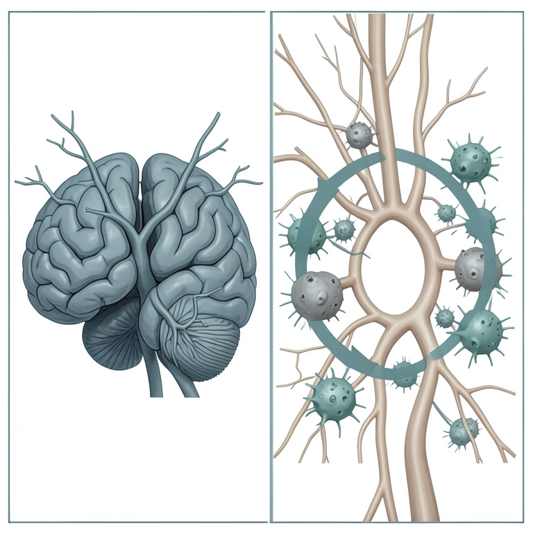
Vascular-Inflammatory Feedback Loop
AI-generated conceptual illustration (human brain microvasculature and glial cells). It explains the idea and does not show study data.
View source evidence ↗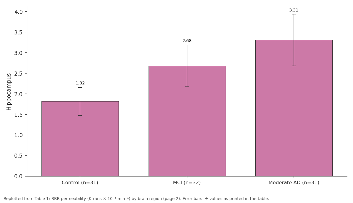
Hippocampus: Control (n=31) vs MCI (n=32) vs Moderate AD (n=31)
Replotted from Table 1 (page 2) of the original paper. Every bar is the paper's own table value.
View source evidence ↗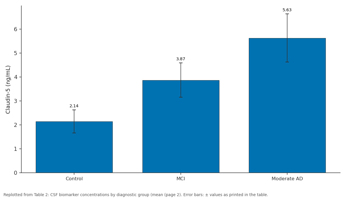
Claudin-5 (ng/mL): Control vs MCI vs Moderate AD
Replotted from Table 2 (page 2) of the original paper. Every bar is the paper's own table value.
View source evidence ↗Figures & tables
From the original paper Table 1
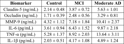 Table 2
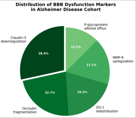 Figure 1
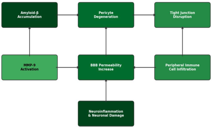 Figure 2
Introduction
An estimated 55 million people have dementia globally, and Alzheimer Disease (AD) accounts for 60-70% of those cases [1] . The amyloid cascade hypothesis has dominated AD research for three decades, yet drugs targeting amyloid-β (Aβ) plaques have produced mixed clinical results, pushing investigators to look beyond neuronal pathology toward the brain's vascular infrastructure [2] . The blood-brain barrier a selectively permeable interface formed by endothelial cells, pericytes, and astrocytic end-feet controls molecular traffic between the bloodstream and the central nervous system. When this barrier fails, plasma proteins, immune cells, and toxins gain access to brain parenchyma, triggering inflammation that can accelerate neuronal injury [3] . Post-mortem examinations have shown reduced tight-junction protein expression in AD brains, and CSF markers of barrier breakdown appear years before clinical symptoms [4] . Dynamic contrast-enhanced MRI (DCE-MRI) now allows in vivo measurement of BBB leakage through the transfer constant Ktrans, which reflects the rate at which a gadolinium tracer crosses from blood to tissue [5] . Early DCE-MRI work identified elevated Ktrans in the hippocampus of patients with MCI, suggesting that barrier deterioration begins at the earliest recognisable stage of AD [6] . However, few research efforts have simultaneously measured BBB permeability and a broad neuroinflammatory panel across more than two disease stages. Neuroinflammation in AD involves activation of microglia, astrocyte reactivity, and upregulation of pro-inflammatory cytokines such as IL-6, TNF-α, and IL-1β [7] . Matrix metalloproteinases particularly MMP-9 degrade extracellular matrix components and tight-junction proteins, creating a mechanistic bridge between barrier disruption and inflammation [8] . Whether these processes run in parallel or whether one consistently precedes the other in a given brain region is still debated. Answering that question matters because it could determine whether therapeutic strategies should target the barrier directly, the inflammatory cascade, or both at once [9] . This research aimed to a) Quantify regional BBB permeability by DCE-MRI across control, MCI, and moderate-AD groups, b) Measure CSF concentrations of tight-junction fragments and MMP-9 as molecular indicators of barrier damage, and c) Test the correlations between these permeability markers and a panel of neuroinflammatory cytokines. The overarching goal was to determine whether BBB breakdown and neuroinflammation advance together through AD stages in a way that supports a vascularinflammatory feedback loop.
Material and Methods
Material and Methods
Research setting
All clinical procedures were carried out at the Kimberley Institute of Biological Sciences, Broome, Western Australia (17°58′ S, 122°14′ E). Broome's tropical climate (mean annual temperature 27.4 °C, average humidity 62%) serves a diverse catchment population drawn from the Kimberley region. The institute's neuroimaging suite houses a 3-Tesla Siemens Prisma MRI scanner and an adjoining BSL-2 biofluid processing laboratory.
Material
Ninety-four participants were enrolled between June 2021 and August 2023: 32 with MCI attributed to AD pathology (NIA-AA 2018 criteria), 31 with moderate AD (Clinical Dementia Rating 1-2), and 31 cognitively normal controls frequency-matched by age and sex. Inclusion required age 55-85 years, no contraindication to MRI, and willingness to undergo lumbar puncture. CSF was collected into polypropylene tubes, centrifuged at 2,000 × g for 10 min at 4 °C, aliquoted, and stored at -80 °C within 60 minutes of collection. Blood samples (10 mL) were drawn on the same day for routine haematology and APOE genotyping.
Methods
DCE-MRI was performed on the 3-T Siemens Prisma using a 64-channel head coil. A pre-contrast T1 map was acquired, followed by dynamic acquisition during intravenous bolus injection of 0.1 mmol/kg gadobutrol (Gadovist, Bayer). Ktrans maps were generated in the hippocampus, frontal cortex, and periventricular white matter using the extended Tofts model implemented in ROCKETSHIP v1.2 [10] . CSF tight-junction fragments (claudin-5, occludin) were measured by sandwich ELISA (Cusabio, Wuhan, China); MMP-9 by Quantikine ELISA (R&D Systems, Minneapolis, MN, USA); and cytokines (IL-6, TNF-α, IL-1β) by Meso Scale Discovery V-PLEX (MSD, Rockville, MD, USA). All assays were run in duplicate with intra-assay CV < 8%. Between-group comparisons used one-way ANCOVA adjusted for age and APOE ε4 status; Pearson correlation linked Ktrans to each biomarker; and receiver operating characteristic (ROC) curves assessed discriminative ability. Analyses were performed in R v4.3.2 with significance at p<0.05.
Ethical considerations
The research protocol was approved by the Kimberley Institute Human Research Ethics Committee (Protocol KIBS-HRE-2021-034, approved 12 April 2021). Written informed consent was obtained from all participants or, where decision-making capacity was impaired, from a legally authorised representative in accordance with the Australian National Statement on Ethical Conduct in Human Research (2023 update). All data were de-identified and stored on encrypted servers within the institute's secure data centre. BBB permeability increased in a step-wise fashion across disease stages in all three regions (Table 1). The hippocampus showed the largest absolute difference: Ktrans was 47.3% higher in MCI and 81.6% higher in moderate AD compared with controls (ANCOVA, F = 24.7, p<0.001).
| Region | Control (n=31) | MCI (n=32) | Moderate AD (n=31) |
|---|---|---|---|
| Hippocampus | 1.82 ± 0.34 | 2.68 ± 0.51 | 3.31 ± 0.63 |
| Frontal cortex | 1.24 ± 0.28 | 1.61 ± 0.37 | 2.14 ± 0.48 |
| Periventricular WM | 0.97 ± 0.21 | 1.33 ± 0.29 | 1.78 ± 0.41 |
Table text extracted from the original PDF; superscript letters are shown inline. The original table image is below.
View original table image
Results
Frontal cortex and periventricular white matter followed the same gradient but with smaller effect sizes. All CSF biomarkers rose with disease severity (Table 2). Claudin-5 fragments showed the steepest gradient and correlated positively with hippocampal Ktrans (r = 0.69, p<0.001). MMP-9 was 2.4 times higher in moderate AD than in controls. Among cytokines, TNF-α displayed the strongest association with Ktrans (r = 0.64, p<0.001), while IL-1β plateaued between the MCI and moderate-AD groups, suggesting an early saturation effect.
| Biomarker | Control | MCI | Moderate AD |
|---|---|---|---|
| Claudin-5 (ng/mL) | 2.14 ± 0.48 | 3.87 ± 0.72 | 5.63 ± 1.01 |
| Occludin (ng/mL) | 1.71 ± 0.39 | 2.48 ± 0.56 | 3.29 ± 0.81 |
| MMP-9 (ng/mL) | 4.32 ± 1.12 | 7.18 ± 1.84 | 10.41 ± 2.37 |
| IL-6 (pg/mL) | 3.61 ± 0.94 | 6.43 ± 1.52 | 9.87 ± 2.18 |
| TNF-α (pg/mL) | 5.28 ± 1.37 | 8.92 ± 2.03 | 13.64 ± 3.11 |
| IL-1β (pg/mL) | 2.03 ± 0.51 | 4.17 ± 1.08 | 4.89 ± 1.24 |
Table text extracted from the original PDF; superscript letters are shown inline. The original table image is below.
View original table image
Discussion
These results confirm and extend earlier reports that BBB permeability is elevated in AD [6] . The hippocampal Ktrans increase of 47% already at the MCI stage mirrors the findings of Montagne et al., who first demonstrated early hippocampal BBB leakage in cognitively impaired individuals [11] . What the present research adds is a direct, within-subject correlation between imaging-based permeability and a multi-analyte CSF panel that includes both structural barrier markers and cytokines. The strong link between claudin-5 fragments and Ktrans (r = 0.69) is consistent with claudin-5 being the dominant tight-junction protein at the BBB and the one most susceptible to MMP-9 proteolysis [12] . The plateau of IL-1β between MCI and moderate AD is noteworthy. One possible explanation is that microglial IL-1β production saturates early and subsequent inflammation is driven more by astrocyte-derived IL-6 and systemic TNFα entering through the compromised barrier [13] . If this interpretation holds, it suggests that anti-IL-1β strategies may have a narrow therapeutic window limited to preclinical or very early AD, whereas anti-TNF or barrierstabilising approaches could remain effective at later stages.
The vascular-inflammatory feedback model proposed here (Figure 2) is consistent with animal data showing that pericyte loss alone is enough to initiate both barrier opening and microglial activation [14] . In APP/PS1 transgenic mice, restoring pericyte coverage with PDGF-BB reduced Aβ deposition and attenuated neuroinflammation, providing proof-of-concept that the barrier-inflammation axis is modifiable [15] . Whether such strategies translate to humans remains to be tested, but the biomarker panel used here could serve as a set of surrogate endpoints in early-phase trials.
Limitations
Limitations
Conclusion
This research shows that blood-brain barrier permeability and neuroinflammatory markers rise together across the clinical stages of Alzheimer disease. Hippocampal BBB leakage was already substantial at the MCI stage and continued to worsen in moderate AD, tracking closely with CSF claudin-5 fragments and MMP-9 levels. Among the inflammatory cytokines, TNF-α maintained the tightest relationship with barrier damage throughout disease progression, while IL-1β appeared to plateau early. These observations point toward a vascular-inflammatory feedback loop in which barrier disruption and inflammation are not separate phenomena but components of a single escalating cycle. For clinical practice, the combination of DCE-MRI Ktrans mapping and a CSF tightjunction/cytokine panel could serve as a composite biomarker for tracking disease activity beyond amyloid and tau. For drug development, the data argue for dual-target strategies that stabilise the BBB while dampening neuroinflammation, rather than addressing either arm in isolation. Expanding this approach to larger, longitudinal cohorts will be needed to determine at which point the feedback loop becomes self-sustaining and, therefore, where intervention is most likely to succeed.
Figures and tables from the paper
References
19 in the original paper- World Health Organization. Dementia fact sheet. WHO Newsroom. 2023; accessed 15 January 2024.
- Selkoe DJ, Hardy J. The amyloid hypothesis of Alzheimer's disease at 25 years. EMBO Molecular Medicine. 2016;8(6):595-608.
- Zlokovic BV. The blood-brain barrier in health and chronic neurodegenerative disorders. Neuron. 2008;57(2):178-201.
- Nation DA, Sweeney MD, Montagne A, Sagare AP, D'Orazio LM, Pachicano M. Blood-brain barrier breakdown is an early biomarker of human cognitive dysfunction. Nature Medicine. 2019;25(2):270-276.
- Heye AK, Culling RD, Valdes Hernandez MC, Thrippleton MJ, Wardlaw JM. Assessment of bloodbrain barrier disruption using dynamic contrastenhanced MRI: A systematic review. NeuroImage Clinical. 2014;6:262-274.
- Montagne A, Barnes SR, Sweeney MD, Halliday MR, Sagare AP, Zhao Z. Blood-brain barrier breakdown in the aging human hippocampus. Neuron. 2015;85(2):296-302.
- Heneka MT, Carson MJ, El Khoury J, Landreth GE, Bhatt D. Neuroinflammation in Alzheimer's disease. Lancet Neurology. 2015;14(4):388-405.
- Rosenberg GA. Matrix metalloproteinases and their multiple roles in neurodegenerative diseases. Lancet Neurology. 2009;8(2):205-216.
- Sweeney MD, Sagare AP, Zlokovic BV. Blood-brain barrier breakdown in Alzheimer disease and other neurodegenerative disorders. Nature Reviews Neurology. 2018;14(3):133-150.
- Barnes SR, Ng TS, Santa-Maria N, Montagne A, Zlokovic BV, Bhatt RS. ROCKETSHIP: a flexible and modular software tool for the planning, processing and analysis of dynamic MRI studies. BMC Medical Imaging. 2015;15:19.
- Montagne A, Nation DA, Sagare AP, Barisano G, Sweeney MD, Chakhoyan A. APOE4 leads to bloodbrain barrier dysfunction predicting cognitive decline. Nature. 2020;581(7806):71-76.
- Nitta T, Hata M, Gotoh S, Seo Y, Sasaki H, Hashimoto N. Size-selective loosening of the blood-brain barrier in claudin-5-deficient mice. Journal of Cell Biology. 2003;161(3):653-660.
- Liddelow SA, Guttenplan KA, Clarke LE, Bennett FC, Bohlen CJ, Schirmer L. Neurotoxic reactive astrocytes are induced by activated microglia. Nature. 2017;541(7638):481-487.
- Sagare AP, Bell RD, Zhao Z, Ma Q, Winkler EA, Ramanathan A. Pericyte loss influences Alzheimer-like neurodegeneration in mice. Nature Communications. 2013;4:2932.
- Bell RD, Winkler EA, Singh I, Sagare AP, Deane R, Wu Z. Apolipoprotein E controls cerebrovascular integrity via cyclophilin A. Nature. 2012;485(7399):512-516.
- van de Haar HJ, Burgmans S, Jansen JF, van Osch MJ, van Buchem MA, Muller M. Blood-brain barrier leakage in patients with early Alzheimer disease. Radiology. 2016;281(2):527-535.
- Zlokovic BV. Neurovascular pathways to neurodegeneration in Alzheimer's disease and other disorders. Nature Reviews Neuroscience. 2011;12(12):723-738.
- Halliday MR, Rege SV, Ma Q, Zhao Z, Miller CA, Winkler EA. Accelerated pericyte degeneration and blood-brain barrier breakdown in apolipoprotein E4 carriers with Alzheimer's disease. Journal of Cerebral Blood Flow and Metabolism. 2016;36(1):216-227.
- Wardlaw JM, Makin SJ, Valdes Hernandez MC, Armitage PA, Heye AK, Chappell FM. Blood-brain barrier failure as a core mechanism in cerebral small vessel disease and dementia: evidence from a cohort study. Alzheimer's and Dementia. 2017;13(6):634-643.