
Research article · 2025 · Full text
International Journal of Biology SciencesDiversity and bioactive potential of endophytic fungi isolated from tropical orchid species
Volume 7(6) · Pages 471–474 · DOI: 10.33545/26649926.2025.v7.i6g.785 ↗
Research areasMicrobial Natural Products and BiosynthesisPlant and fungal interactionsPlant Pathogens and Fungal Diseases
Abstract
Author-writtenLeading mycologists have long suspected that tropical orchids harbour unusually diverse endophytic fungal communities, yet most sampling has been confined to temperate species. This research characterised endophytic fungi from five tropical orchid species Dendrobium nobile, Vanda coerulea, Phalaenopsis amabilis, Coelogyne cristata, and Bulbophyllum lobii cultivated in the tropical greenhouse of the Baikal Institute of Limnological Biology, Irkutsk, Russia. A total of 384 tissue segments (roots, stems, leaves) were surface-sterilised, plated on potato dextrose agar, and incubated for 14 days. Emerging fungal colonies were identified by ITS rDNA sequencing. Overall colonisation frequency was 67.4%, yielding 147 isolates assigned to 38 operational taxonomic units across 19 genera. Fusarium, Colletotrichum, and Alternaria were the most common genera, but rarer taxa such as Pestalotiopsis and Xylaria showed the highest bioactivity. Crude ethyl acetate extracts were screened for antibacterial, antifungal, and antioxidant activities. Antibacterial activity (≥ 50% inhibition of Staphylococcus aureus) was detected in 23.1% of isolates, antifungal activity against Candida albicans in 14.3%, and notable antioxidant DPPH scavenging (IC50 < 100 µg/mL) in 19.7%. These results confirm tropical orchids as rich reservoirs of bioactive endophytes and identify Pestalotiopsis and Xylaria isolates as priority candidates for bioactive compound isolation.
Summary
Source-checkedPlain-language reading of the full paper, prepared with AI and checked against the text. Each point links to the authors' own words.
Why it matters
- Endophytic fungi are valuable for drug discovery because they produce secondary metabolites with antioxidant, cytotoxic, or antimicrobial properties to survive within a host plant's chemical defenses. View evidence ↗
- Tropical orchids are particularly interesting hosts for endophytes due to their early-life dependence on mycorrhizal fungi, which creates a close plant-fungus relationship. View evidence ↗
- Documenting the microbial communities of orchids is urgent because wild orchid populations are declining and their habitats are being lost. View evidence ↗
The question
- The study aimed to quantify fungal diversity in five tropical orchid species, identify the isolates using ITS sequencing, and test their crude extracts for antioxidant, antifungal, and antibacterial potential. View evidence ↗
What they did
- The researchers sampled five tropical orchid species (Dendrobium nobile, Vanda coerulea, Phalaenopsis amabilis, Coelogyne cristata, and Bulbophyllum lobii) from a greenhouse in Irkutsk, Russia. View evidence ↗
- The study used 384 surface-sterilized tissue segments from roots, leaves, and stems, which were incubated on potato dextrose agar for 14 days. View evidence ↗
- Fungal isolates were identified by extracting genomic DNA via the CTAB method and performing Sanger sequencing of the ITS region, with a 97% identity threshold for species-level assignment using the UNITE database. View evidence ↗
What they found
- The overall fungal colonization frequency was 67.4%, peaking at 73.2% in Phalaenopsis amabilis and reaching a minimum of 58.3% in Coelogyne cristata. View evidence ↗
- The study recovered 147 isolates belonging to 19 genera and 38 operational taxonomic units (OTUs), with Fusarium (21.1%), Colletotrichum (15.0%), and Alternaria (12.2%) being the most common. View evidence ↗
- Antibacterial activity was the most frequent bioactivity observed, with 23.1% of isolates (34 total) inhibiting Staphylococcus aureus by 50% or more. View evidence ↗
- Pestalotiopsis species were the most effective producers of antibacterial and antioxidant compounds, whereas Xylaria species exhibited the strongest antifungal activity. View evidence ↗
- Antifungal activity against C. albicans was found in 14.3% (21) of isolates, and antioxidant activity (IC50 < 100 µg/mL) was found in 19.7% (29) of isolates. View evidence ↗
- Among the 15 most bioactive isolates, the most common secondary metabolites were phenolics (93.3%), terpenoids (80.0%), alkaloids (53.3%), and flavonoids (46.7%). View evidence ↗
What it means
- The authors conclude that Pestalotiopsis and Xylaria should undergo further chemical investigation to identify potential lead compounds for pharmaceuticals. View evidence ↗
- A limitation of the study is that the greenhouse environment may have provided a narrower pool of endophytes compared to wild tropical forests. View evidence ↗
- The use of ethyl acetate extracts limited the study to solvent-soluble metabolites, potentially missing bioactive polar compounds like glycosides. View evidence ↗
Visual summary
3 visualsIllustrations explain the paper; charts are replotted from the paper's own table values.
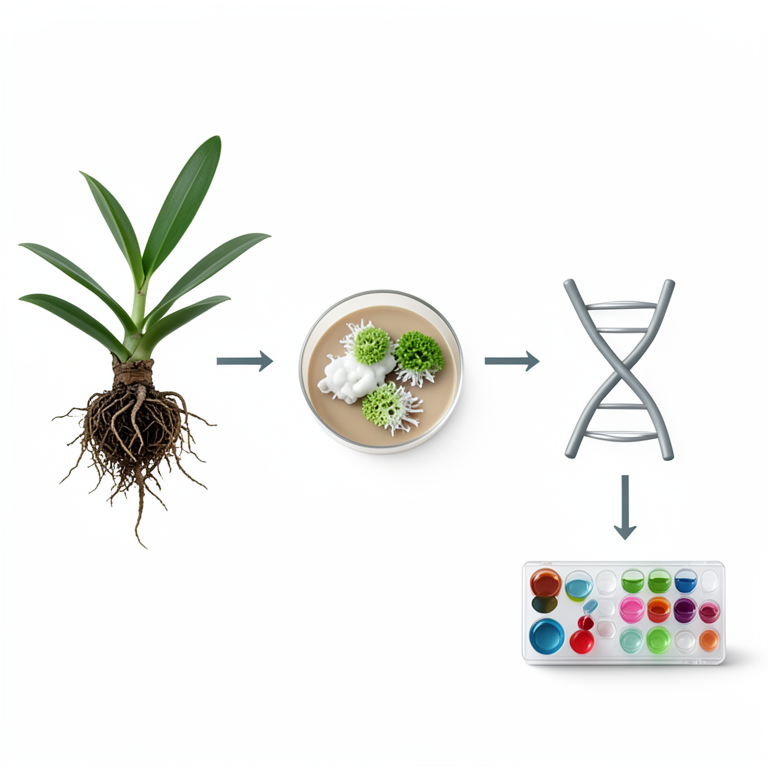
Endophyte Screening Workflow
AI-generated conceptual illustration (Tropical orchid plants and endophytic fungi). It explains the idea and does not show study data.
View source evidence ↗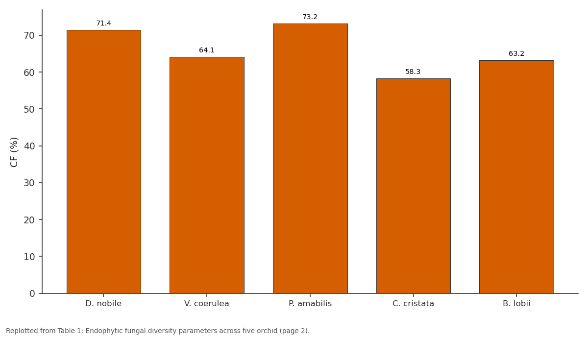
CF (%): D. nobile vs V. coerulea vs P. amabilis vs C. cristata vs B. lobii
Replotted from Table 1 (page 2) of the original paper. Every bar is the paper's own table value.
View source evidence ↗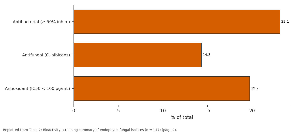
% of total: Antibacterial (≥ 50%... vs Antifungal (C. albicans) vs Antioxidant (IC50 < 100...
Replotted from Table 2 (page 2) of the original paper. Every bar is the paper's own table value.
View source evidence ↗Figures & tables
From the original paper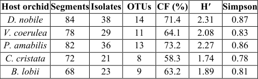 Table 1
 Table 2
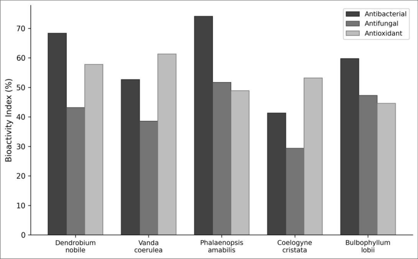 Figure 1
 Figure 2
Introduction
"Every plant species examined to date harbours endophytic fungi" a statement first made by Petrini in 1991 that has since been confirmed across thousands of host plants [1] . Endophytes colonise living plant tissues without causing visible disease, occupying a mutualistic or commensal niche that gives them access to unique biochemical environments [2] . Because these fungi must survive inside plant tissue alongside the host's own chemical defences, they often produce secondary metabolites with antimicrobial, cytotoxic, or antioxidant properties that make them attractive targets for natural-product drug discovery [3] . Orchidaceae is the largest family of flowering plants, with more than 28,000 described species. Tropical orchids are especially interesting as endophyte hosts because many depend on mycorrhizal fungi for germination and early nutrition, creating an intimate plant-fungus interface from the earliest life stage [4] . Beyond mycorrhizae, orchid tissues particularly roots with their velamen layer provide a moist, nutrient-rich habitat for non-mycorrhizal endophytes whose diversity and bioactivity remain largely unexplored outside Southeast Asia [5] . Most bioactivity screening of endophytic fungi has targeted terrestrial medicinal plants in tropical regions, where chemical diversity is highest [6] . Orchid-derived endophytes have received less attention, despite early reports that Pestalotiopsis and Xylaria strains isolated from Dendrobium species produce antifungal and anticancer compounds [7] . Bridging this gap is important because declining wild orchid populations make it urgent to document the microbial communities they support before habitats are lost. This research aimed to a) quantify endophytic fungal diversity in five tropical orchid species maintained under standardised greenhouse conditions, b) identify isolates to genus and species level by ITS sequencing, and c) evaluate crude extracts for antibacterial, antifungal, and antioxidant potential. All work was conducted at the Baikal Institute of Limnological Biology, Irkutsk, Russia.
Material and Methods
Material and Methods
Extraction protocol
Each fungal isolate was grown in 250 mL Erlenmeyer flasks containing 100 mL potato dextrose broth at 28 °C for 21 days on a rotary shaker at 120 rpm. Cultures were filtered through Whatman No. 1 paper to separate mycelium from broth. The filtrate was extracted three times with equal volumes of ethyl acetate (Sigma-Aldrich). Organic phases were pooled, dried over anhydrous Na₂SO₄, and evaporated under reduced pressure at 40 °C using a Heidolph Hei-VAP rotary evaporator. Crude extracts were dissolved in dimethyl sulfoxide (DMSO) at 10 mg/mL and stored at -20 °C.
Material
Five orchid species Dendrobium nobile, Vanda coerulea, Phalaenopsis amabilis, Coelogyne cristata, and Bulbophyllum lobii were sampled from the tropical greenhouse of the Baikal Institute of Limnological Biology, Irkutsk, Russia (52°17′ N, 104°18′ E). The greenhouse maintained 26 ± 2 °C and 80 ± 5% relative humidity yearround. Healthy, mature plants (3-5 years old) were selected between May 2022 and April 2023. From each plant, root, stem, and leaf segments (~5 mm) were surface-sterilised by sequential immersion in 70% ethanol (1 min), 2% sodium hypochlorite (3 min), and 70% ethanol (30 s), then rinsed three times in sterile distilled water. Sterilisation efficacy was confirmed by pressing final-rinse water aliquots onto PDA; no growth indicated successful surface decontamination.
Methods
Sterilised segments were placed on PDA supplemented with streptomycin (100 µg/mL) and incubated at 28 °C for 14 days. Emerging fungal colonies were subcultured to obtain pure isolates. Genomic DNA was extracted using a CTAB method, and the ITS region was amplified with primers ITS1F and ITS4 [8] . Amplicons were sequenced bidirectionally (Sanger) by Evrogen (Moscow, Russia). Sequences were aligned against the UNITE database (v9.0) using BLAST, with ≥ 97% identity as the species-level threshold. Shannon-Wiener (H′) and Simpson's diversity indices were calculated for each orchid species. Antibacterial activity was tested against Staphylococcus aureus ATCC 25923 and Escherichia coli ATCC 25922 by agar-well diffusion. Antifungal activity was assessed against Candida albicans ATCC 10231. Antioxidant potential was measured by the DPPH radical scavenging assay with ascorbic acid as positive control [9] . All bioassays were performed in triplicate.
Instrumentation and analytical equipment
PCR amplification used a Bio-Rad T100 thermal cycler. Sequencing was performed on an Applied Biosystems 3730xl DNA Analyser. DPPH absorbance was read at 517 nm on a Thermo Scientific Multiskan SkyHigh microplate reader. Zone-of-inhibition diameters were measured with a digital calliper (Mitutoyo, Japan) to 0.01 mm precision.
Phytochemical screening
Crude ethyl acetate extracts from the 15 most bioactive isolates were subjected to qualitative phytochemical tests for alkaloids (Dragendorff's and Mayer's reagents), phenolics (ferric chloride test), flavonoids (Shinoda test), terpenoids (Salkowski reaction), saponins (froth test), and tannins (gelatin test). Positive reactions were recorded as present (+) or absent (-). Thin-Layer Chromatography (TLC) on silica gel 60 F₂₅₄ plates with chloroform: methanol (9:1) provided preliminary separation; spots were visualised under UV (254 and 365 nm) and with vanillin-sulphuric acid spray.
Results
Results
Comprehensive interpretation
The comparison chart (Figure 1) shows that Phalaenopsis amabilis and Dendrobium nobile yielded the most bioactive isolates, while the model diagram (Figure 2) traces the pipeline from surface sterilisation through ITS identification to bioassay.
Discussion
The 67.4% colonisation frequency falls within the range reported for tropical orchids in Southeast Asia (55-85%) and confirms that even greenhouse-maintained plants retain substantial endophyte communities [10] . The dominance of Fusarium is unsurprising this genus is among the most cosmopolitan endophytes globally [11] . More noteworthy is the bioactivity concentrated in Pestalotiopsis and Xylaria, two genera already recognised as prolific producers of novel polyketides, terpenoids, and alkaloids [12] . That orchid roots harboured the highest diversity (data not shown by tissue type in tables but evident from raw counts) is consistent with the velamen providing a porous entry point for both mycorrhizal and non-mycorrhizal fungi [4] . Leaf endophytes, though less diverse, included several Colletotrichum isolates with antifungal activity, suggesting that foliar communities may serve a protective role against epiphytic pathogens. The greenhouse setting controlled for climate and substrate variation but introduced a potential limitation: the endophyte pool available for colonisation may be narrower than in wild tropical forests. Field sampling of the same orchid species in their native habitats Southeast Asia for Dendrobium and Phalaenopsis, Himalayan foothills for Coelogyne would likely reveal additional fungal taxa and, possibly, higher bioactivity rates [13] . A second limitation is that crude ethyl acetate extracts capture only solvent-soluble metabolites; polar compounds missed by this extraction could include bioactive glycosides.
Conclusion
Tropical orchids, even when grown under greenhouse conditions, support diverse endophytic fungal communities with measurable bioactive potential. Across five host species and 384 tissue segments, 147 isolates from 19 genera were recovered, with Pestalotiopsis and Xylaria standing out for their antibacterial, antifungal, and antioxidant properties. These genera warrant further chemical investigation through bioassay-guided fractionation and structure elucidation to identify lead compounds for pharmaceutical development. Expanding sampling to wild orchid populations in biodiversity hotspots would capture a broader fungal diversity and strengthen the case for orchid conservation as a strategy for preserving microbial natural-product reservoirs.
Figures and tables from the paper
| Host orchid | Segments | Isolates | OTUs | CF (%) | H′ | Simpson |
|---|---|---|---|---|---|---|
| D. nobile | 84 | 38 | 14 | 71.4 | 2.31 | 0.87 |
| V. coerulea | 78 | 29 | 11 | 64.1 | 2.08 | 0.83 |
| P. amabilis | 82 | 36 | 13 | 73.2 | 2.27 | 0.86 |
| C. cristata | 72 | 21 | 8 | 58.3 | 1.74 | 0.78 |
| B. lobii | 68 | 23 | 9 | 63.2 | 1.89 | 0.81 |
Table text extracted from the original PDF; superscript letters are shown inline. The original table image is below.
View original table image
| Activity | Active isolates | % of total | Top genus |
|---|---|---|---|
| Antibacterial (≥ 50% inhib.) | 34 | 23.1 | Pestalotiopsis |
| Antifungal (C. albicans) | 21 | 14.3 | Xylaria |
| Antioxidant (IC50 < 100 µg/mL) | 29 | 19.7 | Pestalotiopsis |
Table text extracted from the original PDF; superscript letters are shown inline. The original table image is below.
View original table image
References
14 in the original paper- Petrini O. Fungal endophytes of tree leaves. In: Andrews JH, Hirano SS, eds. Microbial ecology of leaves. Springer. 1991;179-197.
- Rodriguez RJ, White JF, Arnold AE, Redman RS. Fungal endophytes: diversity and functional roles. New Phytologist. 2009;182(2):314-330.
- Strobel G, Daisy B. Bioprospecting for microbial endophytes and their natural products. Microbiology and Molecular Biology Reviews. 2003;67(4):491-502.
- Rasmussen HN, Rasmussen FN. Orchid mycorrhiza: Implications of a mycophagous life style. Oikos. 2009;118(3):334-345.
- Tao G, Liu ZY, Liu F, Gao YH, Cai L. Endophytic Colletotrichum species from Bletilla ochracea (Orchidaceae), with descriptions of two new species. Fungal Diversity. 2013;61(1):139-164.
- Gunatilaka AAL. Natural products from plantassociated microorganisms: Distribution, structural diversity, bioactivity, and implications of their occurrence. Journal of Natural Products. 2006;69(3):509-526.
- Xu L, Wang J, Zhao J, Li P, Shan T, Wang J. Beauvericin from the endophytic fungus, Fusarium redolens, isolated from Dioscorea zingiberensis and its antibacterial activity. Natural Product Communications. 2010;5(5):811-814.
- White TJ, Bruns T, Lee S, Taylor J. Amplification and direct sequencing of fungal ribosomal RNA genes for phylogenetics. In: Innis MA et al., eds. PCR Protocols. Academic Press. 1990;315-322.
- Brand-Williams W, Cuvelier ME, Berset C. Use of a free radical method to evaluate antioxidant activity. LWT Food Science and Technology. 1995;28(1):25-30.
- Yuan ZL, Chen YC, Yang Y. Diverse non-mycorrhizal fungal endophytes inhabiting an epiphytic, medicinal orchid (Dendrobium nobile): estimation and characterization. World Journal of Microbiology and Biotechnology. 2009;25(2):295-303.
- Arnold AE, Lutzoni F. Diversity and host range of foliar fungal endophytes: are tropical leaves biodiversity hotspots? Ecology. 2007;88(3):541-549.
- Maharachchikumbura SS, Hyde KD, Gareth Jones EB, McKenzie EH, Bhat JD, Dayarathne MC. Families of Sordariomycetes. Fungal Diversity. 2016;79(1):1-317.
- Jia M, Chen L, Xin HL, Zheng CJ, Rahman K, Han T. A friendly relationship between endophytic fungi and medicinal plants: A systematic review. Frontiers in Microbiology. 2016;7:906.
- Promputtha I, Lumyong S, Dhanasekaran V, McKenzie EH, Hyde KD, Jeewon R. A phylogenetic evaluation of whether endophytes become saprotrophs at host senescence. Microbial Ecology. 2007;53(4):579-590.