
Research article · 2026 · Full text
International Journal of Biology SciencesAdvances in imaging techniques in veterinary sciences
Volume 8(4) · Pages 116–119 · DOI: 10.33545/26649926.2026.v8.i4b.804 ↗
Research areasVeterinary Medicine and SurgeryVeterinary Orthopedics and NeurologyMedical Imaging Techniques and Applications
Abstract
Author-writtenAdvances in imaging techniques have significantly transformed veterinary sciences by enabling accurate, non-invasive diagnosis and improved patient care. Modern imaging modalities such as ultrasonography, radiography, computed tomography (CT), magnetic resonance imaging (MRI), nuclear imaging, and endoscopy provide detailed insights into anatomical and physiological conditions in animals. Ultrasonography offers real-time imaging of soft tissues, while digital radiography enhances image quality and reduces radiation exposure. CT and MRI provide high-resolution cross-sectional and soft tissue imaging, aiding in the diagnosis of complex diseases. Nuclear imaging techniques allow early detection through metabolic visualization, and endoscopy supports minimally invasive diagnostic and therapeutic procedures. Despite these advancements, challenges such as high cost, limited accessibility, and the need for skilled personnel persist, especially in developing regions. Future developments, including artificial intelligence and machine learning, are expected to further enhance diagnostic accuracy, efficiency, and personalized veterinary care.
Summary
Source-checkedPlain-language reading of the full paper, prepared with AI and checked against the text. Each point links to the authors' own words.
Why it matters
- Non-invasive imaging techniques are critical in veterinary medicine because they provide the foundation for treating and predicting the outcome of animal diseases. View evidence ↗
- In developing countries, the availability of advanced imaging equipment is limited by high costs associated with the hardware, its maintenance, and the need for specialized staff. View evidence ↗
The question
- The objective of this review is to examine key developments in veterinary imaging technologies, their clinical importance, and their future potential. View evidence ↗
What they did
- The review analyzes several imaging modalities, including ultrasonography, radiography, CT, MRI, nuclear imaging, and endoscopy. View evidence ↗
What they found
- Ultrasonography is used to provide real-time imaging of soft tissues in animals. View evidence ↗
- The use of high-frequency transducers in ultrasonography has enhanced image quality, allowing for more detailed views of superficial structures. View evidence ↗
- Digital radiography improves upon traditional methods by providing better image quality, faster acquisition, and lower radiation exposure. View evidence ↗
- Computed Tomography (CT) provides superior spatial resolution compared to conventional radiography. View evidence ↗
- Because animal movement creates streak artifacts that reduce image quality, chemical restraint (sedation or anesthesia) is typically required for CT scans. View evidence ↗
- MRI is ideal for imaging joints, soft tissues, and the central nervous system because it offers high soft-tissue contrast without using ionizing radiation. View evidence ↗
- MRI is particularly effective at differentiating between neoplastic masses and inflammatory processes, as well as separating tumors from surrounding edema. View evidence ↗
- Nuclear imaging is specifically valuable for the early-stage diagnosis of diseases by visualizing metabolic activity. View evidence ↗
- The use of high-definition cameras, narrow-band imaging, and miniaturized equipment in endoscopy has improved the accuracy of lesion diagnosis. View evidence ↗
What it means
- The authors conclude that AI and machine learning will likely improve the speed and accuracy of veterinary diagnostics through better image interpretation and lesion detection. View evidence ↗
- The authors state that successfully integrating advanced imaging into veterinary practice requires collaboration between clinicians, researchers, and engineers. View evidence ↗
Visual summary
1 visualsIllustrations explain the paper; they do not show study data.
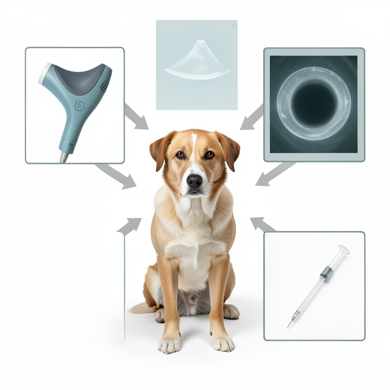
Veterinary Imaging Modalities Overview
AI-generated conceptual illustration (a mixed-breed dog). It explains the idea and does not show study data.
View source evidence ↗Introduction
Non invasive diagnostic techniques are one of the most important aspects in the veterinary clinical as well as research studies. Imaging techniques form the basis of treatment and prognosis of disease conditions in animals. Imaging techniques have revolutionized veterinary medicine, offering unprecedented insights into the anatomical, physiological, and pathological aspects of animal health. Over the years, there have been significant advancements in imaging modalities, enhancing diagnostic accuracy, treatment planning, and overall patient care in veterinary sciences. However availability of these equipments in veterinary field in developing countries is scarce due to high cost of equipments, maintenance and hiring technical staff. This review explores some of the key developments in imaging technologies tailored for veterinary applications, highlighting their clinical relevance and future prospects.
Ultrasonography
Ultrasonography operates on the principle of high-frequency sound wave reflection and transmission through tissues of different densities. It provides excellent resolution of soft tissues, allowing for the visualization of anatomical structures such as the liver, kidneys, heart, gastrointestinal and reproductive organs. Ultrasonography has revolutionized diagnostic imaging in veterinary medicine, offering non-invasive, real-time imaging capabilities that are invaluable in clinical practice. Its application spans across various veterinary disciplines, from small animals to large livestock, equines, and exotic species. The diagnostic ultrasound was first proposed by Dussik in 1942 when head was examined (Dussik et al., 1947) [5] . Later on studies were done on abdomen (Ludwig and Struthers, 1949) [15] , in obstetrics and eyes (Henry et al., 1956) [11] . A mode ultrasound was used for these studies (Wiesauer, 2004) [32] . In and around 1954, time motion or M-Mode ultrasound was developed to examine the organs in motion like movement of heart valve and wall (Edler and Hertz, 1954a, Edler and Hertz, 1954b) [6,7] . In early 1970s the real time or Brightness or B-Mode ultrasonography came in existence which is still the most widely used form of sonography (Griffith and Hennery, 1973) [10] . All the modes of ultrasound developed so far were not capable of studying the flow of blood, therefore in 1842 Johann Christian Doppler hypothesized the principle of Doppler ultrasonography which was proven scientifically in 1845 (White, 1982) [31] . In modern era of USG the Doppler can be used in combination with B-mode ultrasonography making it duplex imaging. The colour Doppler was also became functional in 1990s and used in duplex imaging (Curry et al, 1990) [4] . In this modality a mobile cursor can set the direction of Doppler on B-mode image which can now trace the blood flow in that particular region with more accuracy. Two modes of Doppler were developed i.e. pulse wave (PW) (Well, 1969) and continuous wave (CW) (Light, 1969). The PW Doppler scans a small segment specified by a gate on the cursor line but it can scan blood flow with a limited maximum velocity. On contrary the CW Doppler can scan a larger area (on the cursor line) and blood flow with high speed (King, 2006) [12] . Ultrasonography has undergone significant advancements, allowing non-invasive imaging of soft tissues and internal organs with high resolution. The introduction of highfrequency transducers has improved image quality, enabling detailed examination of superficial structures. Doppler ultrasonography facilitates assessment of blood flow dynamics, aiding in the diagnosis of vascular conditions and guiding interventions like biopsies and fluid aspirations. In addition to diagnostic uses the ultrasonography now a days have aided in therapeutics such as stimulation for tissue healing (Porter, 1991 and Steiss, 2000) [20,26] , vaccination of fish (Zohu et al., 2002) [33] , food preservation (Piyasena et al., 2003) [18] , measuring bone density in humans (Toyras et al., 2002) [28] and animals (Buckingham and Jeffcott, 1991, Toyras et al., 2002, Sandersen et al., 2003) [3,28,22] .
Radiography and Fluroscopy
Radiography remains a cornerstone in veterinary diagnostics, providing detailed images of bones, soft tissues, and internal organs. [13,14,17] . Fluoroscopy, with its real-time imaging capabilities, has become invaluable for dynamic studies such as barium contrast studies and cardiovascular assessments.
Computed Tomography (CT)
CT scanning provides cross-sectional images of anatomical structures, offering superior details as compared to conventional radiography. CT [16,8] . Computed Tomography is an advanced form of X-ray that obtain multi plane images of bone, ligament, tendon etc. used to diagnose problem in the musculo-skeltal system. Whereas conventional radiography produces summed images of an object, tomographic scanners rotate around the object being imaged, dividing the object & organising it into spatially consecutive image slices. The major advantage of CT over conventional radiography is improved spatial resolution. Computed Tomography imaging currently plays a prominent role in the diagnosis and evaluation of many human diseases (Goncalves-Ferreria et al, 2001) [9] . Computed tomography allows cross sectional imaging without bone and soft tissue overlap. Furthermore, three-dimensional rendering of the area of interest and multiplanar reformatting can yield better anatomic orientation of the area of interest and provide for more sensitive detection and characterization of disease extension (Tucker and Sande, 2001 and Bienert and Stadler, 2006) [27,2] . It was not used initially in veterinary medicine because of its limited accessibility and high cost. However, accessibility has improved, which has increased the need of expertise in the use of this technique in animals. CT technology can reach its full potential as an effective diagnostic modality when a normal species-specific crosssectional anatomic reference is provided. The CT Scan studies were performed on Canine (Sammi and Dyce, 2004; Woods et al., 2021) [21] , Cattle (Nuss et al., 2011), Ovine (Vandeweerd et al, 2012; Mao et al., 2017) [29] , Equine (Vekens et al, 2011; Epperly and Whitty, 2020) [30] , camel (Sangwan and Gahlot, 2015) [23] , Rabbit and rodent (Capello, 2016). Recent developments in CT technology have led to faster scanning times, higher spatial resolution, and reduced radiation dose. Multi slice CT scanners allow for rapid image acquisition, making them suitable for imaging of restless or critically ill patients. Advanced software enables 3D reconstruction and virtual endoscopy, enhancing surgical planning and educational purposes. However, CT scanning is a very well recommendable diagnostic modality but there are a few hitches in the use of CT scanning in animals. Firstly, the variable size of animal species ranging from mice to elephant. This great variation hinders the setting of linear attenuation coefficient which is the basic principle of CT scan imaging. Secondly, the movement of animals while scanning, which produces streak artifact reducing quality of images (Schwarz, 2011) [24] . Therefore, chemical restraining ranging from mild sedation to general anesthesia is almost mandatory for CT scanning in veterinary patients. In Veterinary field, the terminology used for CT image anatomical planes is very different as compared to that of human: transversal instead of axial, dorsal instead of coronal, for axial skeleton, the thorax and the abdomen, and frontal, instead of coronal, for the appendicular skeleton; sagittal is used as for human beings (Saunders and Schwarz, 2011) [25] . Contrast media used in Veterinary CT are the same used for human beings. In small animals, a non-ionic iodinated contrast media are usually administered at 400-600 mgI/kg of body weight. Allergic reactions to contrast media are rare in animals. (Pollard and Puchalski, 2011) [19] .
Magnetic Resonance Imaging (MRI)
MRI generates images of the body's organs using powerful magnetic fields, magnetic field gradients, and radio waves. The images are created by using radio waves to stimulate magnetized tissues and then capturing the returned radio waves from the stimulated tissues. Different sequences can be obtained depending on the type of emitted radio wave (duration, phase, and frequency) and the time at which the returning wave is captured. For the diagnosis of most neurologic, orthopedic, oncologic, and other illnesses, it is the imaging modality of choice. MRI investigations often comprise multiple different acquisition planes, including transverse, dorsal, and sagittal planes, in addition to varied sequences. MRI provides detailed soft tissue contrast without ionizing radiation, making it ideal for imaging the central nervous system, joints, and soft tissues. Veterinary MRI has seen advancements in magnet strength, gradient coils, and RF coil designs, improving image resolution and diagnostic accuracy. Functional MRI (fMRI) techniques are being explored for studying brain function in animals, offering insights into neurological diseases and cognitive processes. The MRI in veterinary institutions has been installed in 1990s. MRI is a relatively new imaging modality with considerable potential for visualizing soft tissues. It has a wide spectrum of application. This has enhanced our ability to study anatomy and pathologic changes in vivo. Today, MRI is often available in veterinary medicine and has proven its value in the detection of neurologic, orthopedics, oncologic, and other diseases. Many issues about musculoskeletal diseases in animals, such as the pathophysiology of navicular disease, traumatic arthritis, and osteochondrosis in equines, and wobbler syndrome in dogs, could be addressed with it. Magnetic resonance angiography (MRA) and Magnetic resonance spectroscopy (MRS) are two emerging MRI applications. It is notably useful for distinguishing between an inflammatory process and a neoplastic mass, as well as tumors and peritumoral edema. It is good at detecting osteomyelitis, cellulitis, and abscesses since it is more specific and sensitive. Because of its higher soft-tissue contrast and multiplanar and multislice imaging capabilities, it reveals excellent anatomical information of the canine and feline eye, orbit, and optic nerves. Other areas where MRI is being used for diagnosis in areas such as nasal neoplasia, eye and orbital diseases, and musculoskeletal diseases, including shoulder osteochondrosis, canine elbow dysplasia, and cranial cruciate ligament injuries.
Nuclear Imaging Techniques
Nuclear medicine in veterinary medicine involves the use of radioactive substances (radiopharmaceuticals) to diagnose and treat various conditions in animals. It utilizes imaging techniques such as scintigraphy (gamma camera imaging) and positron emission tomography (PET), similar to those used in human medicine. Techniques such as Scintigraphy, Single Photon Emission Computed Tomography (SPECT), Positron Emission Tomography (PET) and PET-CT are used in veterinary oncology and neurology (Daniel, 2014). These modalities utilize radiopharmaceuticals to visualize metabolic activity and identify abnormal tissue changes not visible with other imaging techniques. Therefore, an important aspect of nuclear imaging is to diagnose the diseases at an early stage. Advances in detector technology and image reconstruction algorithms have improved sensitivity and spatial resolution, expanding their clinical applications. These functional NM images are usually combined with morphologic modalities such as CT or MRI (like PET-CT). However, the availability of all modalities at one place is questionable in veterinary field (LeBlanc and Daniel, 2007).
Endoscopy and Minimally Invasive Imaging
Endoscopic techniques, including gastroscopy, bronchoscopy, and laparoscopy, have become essential for diagnosing and treating gastrointestinal, respiratory, and urogenital disorders in animals. Miniaturization of endoscopic equipment and advances in imaging technology (such as high-definition cameras and narrow-band imaging) have enhanced visualization of lesions and improved diagnostic accuracy.
Future Directions
The future of veterinary imaging holds promise with ongoing research in artificial intelligence (AI) and machine learning. AI algorithms are being developed to assist in image interpretation, lesion detection, and pattern recognition, potentially improving diagnostic speed and accuracy. Furthermore, advancements in imaging contrast agents and molecular imaging techniques are expected to further enhance early disease detection and personalized medicine in veterinary practice.
Conclusion
Advances in imaging techniques have transformed veterinary diagnostics and patient care, enabling precise and non-invasive evaluation of animal health. From digital radiography to advanced MRI and nuclear imaging, each modality offers unique benefits for different clinical scenarios. Continued innovation in imaging technology promises to further refine diagnostic capabilities, ultimately improving outcomes and quality of life for animals worldwide. In conclusion, the integration of cutting-edge imaging modalities into veterinary practice underscores the importance of interdisciplinary collaboration between veterinary clinicians, engineers, and researchers. As technology continues to evolve, so too will our ability to diagnose and treat animal diseases with greater precision and efficacy.
References
31 in the original paper- Arencibia A, Rivero MA, Gil F, Ramirez JA, Corbera JA, Ramirez G, et al. Anatomy of the cranioencephalic structures of the camel (Camelus dromedarius L.) by imaging techniques: a magnetic resonance imaging study. Anatomia Histologia Embryologia. 2005;34(1):52-55.
- Bienert A, Stadler P. Computed tomographic examination of the locomotor apparatus of horses -a review. Pferdeheilkunde. 2006;22:218-222.
- Buckingham SHW, Jeffcott LB. Osteopenic effects of forelimb immobilisation in horses. Veterinary Record. 1991;128:370-373.
- Curry TS, Dowdey JE, Murry RC. Ultrasound. In: Curry TS, Dowdey JE, Murray RC, editors. Christensens physics of diagnostic radiology. 4th ed. Williams & Wilkins; 1990. p. 323-371.
- Dussik KT, Dussik F, Wyt L. Auf dem wege zur hyperphonographic des gehirnes. Wiener Medizinische Wochenschrift. 1947;97:425-429.
- Fike J, LeCouteur R, Cann C. Anatomy of the canine brain using high resolution computed tomography. Veterinary Radiology and Ultrasound. 1981;22:236243.
- Goncalves-Ferreira AJ, Herculano-Carvalho M, Milanica JP, Farias JP, Gomes L. Corpus callosum: microsurgical anatomy and MRI. Surgical Radiologic Anatomy. 2001;23:409-414.
- Griffith JM, Henry WL. A real-time system for twodimensional echocardiography. In: Proceedings of the 26th Annual Conference of Engineering in Medicine and Biology. 1973;15:422.
- Henry JG, Mundt GN, Hughes WF. Ultrasonics in ocular diagnosis. American Journal of Ophthalmology. 1956;41:488.
- King AM. Development, advances and applications of diagnostic ultrasound in animals. The Veterinary Journal. 2006;171:408-420.
- Kraft SL, Gavin P. Physical principles and technical considerations for equine computed tomography and magnetic resonance imaging. Veterinary Clinics of North America Equine Practice. 2001;17:115-130.
- Latorre R, Arencibia A, Gil F, Rivero M, Henry RW, Ramirez G, et al. Correlation of magnetic resonance images with anatomic features of the equine tarsus. American Journal of Veterinary Research. 2006;67:756-761.
- Ludwig GD, Struthers FW. Considerations underlying the use of ultrasound to detect gall stones and foreign bodies in the tissues. United States Navy Medical Research Institute Report. 1949;4:1-27.
- Marincek B, Young SW. Computed tomography of spontaneous canine neoplasms. Veterinary Radiology. 1980;21(4):181-184.
- Park RD, Nelson TR, Hoopes J. Magnetic resonance imaging of the normal equine digit and metacarpophalangeal joint. Veterinary Radiology. 1987;28:105-116.
- Piyasena P, Mohareb E, Mckellar RC. Inactivation of microbes using ultrasound: a review. International Journal of Food Microbiology. 2003;87:207-216.
- Pollard R, Puchalski S. CT contrast media and application. In: Veterinary computed tomography. 1st ed. Wiley Blackwell; 2011. p. 57-65.
- Porter M. Therapeutic ultrasound. Journal of Equine Veterinary Science. 1991;11:357-362.
- Samii VF, Dyce J. Computed tomographic arthrography of the normal canine stifle. Veterinary Radiology and Ultrasound. 2004;45(5):402-406.
- Sandersen C, Guyot H, Vandeputte S, Carstanjen B, Amory H, Rollin F. Speed of sound measurements in the evaluation of bone properties in Holstein-Friesian cows: a preliminary study. Acta Veterinaria Scandinavica Supplementum. 2003;2:149.
- Sangwan A, Gahlot TK. Advanced imaging studies on normal stifle joint of camel (Camelus dromedarius). Journal of Camel Practice and Research. 2015;22(1):133-139.
- Schwarz T. Artifacts in CT. In: Schwarz T, Saunders J, editors. Veterinary computed tomography. 1st ed. Wiley Blackwell; 2011. p. 35-55.
- Saunders J, Schwarz T. Principles of CT images interpretation. In: Veterinary computed tomography. 1st ed. Wiley Blackwell; 2011. p. 29-34.
- Steiss JE. Physical therapy modalities: therapeutic ultrasound and phonophoresis. Compendium on Continuing Education for the Practicing Veterinarian. 2000;22:690-693.
- Tucker RL, Sande RD. Computed tomography and magnetic resonance imaging of the equine musculoskeletal conditions. Veterinary Clinics of North America Equine Practice. 2001;17:145-157.
- Toyras J, Nieminen MT, Kroger H, Jurvelin JS. Bone mineral density, ultrasound velocity, and broadband attenuation predict mechanical properties of trabecular bone differently. Bone. 2002;31:503-507.
- Vandeweerd JM, Kirschvink N, Muylkens B, Depiereux E, Clegg P, Herteman N, et al. A study of the anatomy and injection techniques of the ovine stifle by positive contrast arthrography, computed tomography arthrography and gross anatomical dissection. The Veterinary Journal. 2012;193:426-432.
- Vekens EV, Bergman EH, Vanderperren K, Raes EV, Puchalski SM, Bree HJ, et al. Computed tomographic anatomy of equine stifle joint. American Journal of Veterinary Research. 2011;72(4):512-521.
- White DN. Johann Christian Doppler and his effect: a brief history. Ultrasound in Medicine and Biology. 1982;8:583.
- Wiesauer F. Kretz museum tour -the history of ultrasound. 2004. Available from: http://www.obgyn.net/avtranscripts/RSNA/transducers. htm
- Zhou YC, Wang J, Zhang B, Su YQ. Ultrasonic immunization of sea bream, Pagrus major (Temminck and Schlegel), with a mixed vaccine against Vibrio alginolyticus and Vibrio anguillarum. Journal of Fish Diseases. 2002;25:325-331.