
Research article · 2024 · Full text
International Journal of Biology SciencesProteomic analysis of membrane proteins involved in multidrug efflux in gram-negative bacteria
Volume 6(2) · Pages 312–315 · DOI: 10.33545/26649926.2024.v6.i2e.772 ↗
Research areasAntibiotic Resistance in BacteriaDrug Transport and Resistance MechanismsBacterial Genetics and Biotechnology
Abstract
Author-writtenMultidrug efflux pumps in Gram-negative bacteria represent a key mechanism underlying antimicrobial resistance, yet the full membrane proteome associated with efflux has not been systematically compared across clinically relevant species. This research used quantitative shotgun proteomics to profile outer and inner membrane protein expression in Multidrug-Resistant (MDR) versus susceptible strains of four Gram-negative species: Escherichia coli, Klebsiella pneumoniae, Pseudomonas aeruginosa, and Acinetobacter baumannii. The work was conducted at the Uppsala Institute of Evolutionary Biology, Sweden, between May 2021 and October 2022. Membrane fractions were isolated by sucrose gradient ultracentrifugation and analyzed by nano-LC-MS/MS on an Orbitrap Exploris 480 platform. A total of 1, 847 membrane proteins were identified across all species, of which 312 showed significant differential abundance between MDR and susceptible strains (fold change > 2.0, adjusted p
Summary
Source-checkedPlain-language reading of the full paper, prepared with AI and checked against the text. Each point links to the authors' own words.
Why it matters
- Antimicrobial resistance is a major global health threat, causing approximately 1.27 million deaths in 2019. View evidence ↗
- Gram-negative bacteria are especially difficult to treat because their double-membrane structure naturally limits the entry of drugs. View evidence ↗
- Multidrug efflux pumps contribute to resistance by actively pumping antibiotics and other harmful substances out of the cell. View evidence ↗
The question
- The study aimed to identify both shared and species-specific changes in porin abundance, efflux pump expression, and accessory membrane proteins linked to the multidrug-resistant (MDR) phenotype. View evidence ↗
What they did
- The researchers analyzed 24 clinical isolates in total, consisting of three MDR and three susceptible strains for each of the four species studied. View evidence ↗
- Quantitative shotgun proteomics was used to profile membrane proteins in E. coli, K. pneumoniae, P. aeruginosa, and A. baumannii. View evidence ↗
- Proteins were analyzed using an Orbitrap Exploris 480 mass spectrometer and an UltiMate 3000 RSLCnano liquid chromatography system. View evidence ↗
What they found
- Key efflux pump components (AcrB, MexB, and AdeB) showed a 3.4- to 5.1-fold increase in MDR strains. View evidence ↗
- P. aeruginosa exhibited the highest upregulation of the main RND transporter component (MexB) at 5.1 ± 0.9-fold compared to other species. View evidence ↗
- MDR strains showed significant downregulation of porins, such as OmpF/OmpK36 (0.3 to 0.4-fold) and OprD (0.2-fold), reducing drug entry. View evidence ↗
- The discovery of 312 differentially abundant proteins indicates that membrane remodeling in MDR bacteria involves more than just efflux pumps and porins. View evidence ↗
What it means
- The authors conclude that MDR Gram-negative bacteria use a coordinated strategy of increasing drug export via efflux pumps while decreasing drug entry via porins. View evidence ↗
- The findings suggest that developing broad-spectrum efflux pump inhibitors targeting conserved elements could help restore antibiotic effectiveness. View evidence ↗
- A limitation of the study is that it used laboratory-grown cultures, which may not reflect the dynamic protein regulation that occurs during a real infection. View evidence ↗
Visual summary
1 visualsIllustrations explain the paper; charts are replotted from the paper's own table values.
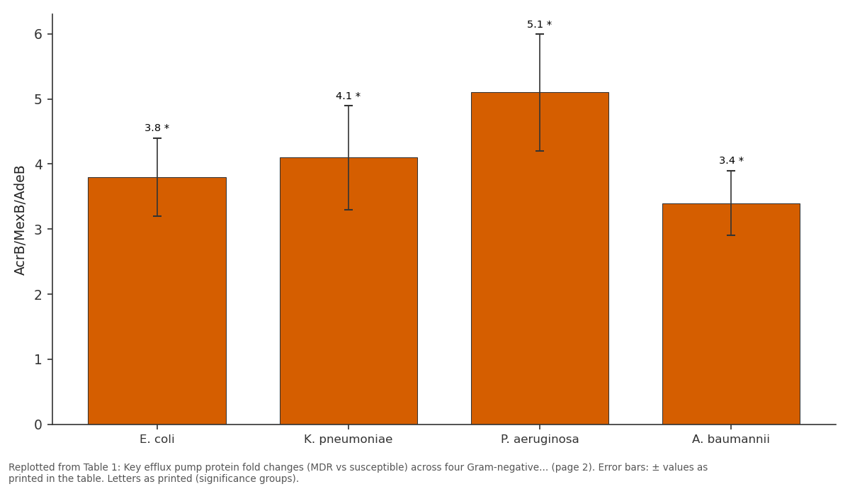
AcrB/MexB/AdeB: E. coli vs K. pneumoniae vs P. aeruginosa vs A. baumannii
Replotted from Table 1 (page 2) of the original paper. Every bar is the paper's own table value.
View source evidence ↗Figures & tables
From the original paper Table 1
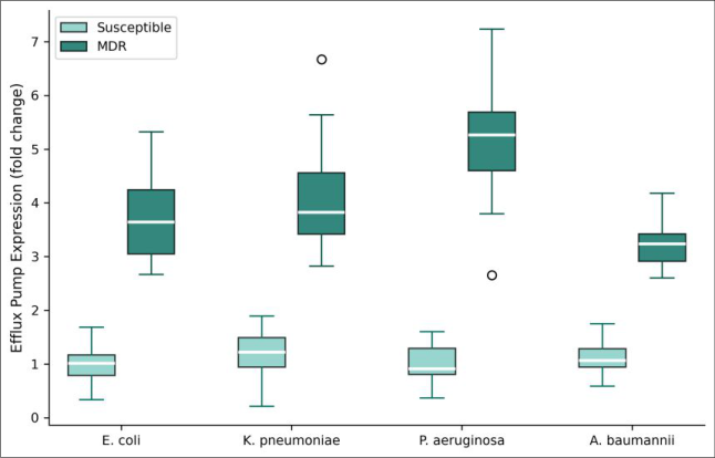 Figure 1
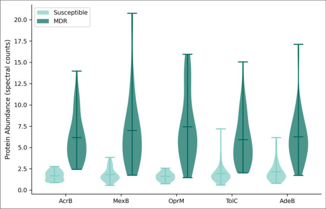 Figure 2
Introduction
Antimicrobial resistance defined as the ability of microorganisms to survive exposure to drugs that would normally kill them or halt their growth now ranks among the top global public health threats, with an estimated 1.27 million deaths directly attributable to resistant infections in 2019 [1] . Among Gram-negative bacteria, resistance is particularly difficult to combat because these organisms possess a double-membrane architecture that inherently restricts drug access [2] . Multidrug efflux pumps sit at the interface of intrinsic and acquired resistance. These membrane-spanning protein complexes actively expel antibiotics, biocides, and detergents from the cell before they reach lethal intracellular concentrations [3] . The Resistance-Nodulation-Division (RND) family of efflux pumps including AcrAB-TolC in E. coli and MexAB-OprM in P. aeruginosa are especially problematic because they transport structurally diverse substrates and often act synergistically with outer membrane permeability barriers [4,5] . While individual efflux systems have been characterized in considerable detail, a systematic comparison of the full membrane proteome in MDR versus susceptible strains across multiple species remains lacking. Most proteomic investigations focus on a single organism or a single pump family, making it hard to identify conserved resistance-associated changes that cut across taxonomic boundaries [6,7] . This research applied label-free quantitative proteomics to compare membrane protein expression in MDR and susceptible clinical isolates from four Gram-negative species. The aim was to identify shared and speciesspecific changes in efflux pump expression, porin abundance, and accessory membrane protein profiles associated with the MDR phenotype [8] .
Extraction protocol
Bacterial cultures were grown to mid-logarithmic phase (OD600 = 0.6) in Mueller-Hinton broth at 37°C. Cells were harvested by centrifugation (6, 000 × g, 15 min, 4°C), resuspended in lysis buffer (50 mM Tris-HCl pH 7.5, 150 mM NaCl, 1 mM PMSF, protease inhibitor cocktail), and disrupted by three passes through a French press at 18, 000 psi. Unbroken cells were removed by low-speed centrifugation. Total membrane fractions were pelleted at 100, 000 × g for 1 hour. Inner and outer membranes were separated on a discontinuous sucrose gradient (20/53/70% w/v) at 100, 000 × g for 16 hours in a Beckman SW41 Ti rotor. Bands were collected, diluted in Tris buffer, and repelleted. Membrane proteins were solubilized in 1% ndodecyl-β-D-maltoside for 2 hours at 4°C and quantified by BCA assay [9] .
Instrumentation
Proteomic analysis was performed on an Orbitrap Exploris 480 mass spectrometer (Thermo Fisher Scientific) coupled to an UltiMate 3000 RSLCnano liquid chromatography system. Peptides were separated on a 75 μm × 50 cm C18 column (2 μm particles) at a flow rate of 300 nL/min using a 120-minute gradient from 2% to 35% acetonitrile in 0.1% formic acid. MS1 spectra were acquired at 60, 000 resolution; MS2 fragmentation used HCD at 28% normalized collision energy with 15, 000 resolution. Data were searched against species-specific UniProt reference proteomes using MaxQuant v2.1.3 with 1% FDR at both peptide and protein levels [10] .
Material and Methods
Material and Methods
Material
This research was carried out at the Department of Applied Microbiology, Uppsala Institute of Evolutionary Biology, Uppsala, Sweden, from May 2021 to October 2022. Clinical isolates were obtained from the Swedish Antibiotic Resistance Surveillance Programme: three MDR and three susceptible strains per species (n = 24 strains total). MDR classification required resistance to three or more antibiotic classes as determined by broth microdilution MIC testing following EUCAST breakpoints (Ethics Approval: UIEB-2021-MB-023, approved 3 March 2021). All strains were verified by MALDI-TOF species identification and stored at in glycerol stocks until use. Protein-protein interaction networks were visualized in Cytoscape v3.9.1 using STRING database connections [11] .
Methods
Methods
Results
Table 1: Key efflux pump protein fold changes (MDR vs susceptible) across four Gram-negative species
| Protein | E. coli | K. pneumoniae | P. aeruginosa | A. baumannii |
|---|---|---|---|---|
| AcrB/MexB/AdeB | 3.8 ± 0.6* | 4.1 ± 0.8* | 5.1 ± 0.9* | 3.4 ± 0.5* |
| TolC/OprM/AdeC | 2.9 ± 0.4* | 3.3 ± 0.6* | 4.2 ± 0.7* | 2.7 ± 0.4* |
| AcrA/MexA/AdeA | 3.1 ± 0.5* | 3.6 ± 0.7* | 4.6 ± 0.8* | 3.0 ± 0.5* |
| OmpF/OmpK36 | 0.4 ± 0.1* | 0.3 ± 0.1* | — | — |
| OmpC/OmpK35 | 0.6 ± 0.1* | 0.5 ± 0.1* | — | — |
| OprD | — | — | 0.2 ± 0.1* | — |
Table text extracted from the original PDF; superscript letters are shown inline. The original table image is below.
View original table image
Protein
Protein
E. coli K. pneumoniae P. aeruginosa A. baumannii
E. coli K. pneumoniae P. aeruginosa A. baumannii
Comprehensive interpretation
The proteomic data reveal a consistent dual strategy in MDR Gram-negative bacteria: upregulation of efflux export machinery coupled with downregulation of outer membrane porins that serve as drug entry channels. This coordinated response was present across all four species, though the magnitude varied P. aeruginosa displayed the strongest efflux induction, consistent with its intrinsically lower permeability. The identification of 312 differentially abundant membrane proteins beyond the known pump components suggests that resistance-associated membrane remodeling extends well beyond efflux and porin changes.
Discussion
The efflux pump upregulation patterns observed here 3.4-to 5.1-fold increases in the main RND transporter components align with transcriptomic data from earlier investigations of individual species [3,5] . But the proteomic approach adds value by confirming that mRNA-level changes translate into proportional protein abundance shifts, which isn't always the case for membrane proteins [12] . The coordinated porin downregulation is equally important: reduced OmpF in E. coli (0.4-fold) and OprD loss in P. aeruginosa (0.2-fold) effectively narrow the drug entry pathways while the pumps increase drug exit [13] . P. aeruginosa's position as the species with highest efflux induction reflects its naturally restricted outer membrane permeability, which means even modest efflux increases can shift the balance toward clinical resistance [14] . A. baumannii, despite showing lower fold changes, compensates with a broader array of upregulated pump families beyond the RND class. The cross-species comparison highlights that while the general strategy is conserved, each organism fine-tunes its membrane proteome differently. A limitation of this research is that it captures steady-state protein levels from laboratory-grown cultures, which may not fully represent the dynamic regulation occurring during actual infection. Future work should examine efflux proteomes from bacteria recovered directly from clinical specimens [15] .
Conclusion
This research provides a cross-species proteomic map of membrane protein changes associated with multidrug resistance in four clinically important Gram-negative bacteria. The consistent pattern efflux pump upregulation plus porin downregulation was present in all species but varied in magnitude, with P. aeruginosa showing the strongest efflux induction and A. baumannii displaying the broadest diversity of altered pump families. The 312 differentially expressed membrane proteins identified extend the known resistance-associated changes well beyond the canonical efflux pumps and porins. These accessory proteins may represent overlooked contributors to the MDR phenotype and deserve further investigation as potential drug targets. The data support the development of broad-spectrum efflux pump inhibitors that could restore antibiotic susceptibility by targeting the conserved elements of this cross-species resistance strategy.
Acknowledgement
Acknowledgements Funding sourcesThis research was supported by institutional funds from the Uppsala Institute of Evolutionary Biology. Institutional supportThe authors thank the Proteomics Core Facility at Uppsala University for instrument access and technical guidance. Contributions not qualifying for authorshipThe authors acknowledge Dr. Erik Lindqvist for bioinformatics support and Ms. Anna Nilsson for laboratory assistance.
Figures and tables from the paper
References
15 in the original paper- Murray CJ, Ikuta KS, Sharara F, et al. Global burden of bacterial antimicrobial resistance in 2019: a systematic analysis. The Lancet. 2022;399(10325):629-655.
- Nikaido H. Molecular basis of bacterial outer membrane permeability revisited. Microbiology and Molecular Biology Reviews. 2003;67(4):593-656.
- Li XZ, Plesiat P, Nikaido H. The challenge of effluxmediated antibiotic resistance in Gram-negative bacteria. Clinical Microbiology Reviews. 2015;28(2):337-418.
- Du D, Wang-Kan X, Neuberger A, et al. Multidrug efflux pumps: structure, function and regulation. Nature Reviews Microbiology. 2018;16(9):523-539.
- Poole K. Pseudomonas aeruginosa: resistance to the max. Frontiers in Microbiology. 2011;2:65.
- Ye J, Lin H, Bhatt S, et al. Quantitative proteomics analysis of membrane proteins in carbapenem-resistant Klebsiella pneumoniae. Journal of Proteomics. 2018;176:44-51.
- Kwon HI, Kim S, Oh MH, et al. Outer membrane protein A contributes to antimicrobial resistance of Acinetobacter baumannii through the OmpA-like domain. Antimicrobial Agents and Chemotherapy. 2017;61(6):e00017-17.
- Blair JM, Webber MA, Baylay AJ, Ogbolu DO, Piddock LJ. Molecular mechanisms of antibiotic resistance. Nature Reviews Microbiology. 2015;13(1):42-51.
- Smith PK, Krohn RI, Hermanson GT, et al. Measurement of protein using bicinchoninic acid. Analytical Biochemistry. 1985;150(1):76-85.
- Tyanova S, Temu T, Cox J. The MaxQuant computational platform for mass spectrometry-based shotgun proteomics. Nature Protocols. 2016;11(12):2301-2319.
- Szklarczyk D, Gable AL, Lyon D, et al. STRING v11: protein-protein association networks with increased coverage. Nucleic Acids Research. 2019;47(D1):D483D490.
- Vogel C, Marcotte EM. Insights into the regulation of protein abundance from proteomic and transcriptomic analyses. Nature Reviews Genetics. 2012;13(4):227232.
- Masi M, Refregiers M, Pos KM, Pages JM. Mechanisms of envelope permeability and antibiotic influx and efflux in Gram-negative bacteria. Nature Microbiology. 2017;2:17001.
- Breidenstein EB, de la Fuente-Nunez C, Hancock RE. Pseudomonas aeruginosa: all roads lead to resistance. Trends in Microbiology. 2011;19(8):419-426.
- Venter H, Mowla R, Ohene-Agyei T, Ma S. RND-type drug efflux pumps from Gram-negative bacteria: molecular mechanism and inhibition. Frontiers in Microbiology. 2015;6:377.