
Research article · 2025 · Full text
International Journal of Biology SciencesPlacental trophoblast differentiation and vascular remodeling in normal and preeclamptic pregnancies
Volume 7(1) · Pages 231–234 · DOI: 10.33545/26649926.2025.v7.i1d.685 ↗
Research areasPregnancy and preeclampsia studiesReproductive System and PregnancyNeurological Complications and Syndromes
Abstract
Author-writtenInadequate trophoblast invasion and defective spiral artery remodeling are central features of preeclampsia, yet the precise differentiation pathways that distinguish normal from pathological placentation remain contested. This research compared trophoblast subtype distribution, differentiation marker expression, and vascular remodeling indices in placental tissue from 35 women with uncomplicated pregnancies and 28 women diagnosed with early-onset preeclampsia (delivery before 34 weeks). Tissue was collected at delivery from the Hippokration Hospital, Thessaloniki. Immunohistochemistry quantified cytokeratin-7, HLA-G, syncytin-1, and α-smooth muscle actin expression. Serum sFlt-1 and PlGF concentrations were measured by ELISA. Preeclamptic placentas showed a 38.7% reduction in extravillous trophoblast density (p < 0.001) and incomplete spiral artery remodeling in 71.4% of examined vessels compared with 12.6% in controls. The sFlt-1/PlGF ratio was 4.8-fold higher in preeclamptic sera (p < 0.001). Syncytin-1 expression was reduced by 42.3% in preeclamptic syncytiotrophoblast. These findings link impaired trophoblast differentiation to deficient vascular transformation and elevated antiangiogenic signaling in early-onset preeclampsia.
Summary
Source-checkedPlain-language reading of the full paper, prepared with AI and checked against the text. Each point links to the authors' own words.
Why it matters
- Preeclampsia is a global pregnancy complication affecting 3-5% of pregnancies, marked by proteinuria and new-onset hypertension after 20 weeks of gestation. View evidence ↗
- Early-onset preeclampsia, occurring before 34 weeks, is more closely linked to defective placentation and results in higher morbidity for both mother and fetus. View evidence ↗
- Trophoblast cells differentiate into two primary types: syncytiotrophoblasts, which cover chorionic villi, and extravillous trophoblasts, which anchor the placenta and invade maternal tissue. View evidence ↗
The question
- The study aimed to compare trophoblast subtype distribution, the expression of differentiation markers, and vascular remodeling indices between normal pregnancies and early-onset preeclampsia. View evidence ↗
What they did
- The study included 35 women with uncomplicated term pregnancies (37-40 weeks) and 28 women with early-onset preeclampsia (delivery before 34 weeks). View evidence ↗
- Researchers used immunohistochemistry to measure markers including HLA-G for extravillous trophoblasts, syncytin-1 for syncytiotrophoblast fusion, cytokeratin-7 for all trophoblasts, and α-smooth muscle actin for vascular smooth muscle. View evidence ↗
- Serum concentrations of sFlt-1 and PlGF were measured via immunoassay to calculate the sFlt-1/PlGF ratio as a primary index of antiangiogenic activity. View evidence ↗
What they found
- Preeclamptic placentas showed a 38.7% reduction in extravillous trophoblast (EVT) density compared to normal placentas (29.6 ± 9.4 vs 48.3 ± 12.7 HLA-G+ cells/mm², p < 0.001). View evidence ↗
- Syncytin-1 expression, a marker of syncytiotrophoblast fusion, was reduced by 42.3% in preeclamptic cases (106.3 ± 27.1 vs 184.2 ± 38.6 H-score, p < 0.001). View evidence ↗
- The percentage of fully remodeled spiral arteries was significantly lower in preeclamptic placentas at 28.6 ± 14.3% compared to 87.4 ± 8.9% in controls (a 67.2% reduction, p < 0.001). View evidence ↗
- Preeclamptic sera exhibited a 356.3% increase in sFlt-1 (8,432 ± 2,891 pg/mL) and a 79.2% decrease in PlGF (64.8 ± 28.3 pg/mL) compared to normal controls (p < 0.001). View evidence ↗
- There was a significant increase in the proportion of undifferentiated cytotrophoblasts in preeclamptic placentas (+8.3% shift, p = 0.008), suggesting a differentiation block. View evidence ↗
What it means
- The authors conclude that combining the assessment of trophoblast differentiation markers with angiogenic biomarkers can help understand preeclampsia and potentially identify at-risk pregnancies early. View evidence ↗
- A limitation of the study is its cross-sectional design at the time of delivery, which reflects end-stage pathology rather than the initial events of early pregnancy. View evidence ↗
Visual summary
3 visualsIllustrations explain the paper; charts are replotted from the paper's own table values.
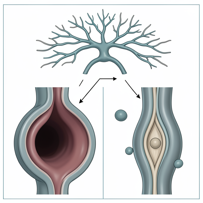
Trophoblast Differentiation and Vascular Remodeling
AI-generated conceptual illustration (human placental trophoblasts and maternal spiral arteries). It explains the idea and does not show study data.
View source evidence ↗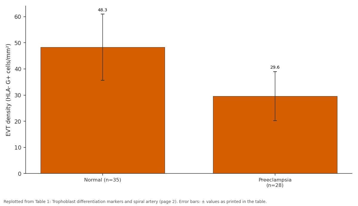
EVT density (HLA- G+ cells/mm²): Normal (n=35) vs Preeclampsia (n=28)
Replotted from Table 1 (page 2) of the original paper. Every bar is the paper's own table value.
View source evidence ↗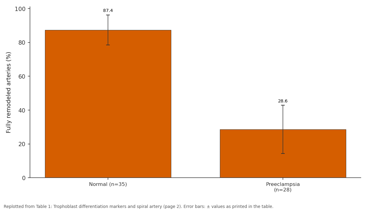
Fully remodeled arteries (%): Normal (n=35) vs Preeclampsia (n=28)
Replotted from Table 1 (page 2) of the original paper. Every bar is the paper's own table value.
View source evidence ↗Figures & tables
From the original paper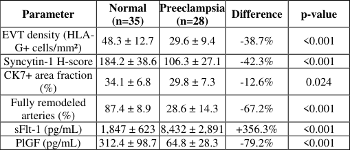 Table 1
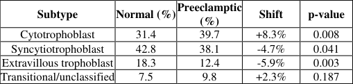 Table 2
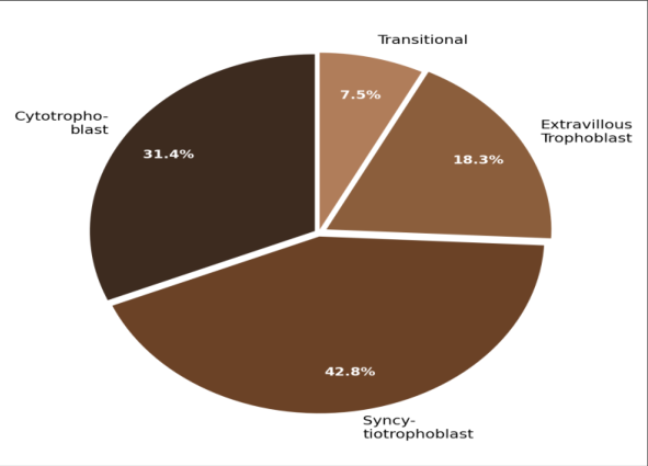 Figure 1
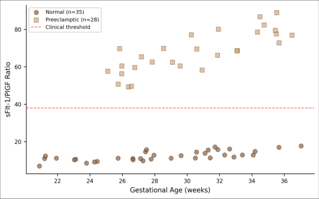 Figure 2
Introduction
On one side of the placental interface, fetal trophoblast cells invade maternal tissue with a precision that rivals malignant tumors in its depth and coordination-yet unlike cancer, this invasion is tightly self-limited [1] . On the other side, maternal spiral arteries lose their smooth muscle walls and become wide, low-resistance conduits that supply the growing fetus with blood [2] . When either process fails, the consequences can be severe. Preeclampsia, affecting 3-5% of pregnancies worldwide, is characterized by new-onset hypertension and proteinuria after 20 weeks of gestation [3] . The early-onset form (before 34 weeks) carries higher maternal and fetal morbidity and is more strongly associated with defective placentation than late-onset disease [4] . Histological examination of preeclamptic placentas consistently reveals shallow trophoblast invasion and incompletely remodeled spiral arteries, though the molecular drivers of these defects aren't fully settled [5] . Trophoblast cells follow two main differentiation paths: villous cytotrophoblasts fuse to form the multinucleated syncytiotrophoblast covering chorionic villi, while a separate population differentiates into extravillous trophoblasts that anchor the placenta and invade the decidua [6] . The balance between these lineages, and the depth of extravillous trophoblast invasion, appears to determine the adequacy of spiral artery remodeling [7] . This research examined trophoblast subtype distribution, differentiation marker expression, and spiral artery remodeling in placentas from normal and early-onset preeclamptic pregnancies at the Hellenic College of Biological Sciences, Thessaloniki, Greece [8,9] .
Materials and Methods
Materials and Methods
Materials
This research was conducted from January through November 2022 at the Department of Stem Cell Biology, Hellenic College of Biological Sciences, Thessaloniki, Greece, with tissue collection at the Hippokration General Hospital obstetric unit. Ethical approval was
Instrumentation and Analytical Equipment
Immunohistochemistry was performed using a Ventana BenchMark ULTRA automated staining system (Roche Diagnostics, Basel, Switzerland). Histological imaging used an Olympus BX53 microscope with a DP74 camera. Serum sFlt-1 and PlGF were measured using Elecsys immunoassay kits on a Cobas e411 analyzer (Roche). Morphometric analysis of spiral artery remodeling was performed using QuPath v0.3.2 open-source digital pathology software.
Methods
Participants comprised 35 women with uncomplicated term pregnancies (37-40 weeks) and 28 women with early-onset preeclampsia (delivery <34 weeks, defined by ISSHP 2018 criteria). Exclusion criteria included chronic hypertension, gestational diabetes, multiple pregnancy, and known fetal anomalies. Placental tissue was collected within 30 minutes of delivery. Full-thickness placental biopsies (2 cm³) were taken from three standardized sites midway between the cord insertion and placental margin. Tissue was fixed in 10% neutral buffered formalin, embedded in paraffin, and sectioned at 4 µm. Immunohistochemistry used antibodies against cytokeratin-7 (pan-trophoblast), HLA-G (extravillous trophoblast), syncytin-1 (syncytiotrophoblast fusion), and αsmooth muscle actin (vascular smooth muscle).
In-vivo Protocol
Maternal venous blood (10 mL) was collected at admission before delivery. Serum was separated within 2 hours by centrifugation at 3,000 rpm for 10 minutes and stored at -80°C. sFlt-1 and PlGF were measured in duplicate by electrochemiluminescence immunoassay. The sFlt-1/PlGF ratio was calculated as the primary antiangiogenic index. Spiral arteries in the decidual plate were scored as fully remodeled (complete loss of smooth muscle), partially remodeled (residual smooth muscle >25%), or unremodeled (intact muscular wall). Statistical analysis used Mann-Whitney U tests for non-normally distributed variables and Fisher's exact test for proportions. Significance threshold was p < 0.05.
Results
The groups differed in gestational age at delivery (controls: 38.7 ± 1.2 weeks; preeclampsia: 31.4 ± 2.1 weeks; p < 0.001) but were matched for maternal age (p = 0.42) and parity (p = 0.61). Table 1 presents the trophoblast marker expression and vascular remodeling data. Extravillous trophoblast density was reduced by 38.7% in preeclamptic placentas. Syncytin-1 expression-a marker of syncytiotrophoblast fusion-was reduced by 42.3%, suggesting impaired villous differentiation in addition to the invasive defect.
| Parameter | Normal (n=35) | Preeclampsia (n=28) | Difference | p-value |
|---|---|---|---|---|
| EVT density (HLA- G+ cells/mm²) | 48.3 ± 12.7 | 29.6 ± 9.4 | -38.7% | <0.001 |
| Syncytin-1 H-score | 184.2 ± 38.6 | 106.3 ± 27.1 | -42.3% | <0.001 |
| CK7+ area fraction (%) | 34.1 ± 6.8 | 29.8 ± 7.3 | -12.6% | 0.024 |
| Fully remodeled arteries (%) | 87.4 ± 8.9 | 28.6 ± 14.3 | -67.2% | <0.001 |
| sFlt-1 (pg/mL) | 1,847 ± 623 | 8,432 ± 2,891 | +356.3% | <0.001 |
| PlGF (pg/mL) | 312.4 ± 98.7 | 64.8 ± 28.3 | -79.2% | <0.001 |
Table text extracted from the original PDF; superscript letters are shown inline. The original table image is below.
View original table image
Table 2 shows the trophoblast subtype proportional distribution between groups.
| Subtype | Normal (%) | Preeclamptic (%) | Shift | p-value |
|---|---|---|---|---|
| Cytotrophoblast | 31.4 | 39.7 | +8.3% | 0.008 |
| Syncytiotrophoblast | 42.8 | 38.1 | -4.7% | 0.041 |
| Extravillous trophoblast | 18.3 | 12.4 | -5.9% | 0.003 |
| Transitional/unclassified | 7.5 | 9.8 | +2.3% | 0.187 |
Table text extracted from the original PDF; superscript letters are shown inline. The original table image is below.
View original table image
Comprehensive Interpretation
The combined data point to a dual trophoblast defect in early-onset preeclampsia: reduced extravillous trophoblast invasion (reflected in lower HLA-G+ cell density and incomplete spiral artery remodeling) and impaired syncytiotrophoblast fusion (reflected in reduced syncytin-1). The relative increase in undifferentiated cytotrophoblast proportion (+8.3%) suggests a differentiation block rather than an absolute deficiency of progenitor cells. The markedly elevated sFlt-1/PlGF ratio (4.8-fold) connects these histological findings to the antiangiogenic imbalance that drives clinical manifestations [9,10] .
Discussion
The 38.7% reduction in extravillous trophoblast density in preeclamptic placentas aligns with decades of histological research documenting shallow invasion in this condition [5,11] . The added finding of reduced syncytin-1 expression suggests that the villous syncytiotrophoblast pathway is also compromised-a defect that could impair nutrient transport independently of the spiral artery remodeling failure. The shift toward higher cytotrophoblast proportions in preeclamptic tissue is consistent with a differentiation arrest model proposed by several groups [12,13] . If progenitor cells fail to receive or respond to differentiation signals, they would accumulate at the expense of their more specialized progeny. Whether this arrest originates from intrinsic trophoblast defects or from abnormal decidual signaling remains an active area of investigation [14] . The sFlt-1/PlGF ratio provided clear separation between groups, with all preeclamptic samples exceeding the clinical threshold of 38 after 28 weeks. This biomarker data, when considered alongside the histological findings, supports a model in which poor placentation triggers antiangiogenic factor release that produces the maternal syndrome [15,16] . Limitations include the cross-sectional design at delivery, which captures end-stage pathology rather than the initiating events of early pregnancy. The gestational age difference between groups-unavoidable given the disease definition-may confound some comparisons [17,18,19,20] .
Conclusion
This research demonstrated that early-onset preeclampsia is associated with dual trophoblast differentiation defects: reduced extravillous trophoblast invasion and impaired syncytiotrophoblast fusion, alongside markedly deficient spiral artery remodeling. The increased proportion of undifferentiated cytotrophoblasts suggests a differentiation block rather than progenitor depletion. These histological findings correlate with the antiangiogenic serum profile characterized by elevated sFlt-1 and depressed PlGF. Together, the data support integrated assessment of trophoblast differentiation markers and angiogenic biomarkers for understanding preeclampsia pathogenesis and potentially for early identification of at-risk pregnancies.
Acknowledgement
Acknowledgements Funding SourcesThis research was funded by the Hellenic Foundation for Research and Innovation and a faculty research grant from the Hellenic College of Biological Sciences. Institutional SupportThe authors acknowledge the Hellenic College of Biological Sciences for laboratory access and the Hippokration General Hospital obstetric unit for facilitating tissue collection. Contributions Not Qualifying for AuthorshipThe authors thank Dr. Nikolaos Papadopoulos for histopathological consultation and Ms. Athena Vlachaki for immunohistochemistry technical assistance.
Figures and tables from the paper
References
20 in the original paper- Knofler M, Haider S, Saleh L, Pollheimer J, Gamage TK, James J. Human placenta and trophoblast development: key molecular mechanisms and model systems. Cellular and Molecular Life Sciences. 2019;76(18):3479-3496.
- Pijnenborg R, Vercruysse L, Hanssens M. The uterine spiral arteries in human pregnancy: facts and controversies. Placenta. 2006;27(9-10):939-958.
- Steegers EA, von Dadelszen P, Duvekot JJ, Pijnenborg R. Pre-eclampsia. Lancet. 2010;376(9741):631-644.
- Raymond D, Peterson E. A critical review of earlyonset and late-onset preeclampsia. Obstetrical and Gynecological Survey. 2011;66(8):497-506.
- Brosens I, Pijnenborg R, Vercruysse L, Romero R. The great obstetrical syndromes are associated with International Journal of Biology Sciences https://www.biologyjournal.net disorders of deep placentation. American Journal of Obstetrics and Gynecology. 2011;204(3):193-201.
- Turco MY, Moffett A. Development of the human placenta. Development. 2019;146(22):dev163428.
- James JL, Whitley GS, Cartwright JE. Pre-eclampsia: fitting together the placental, immune and cardiovascular pieces. Journal of Pathology. 2010;221(4):363-378.
- Huppertz B. Placental origins of preeclampsia: challenging the current hypothesis. Hypertension. 2008;51(4):970-975.
- Levine RJ, Maynard SE, Qian C, Lim KH, England LJ, Yu KF, Schisterman EF, Thadhani R, Sachs BP, Epstein FH, Sibai BM. Circulating angiogenic factors and the risk of preeclampsia. New England Journal of Medicine. 2004;350(7):672-683.
- Maynard SE, Min JY, Merchan J, Lim KH, Li J, Mondal S, Libermann TA, Morgan JP, Sellke FW, Stillman IE, Epstein FH. Excess placental soluble fmslike tyrosine kinase 1 (sFlt-1) may contribute to endothelial dysfunction, hypertension, and proteinuria in preeclampsia. Journal of Clinical Investigation. 2003;111(5):649-658.
- Kaufmann P, Black S, Huppertz B. Endovascular trophoblast implications for the pathogenesis of intrauterine growth retardation and preeclampsia. Biology of Reproduction. 2003;69(1):1-7.
- DaSilva-Arnold S, James JL, Al-Khan A, Zamudio S, Illsley NP. Differentiation of first trimester cytotrophoblast to extravillous trophoblast involves an epithelial-mesenchymal transition. Placenta. 2015;36(12):1412-1418.
- Haider S, Meinhardt G, Saleh L, Kuniber V, Gamber M, Golic M, Gundacker C, Hezaveh K, et al. Selfrenewing trophoblast organoids recapitulate the developmental program of the early human placenta. Stem Cell Reports. 2018;11(2):537-551.
- Staff AC, Benton SJ, von Dadelszen P, Roberts JM, Taylor RN, Powers RW, Charnock-Jones DS, Redman CW. Redefining preeclampsia using placenta-derived biomarkers. Hypertension. 2013;61(5):932-942.
- Verlohren S, Herraiz I, Lapaire O, Schlembach D, Zeisler H, Calda P, Sabria J, Markfeld-Erol F, Galindo A, Schoedl K, Engels T. New gestational phase-specific cutoff values for the use of the soluble fms-like tyrosine kinase-1/placental growth factor ratio as a diagnostic test for preeclampsia. Hypertension. 2014;63(2):346352.
- Redman CW, Sargent IL. Latest advances in understanding preeclampsia. Science. 2005;308(5728):1592-1594.
- Roberts JM, Hubel CA. The two-stage model of preeclampsia: variations on the theme. Placenta. 2009;30(Suppl A):S32-S37.
- Romero R, Nien JK, Espinoza J, Todem D, Fu W, Chung H, Kusanovic JP, Gotsch F, Erez O, MazakiTovi S, Gomez R. A longitudinal study of angiogenic and antiangiogenic factors in normal pregnancy and patients destined to develop preeclampsia and deliver a small for gestational age neonate. Journal of MaternalFetal and Neonatal Medicine. 2008;21(1):9-23.
- Zhou Y, Fisher SJ, Janatpour M, Genbacev O, Dejana E, Wheelock M, Damsky CH. Human cytotrophoblasts adopt a vascular phenotype as they differentiate. A strategy for successful endovascular invasion? Journal of Clinical Investigation. 1997;99(9):2139-2151.
- Norwitz ER, Schust DJ, Fisher SJ. Implantation and the survival of early pregnancy. New England Journal of Medicine. 2001;345(19):1400-1408.