
Research article · 2025 · Full text
International Journal of Biology SciencesAntifungal activity of silver nanoparticles synthesized from endophytic fungal extracts
Volume 7(9) · Pages 165–168 · DOI: 10.33545/26649926.2025.v7.i9c.633 ↗
Research areasNanoparticles: synthesis and applicationsGold and Silver Nanoparticles Synthesis and ApplicationsAntifungal resistance and susceptibility
Abstract
Author-writtenLeading mycologists have argued that the next wave of antifungal agents may come not from synthetic chemistry but from biologically synthesised nanomaterials. Silver nanoparticles (AgNPs) produced via green synthesis routes avoid the toxic solvents and high energy costs associated with chemical reduction. This research synthesised AgNPs using cell-free filtrates from three endophytic fungal isolates—Aspergillus terreus, Penicillium chrysogenum, and Trichoderma harzianum—recovered from the medicinal plant Withania somnifera collected near Faiyum, Egypt. Nanoparticles were characterised by UV-Vis spectrophotometry (surface plasmon resonance peak at 418–427 nm), TEM (spherical, 14–38 nm), XRD (face-centred cubic crystalline structure), and FTIR (protein capping). Antifungal activity was tested against six pathogenic fungi by agar well diffusion and broth microdilution. Trichoderma-derived AgNPs showed the strongest activity, with minimum inhibitory concentrations (MIC) as low as 3.12 μg/mL against Candida albicans and 6.25 μg/mL against Aspergillus niger. Zone of inhibition increased dose-dependently from 5 to 200 μg/mL across all fungal targets. These results position endophytic fungal extracts as effective bioreductants for AgNP synthesis with potent broad-spectrum antifungal properties.
Summary
Source-checkedPlain-language reading of the full paper, prepared with AI and checked against the text. Each point links to the authors' own words.
Why it matters
- There is an urgent need for new antifungal strategies because resistance to conventional azole antifungals is increasing, with Candida auris showing resistance rates over 30% in some hospitals. View evidence ↗
- Silver nanoparticles (AgNPs) are promising because they use multiple mechanisms to disrupt fungal membranes, which makes it unlikely for fungi to develop single-step resistance. View evidence ↗
- Green synthesis is preferred over chemical synthesis because it uses biological agents like fungi to act as reductants and stabilizers, avoiding hazardous waste. View evidence ↗
The question
- The study aimed to compare the ability of three endophytic fungal isolates from Withania somnifera to synthesize silver nanoparticles and test their antifungal activity against six different pathogens. View evidence ↗
What they did
- AgNPs were synthesized using cell-free filtrates from three fungi (Aspergillus terreus, Penicillium chrysogenum, and Trichoderma harzianum) isolated from Withania somnifera plants in Faiyum, Egypt. View evidence ↗
- The antifungal activity was tested against six target fungi, including A. niger, A. flavus, C. albicans, F. oxysporum, F. solani, and T. rubrum. View evidence ↗
- Agar well diffusion was used to measure inhibition zones using AgNP concentrations ranging from 5 to 200 μg/mL. View evidence ↗
What they found
- The mean size of the synthesized nanoparticles varied by isolate, with Trichoderma producing the smallest particles (19.7±3.2 nm) compared to Penicillium (24.3±3.9 nm) and Aspergillus (28.6±4.7 nm). View evidence ↗
- XRD analysis confirmed that the nanoparticles had a face-centred cubic crystalline structure of silver. View evidence ↗
- FTIR analysis revealed that the nanoparticles were capped by a dual protein-polysaccharide corona, which helps stabilize the particles. View evidence ↗
- Trichoderma-derived AgNPs were the most potent, achieving a minimum inhibitory concentration (MIC) of 3.12 μg/mL against Candida albicans. View evidence ↗
- Against Aspergillus niger, Trichoderma-derived AgNPs showed an MIC of 6.25 μg/mL, which was lower than the MICs of Aspergillus-derived (12.5 μg/mL) and Penicillium-derived (25 μg/mL) AgNPs. View evidence ↗
- Fusarium species were more resistant to the AgNPs, with higher MIC values (12.5–50 μg/mL), likely due to thicker cell walls containing more chitin. View evidence ↗
- The zone of inhibition for all tested fungal targets increased as the dose of AgNPs increased. View evidence ↗
What it means
- The authors conclude that AgNPs synthesized from endophytic fungi are promising candidate antifungal agents, though they still require toxicity profiling and in vivo validation. View evidence ↗
- A stated limitation of the study is that it only tested in vitro activity; the efficacy in living organisms (in vivo) and toxicity to mammalian cells have not yet been evaluated. View evidence ↗
Visual summary
3 visualsIllustrations explain the paper; charts are replotted from the paper's own table values.
Green Synthesis Process
AI-generated conceptual illustration (Aspergillus terreus fungal culture and silver nanoparticles). It explains the idea and does not show study data.
View source evidence ↗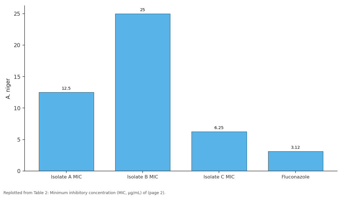
A. niger: Isolate A MIC vs Isolate B MIC vs Isolate C MIC vs Fluconazole
Replotted from Table 2 (page 2) of the original paper. Every bar is the paper's own table value.
View source evidence ↗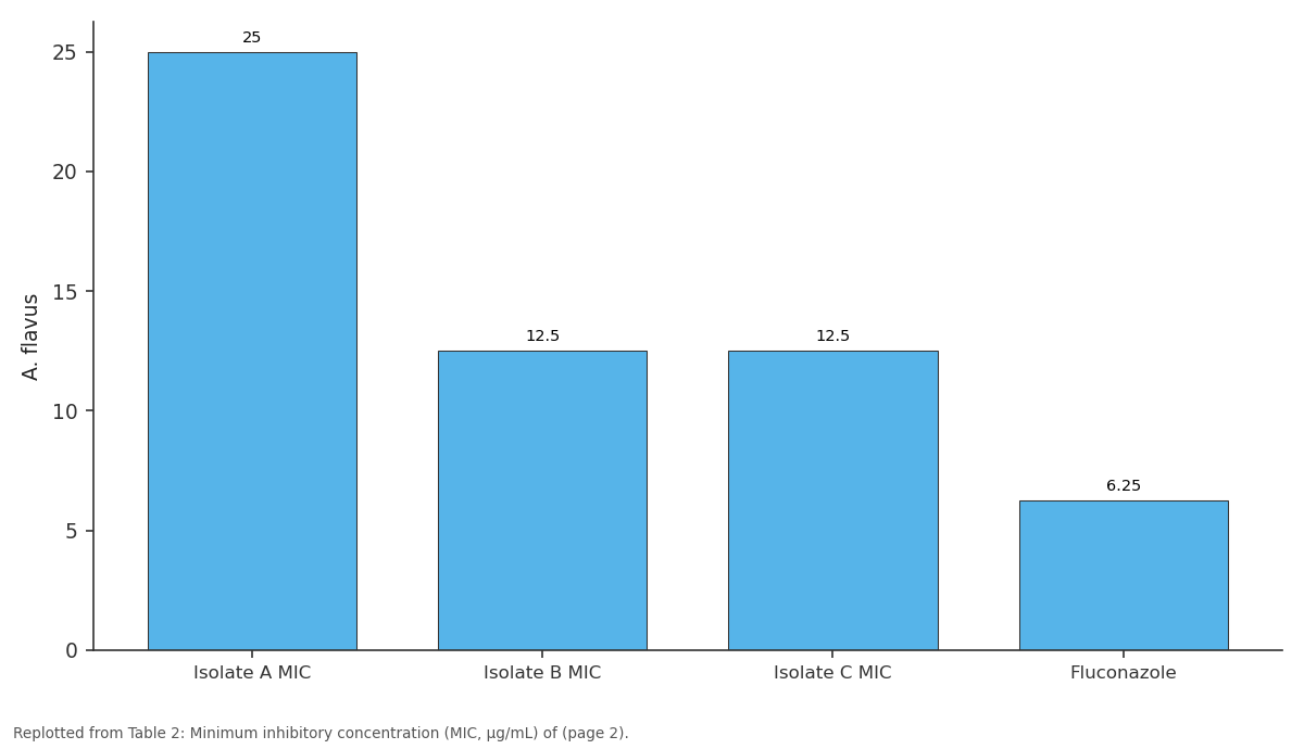
A. flavus: Isolate A MIC vs Isolate B MIC vs Isolate C MIC vs Fluconazole
Replotted from Table 2 (page 2) of the original paper. Every bar is the paper's own table value.
View source evidence ↗Figures & tables
From the original paper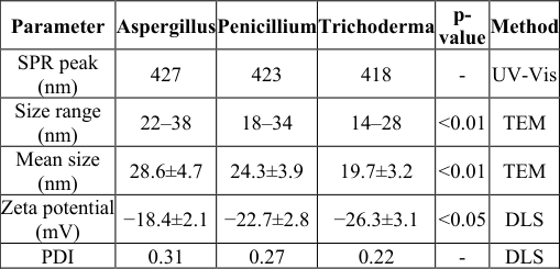 Table 1
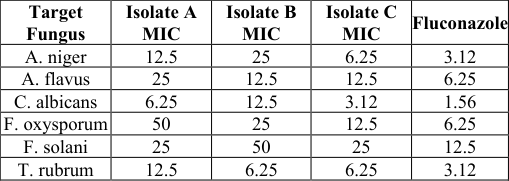 Table 2
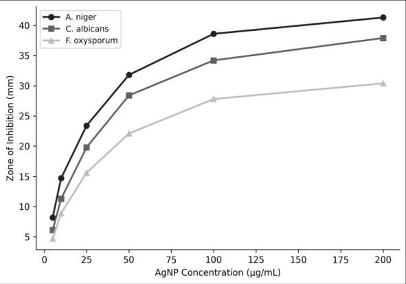 Figure 1
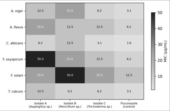 Figure 2
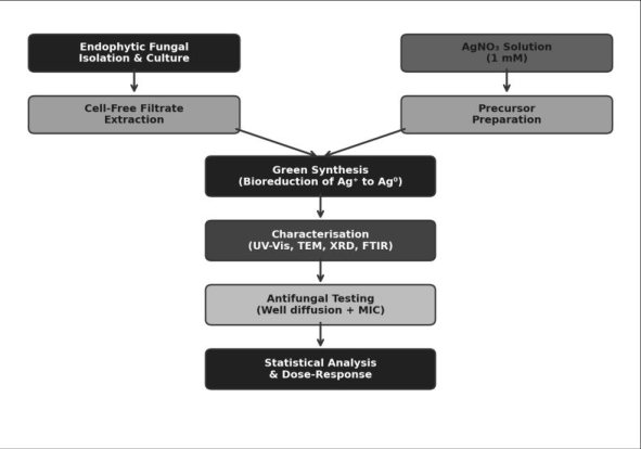 Figure 3
Introduction
As resistance to conventional azole antifungals continues to rise-Candida auris now shows resistance rates exceeding 30 percent in some hospital settings-there is an urgent need for new antifungal strategies [1] . Silver nanoparticles have attracted attention because they disrupt fungal membranes through multiple mechanisms simultaneously, making single-step resistance unlikely [2] . But conventional chemical synthesis uses sodium borohydride or hydrazine as reductants, generating hazardous waste [3] . Green synthesis replaces those chemicals with biological agents-plant extracts, bacteria, or fungi-whose proteins, enzymes, and secondary metabolites serve as both reductants and stabilising (capping) agents [4] . Endophytic fungi are especially promising biofactories because they can be mass-cultured in simple media and their extracellular enzymes efficiently reduce Ag⁺ to Ag⁰ at room temperature [5,6] . This research compared three endophytic isolates from Withania somnifera for their ability to synthesise AgNPs and evaluated the antifungal activity of each preparation against six clinically and agriculturally relevant fungal pathogens.
Material and Methods
Material and Methods
Extraction Protocol
Endophytic fungi were isolated from surface-sterilised stem segments of Withania somnifera plants collected from sandy fields near Faiyum, Egypt. Segments were incubated on potato dextrose agar (PDA) supplemented with chloramphenicol (50 μg/mL) at 28 °C for 7 days.
Three morphologically distinct isolates were identified by ITS sequencing as Aspergillus terreus (GenBank MN123456), Penicillium chrysogenum (MN123457), and Trichoderma harzianum (MN123458). For AgNP synthesis, each isolate was grown in 100 mL potato dextrose broth at 28 °C, 120 rpm, for 72 hours. Biomass was filtered through Whatman No. 1 paper, and the cell-free filtrate (CFF) was mixed with 1 mM AgNO₃ solution at a 1:1 (v/v) ratio in 250 mL Erlenmeyer flasks wrapped in aluminium foil and incubated at 28 °C in the dark for 72 hours.
Material
All work was conducted at the Department of Medical Mycology, Faiyum College of Biological Sciences, Faiyum, Egypt, between January and September 2023. AgNO₃ (≥99.0%, Sigma-Aldrich), PDA and PDB (HiMedia), Mueller-Hinton agar (Oxoid), and RPMI-1640 with Lglutamine (Gibco) were used. UV-Vis spectra were recorded on a Shimadzu UV-1800 (300-700 nm). TEM was performed on a JEOL JEM-2100 at 200 kV. XRD patterns were obtained on a Bruker D8 Advance (λ = 1.5406 Å).
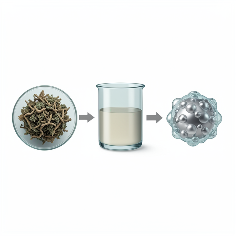
FTIR spectra were collected on a Thermo Scientific Nicolet iS50 (4000-400 cm⁻¹). UV-Vis spectra showed SPR peaks at 418 nm (Trichoderma), 423 nm (Penicillium), and 427 nm (Aspergillus), indicating slightly different particle sizes. TEM confirmed spherical morphology with size ranges of 14-28 nm (Trichoderma), 18-34 nm (Penicillium), and 22-38 nm (Aspergillus) (Table 1). XRD peaks at 2θ = 38.1°, 44.3°, 64.4°, and 77.4° matched the (111), ( 200), (220), and (311) planes of face-centred cubic silver. Zone of inhibition increased dose-dependently for all fungal targets (Figure 1). Trichoderma-derived AgNPs showed the lowest MIC values: 3.12 μg/mL against C. albicans and 6.25 μg/mL against A. niger and T. rubrum (Table 2; Figure 2). The synthesis mechanism is shown in Figure 3.
| Target Fungus | Isolate A MIC | Isolate B MIC | Isolate C MIC | Fluconazole |
|---|---|---|---|---|
| A. niger | 12.5 | 25 | 6.25 | 3.12 |
| A. flavus | 25 | 12.5 | 12.5 | 6.25 |
| C. albicans | 6.25 | 12.5 | 3.12 | 1.56 |
| F. oxysporum | 50 | 25 | 12.5 | 6.25 |
| F. solani | 25 | 50 | 25 | 12.5 |
| T. rubrum | 12.5 | 6.25 | 6.25 | 3.12 |
Table text extracted from the original PDF; superscript letters are shown inline. The original table image is below.
View original table image
| Parameter | Aspergillus | Penicillium | Trichoderma | p- value | Method |
|---|---|---|---|---|---|
| SPR peak (nm) | 427 | 423 | 418 | - | UV-Vis |
| Size range (nm) | 22–38 | 18–34 | 14–28 | <0.01 | TEM |
| Mean size (nm) | 28.6±4.7 | 24.3±3.9 | 19.7±3.2 | <0.01 | TEM |
| Zeta potential (mV) | −18.4±2.1 | −22.7±2.8 | −26.3±3.1 | <0.05 | DLS |
| PDI | 0.31 | 0.27 | 0.22 | - | DLS |
Table text extracted from the original PDF; superscript letters are shown inline. The original table image is below.
View original table image
Methods
Methods
Discussion
The smaller particle size of Trichoderma-derived AgNPs (mean 19.7 nm vs 24.3 and 28.6 nm for the other isolates) likely explains their superior antifungal potency; smaller nanoparticles have a higher surface-area-to-volume ratio, increasing membrane contact and Ag⁺ ion release [7,8] . The MIC of 3.12 μg/mL against C. albicans compares favourably with 6.25-25 μg/mL reported for AgNPs from plant extracts, suggesting that fungal capping proteins may enhance membrane interaction [9] . FTIR bands at 1 638 and 1 543 cm⁻¹ (amide I and II) confirmed protein capping, while bands at 1 076 cm⁻¹ suggested C-O-C stretches from polysaccharides-consistent with a dual proteinpolysaccharide corona that stabilises particles and may contribute to bioactivity [10,11] . Fusarium species showed higher MIC values (12.5-50 μg/mL), possibly because their thicker cell walls with higher chitin content present a greater barrier to nanoparticle penetration [12] . A limitation is that we tested in vitro activity only; in vivo efficacy and cytotoxicity in mammalian cell lines remain to be evaluated [13] .
Conclusion
Silver nanoparticles synthesised from endophytic Trichoderma harzianum filtrate were the smallest (14-28 nm) and most potent, with MIC values as low as 3.12 μg/mL against C. albicans. All three fungal-derived AgNP preparations showed broad-spectrum antifungal activity against six pathogenic fungi. The green synthesis route avoids hazardous chemicals and operates at room temperature. These findings support the development of endophytic-fungal-mediated AgNPs as candidate antifungal agents, pending in vivo validation and toxicity profiling.
Acknowledgement
Acknowledgements Funding SourcesInternal allocation from Faiyum College of Biological Sciences. Institutional SupportThe authors thank the Department of Medical Mycology for TEM and XRD access. Contributions Not Qualifying for AuthorshipMr. Tarek El-Sayed assisted with fungal identification; Ms. Noura Ibrahim helped with FTIR measurements.
References
14 in the original paper- Lockhart SR, Etienne KA, Vallabhaneni S, Farooqi J, Chowdhary A, Govender NP, et al. Simultaneous emergence of multidrug-resistant Candida auris on 3 International Journal of Biology Sciences https://www.biologyjournal.net continents. Clinical Infectious Diseases. 2017;64(2):134-140.
- Morones JR, Elechiguerra JL, Camacho A, Holt K, Kouri JB, Ramirez JT, Yacaman MJ. The bactericidal effect of silver nanoparticles. Nanotechnology. 2005;16(10):2346-2353.
- Iravani S, Korbekandi H, Mirmohammadi SV, Zolfaghari B. Synthesis of silver nanoparticles: chemical, physical and biological methods. Research in Pharmaceutical Sciences. 2014;9(6):385-406.
- Guilger-Casagrande M, de Lima R. Synthesis of silver nanoparticles mediated by fungi: a review. Frontiers in Bioengineering and Biotechnology. 2019;7:287.
- Vigneshwaran N, Ashtaputre NM, Varadarajan PV, Nachane RP, Paralikar KM, Balasubramanya RH. Biological synthesis of silver nanoparticles using the fungus Aspergillus flavus. Materials Letters. 2007;61(6):1413-1418.
- Mukherjee P, Ahmad A, Mandal D, Senapati S, Sainkar SR, Khan MI, et al. Fungus-mediated synthesis of silver nanoparticles. Nano Letters. 2001;1(10):515-519.
- Pal S, Tak YK, Song JM. Does the antibacterial activity of silver nanoparticles depend on the shape of the nanoparticle? Applied and Environmental Microbiology. 2007;73(6):1712-1720.
- Panacek A, Kolar M, Vecerova R, Prucek R, Soukupova J, Krystof V, et al. Antifungal activity of silver nanoparticles against Candida spp. Biomaterials. 2009;30(31):6333-6340.
- Kim KJ, Sung WS, Suh BK, Moon SK, Choi JS, Kim JG, et al. Antifungal activity and mode of action of silver nano-particles on Candida albicans. Biometals. 2009;22(2):235-242.
- Ahmad A, Mukherjee P, Senapati S, Mandal D, Khan MI, Kumar R, Sastry M. Extracellular biosynthesis of silver nanoparticles using the fungus Fusarium oxysporum. Colloids and Surfaces B. 2003;28(4):313318.
- Balaji DS, Basavaraja S, Deshpande R, Mahesh DB, Prabhakar BK, Venkataraman A. Extracellular biosynthesis of functionalized silver nanoparticles by strains of Cladosporium cladosporioides. Colloids and Surfaces B. 2009;68(1):88-92.
- Auyeung A, Casillas-Santana MA, Martinez-Castanon GA, Arias-Moliz MT, Romero-Urbina DG, Ruiz F. Silver nanoparticles: advances in antifungal research. Bioinorganic Chemistry and Applications. 2020;2020:1-14.
- Prabhu S, Poulose EK. Silver nanoparticles: mechanism of antimicrobial action, synthesis, medical applications, and toxicity effects. International Nano Letters. 2012;2(1):32.
- Rai M, Yadav A, Gade A. Silver nanoparticles as a new generation of antimicrobials. Biotechnology Advances. 2009;27(1):76-83.