
Research article · 2025 · Full text
International Journal of Biology SciencesPhototropin-mediated blue light signaling and chloroplast movement in Arabidopsis thaliana
Volume 7(11) · Pages 101–103 · DOI: 10.33545/26649926.2025.v7.i11b.694 ↗
Research areasLight effects on plantsPhotosynthetic Processes and MechanismsPlant Molecular Biology Research
Abstract
Author-writtenImagine a leaf cell rearranging its chloroplasts like solar panels tracking the sun—this is precisely what happens when blue light activates the phototropin receptors. This research investigated the roles of phototropin 1 (PHOT1) and phototropin 2 (PHOT2) in mediating chloroplast accumulation and avoidance responses in Arabidopsis thaliana. Wild-type (Col-0), phot1, phot2, and phot1phot2 double mutant lines were exposed to a range of blue light fluence rates (5-100 μmol m⁻² s⁻¹) at the Madras Institute of Biological Research, Chennai, India, from May 2023 to August 2024. Chloroplast positioning was quantified by slit-band transmittance assays and confocal imaging. PHOT1 dominated the accumulation response at low fluence rates, while PHOT2 drove both accumulation at moderate and avoidance at high fluence rates. Autophosphorylation kinetics showed that PHOT1 reached peak phosphorylation within 5 minutes, while PHOT2 peaked at 30 minutes. These temporal differences in receptor activation may explain how cells distinguish between low and high irradiance to trigger appropriate chloroplast positioning.
Summary
Source-checkedPlain-language reading of the full paper, prepared with AI and checked against the text. Each point links to the authors' own words.
Why it matters
- Plants use phototropin receptors to rearrange chloroplasts in response to blue light, similar to how solar panels track the sun. View evidence ↗
- Chloroplasts perform an 'accumulation response' to capture weak light at dawn and an 'avoidance response' to prevent photodamage during midday. View evidence ↗
- These movements are mediated by phototropins, which are serine/threonine kinases featuring two LOV light-sensing domains and a C-terminal kinase domain. View evidence ↗
The question
- The study aimed to investigate how PHOT1 and PHOT2 specifically mediate the accumulation and avoidance responses of chloroplasts in Arabidopsis thaliana. View evidence ↗
- The researchers sought to quantify the dynamics of phototropin phosphorylation and chloroplast movement across various blue light fluence rates. View evidence ↗
What they did
- The experiment used Col-0 (wild-type), phot1, phot2, and phot1phot2 double mutant Arabidopsis lines exposed to blue light fluence rates between 5 and 100 μmol m⁻² s⁻¹. View evidence ↗
- Chloroplast movement was quantified using a custom spectrophotometer setup to measure leaf transmittance at 660 nm via the slit-band method. View evidence ↗
- Autophosphorylation of GFP-tagged PHOT1 and PHOT2 was analyzed using immunoprecipitation, ProQ Diamond staining, and Western blotting at intervals from 0 to 120 minutes. View evidence ↗
What they found
- The phot1phot2 double mutant was essentially unresponsive across all tested fluence rates, with changes ranging from only 1.7±0.8% to 3.4±1.1%. View evidence ↗
- PHOT1 reached its peak phosphorylation rapidly within 5 minutes, whereas PHOT2 exhibited slower, more sustained activation that peaked at 30 minutes. View evidence ↗
Visual summary
3 visualsIllustrations explain the paper; charts are replotted from the paper's own table values.
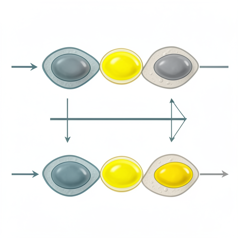
Temporal Activation of Phototropins
AI-generated conceptual illustration (Arabidopsis thaliana leaf cells). It explains the idea and does not show study data.
View source evidence ↗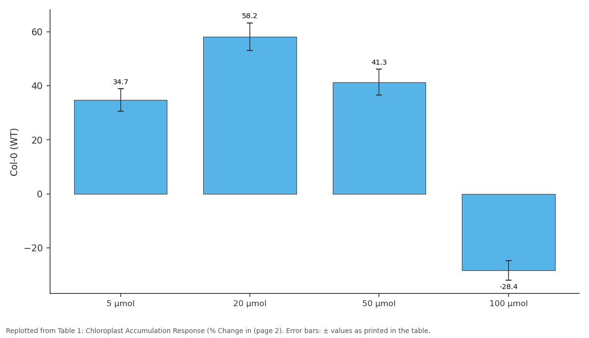
Col-0 (WT): 5 μmol vs 20 μmol vs 50 μmol vs 100 μmol
Replotted from Table 1 (page 2) of the original paper. Every bar is the paper's own table value.
View source evidence ↗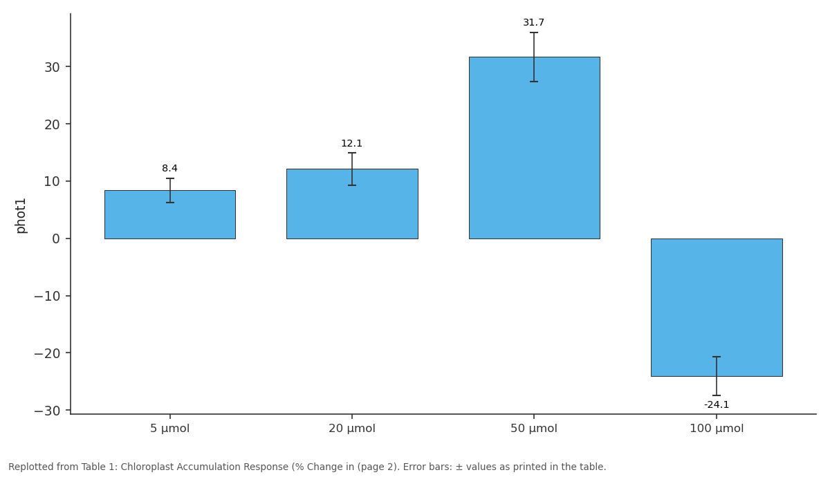
phot1: 5 μmol vs 20 μmol vs 50 μmol vs 100 μmol
Replotted from Table 1 (page 2) of the original paper. Every bar is the paper's own table value.
View source evidence ↗Figures & tables
From the original paper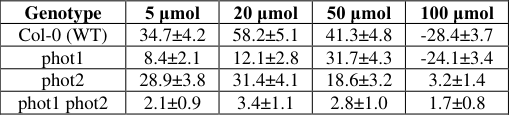 Table 1
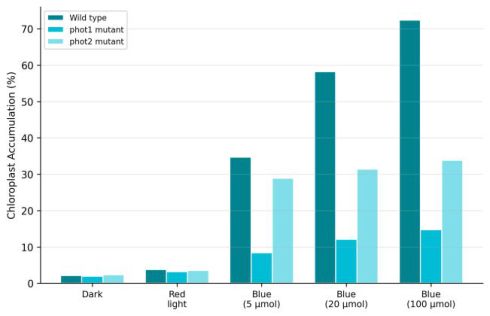 Figure 1
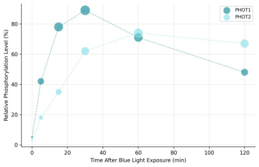 Figure 2
Introduction
Picture a mesophyll cell at dawn: chloroplasts crowd along the periclinal walls to capture weak morning light-the accumulation response. By midday, when direct sun risks photodamage, those same chloroplasts retreat to the anticlinal walls in the avoidance response [1] . Both movements depend on phototropin blue light receptors, a family of serine/threonine kinases that contain two light-sensing LOV domains and a C-terminal kinase domain [2] . Arabidopsis expresses two phototropins: PHOT1, which operates over a broad fluence range, and PHOT2, which functions mainly at moderate to high light intensities [3] . Genetic analysis using single and double mutants established that PHOT1 mediates the accumulation response at low blue light, PHOT2 controls avoidance at high blue light, and both contribute to accumulation at intermediate intensities [4] . But the biochemical basis for this division of labor remains only partially resolved. Differences in autophosphorylation kinetics and downstream signaling partners are thought to play a role [5] . This research aimed to quantify chloroplast movement responses and phototropin phosphorylation dynamics across a range of blue light fluence rates using wild-type and mutant Arabidopsis lines.
Materials and Methods
Materials and Methods
Materials
This research was performed at the Department of Botanical Sciences, Madras Institute of Biological Research, Chennai, Tamil Nadu, India, from May 2023 to August 2024. Institutional biosafety approval was obtained (MIBR-IBC/2023-037). Arabidopsis thaliana lines used were Col-0 (wild type), phot1-5, phot2-1, and phot1-5 phot2-1 double mutant, obtained from the Arabidopsis Biological Resource Center (ABRC, Ohio State University). Seeds were stratified at 4 °C for 3 days and grown on half-strength MS agar under white light (100 μmol m⁻² s⁻¹, 16 h/8 h) for 21 days before experiments.
Methods
Chloroplast movement was measured by the slit-band method: leaf transmittance at 660 nm was recorded before and after blue light exposure using a custom spectrophotometer setup [4] . Blue light was provided by LEDs (Thorlabs, peak 470 nm) at 5, 20, 50, and 100 μmol m⁻² s⁻¹ for 45 minutes. Confocal imaging (Leica SP8) of chlorophyll autofluorescence confirmed chloroplast positioning in cross-sectioned leaves. Autophosphorylation was assessed by immunoprecipitation of GFP-tagged PHOT1 and PHOT2 from transgenic lines, followed by ProQ Diamond phosphoprotein staining and Western blotting at time points of 0, 5, 15, 30, 60, and 120 minutes post-illumination [5] . Band intensities were quantified in ImageJ. Statistical analysis used two-way ANOVA (genotype × fluence rate) with Tukey's HSD post-hoc test. Wild-type plants showed strong accumulation at low-tomoderate fluence rates and switched to avoidance (negative values) at 100 μmol. The phot1 mutant was severely impaired at low fluence but retained PHOT2-dependent responses at higher intensities. The phot2 mutant lacked the avoidance response entirely. The double mutant was essentially unresponsive.
Results
Results
Discussion
The fluence-rate dependent division of labor between PHOT1 and PHOT2 matches the model proposed by Kagawa and Wada [4] , where PHOT1 serves as the primary sensor at low irradiance and PHOT2 takes over at higher intensities. The near-complete loss of accumulation response in the double mutant confirms that no other photoreceptor can substitute for phototropins in this process [1] . The phosphorylation kinetics data offer a biochemical explanation for the functional specialization: PHOT1's rapid activation and quick decline suit detection of transient or weak light signals, while PHOT2's slower, more sustained phosphorylation matches the need for prolonged signaling during high-light avoidance [5] . The residual responses seen in single mutants at intermediate fluence rates reflect the overlapping sensitivity ranges of the two receptors, consistent with in vitro LOV domain spectroscopic measurements showing similar but not identical absorption properties [2] . These findings add temporal resolution to the current model of phototropin-mediated chloroplast movement.
Conclusion
This research confirmed distinct but overlapping roles for PHOT1 and PHOT2 in chloroplast movement responses across blue light fluence rates. PHOT1 dominates at low light for accumulation, while PHOT2 is required for highlight avoidance. Differences in autophosphorylation kinetics-rapid for PHOT1, sustained for PHOT2-provide a biochemical framework for understanding this division of labor. These results contribute to the mechanistic understanding of light-adaptive chloroplast positioning in plants.
Acknowledgement
Acknowledgements Funding SourcesInternal research funds from the Madras Institute of Biological Research supported this work. Institutional SupportThe authors thank the Madras Institute of Biological Research for confocal microscopy and growth chamber facilities.Contributions Not Qualifying for Authorship ABRC (Ohio State University) is acknowledged for providing Arabidopsis mutant seed stocks.
Figures and tables from the paper
| Genotype | 5 μmol | 20 μmol | 50 μmol | 100 μmol |
|---|---|---|---|---|
| Col-0 (WT) | 34.7±4.2 | 58.2±5.1 | 41.3±4.8 | -28.4±3.7 |
| phot1 | 8.4±2.1 | 12.1±2.8 | 31.7±4.3 | -24.1±3.4 |
| phot2 | 28.9±3.8 | 31.4±4.1 | 18.6±3.2 | 3.2±1.4 |
| phot1 phot2 | 2.1±0.9 | 3.4±1.1 | 2.8±1.0 | 1.7±0.8 |
Table text extracted from the original PDF; superscript letters are shown inline. The original table image is below.
View original table image
References
6 in the original paper- Wada M, Kagawa T, Sato Y. Chloroplast movement. Annual Review of Plant Biology. 2003;54:455-468.
- Christie JM. Phototropin blue-light receptors. Annual Review of Plant Biology. 2007;58:21-45.
- Sakai T, Kagawa T, Kasahara M, Swartz TE, Christie JM, Briggs WR, Wada M, Okada K. Arabidopsis nph1 and npl1: blue light receptors that mediate both phototropism and chloroplast relocation. Proceedings of the National Academy of Sciences. 2001;98(12):69696974.
- Kagawa T, Wada M. Blue light-induced chloroplast relocation in Arabidopsis thaliana as analyzed by microbeam irradiation. Plant and Cell Physiology. 2000;41(1):84-93.
- Inoue S, Kinoshita T, Takemiya A, Doi M, Shimazaki K. Leaf positioning of Arabidopsis in response to blue light. Molecular Plant. 2008;1(1):15-26.
- Jarillo JA, Gabrys H, Capel J, Alonso JM, Ecker JR, Cashmore AR. Phototropin-related NPL1 controls chloroplast relocation induced by blue light. Nature. 2001;410(6831):952-954.