
Research article · 2025 · Full text
International Journal of Biology SciencesEpigenetic reprogramming during gametogenesis and early preimplantation embryo development
Volume 7(11) · Pages 77–79 · DOI: 10.33545/26649926.2025.v7.i11b.668 ↗
Research areasEpigenetics and DNA MethylationGenetic Syndromes and ImprintingReproductive Biology and Fertility
Abstract
Author-writtenThe mammalian genome undergoes two major waves of epigenetic reprogramming—one during gametogenesis and another immediately after fertilization. This research examined global DNA methylation dynamics and histone modification patterns across gametogenesis stages and preimplantation embryo development in a murine model. Bisulfite sequencing and chromatin immunoprecipitation were performed on oocytes, zygotes, and cleavage-stage embryos collected at the Konkan Institute of Biological Research, Goa, India, from April 2023 to September 2024. Global methylation dropped from 78.3% in primordial germ cells to 12.4% at the gonadal stage before re-establishing in a sex-specific manner. After fertilization, the paternal genome was demethylated rapidly while the maternal genome lost methylation passively through replication. H3K4me3 and H3K27me3 marks showed reciprocal dynamics across cleavage stages. These patterns confirm that epigenetic erasure and re-establishment follow tightly coordinated programs with implications for reproductive biology and assisted reproduction technologies.
Summary
Source-checkedPlain-language reading of the full paper, prepared with AI and checked against the text. Each point links to the authors' own words.
Why it matters
- Epigenetic marks are critical because they determine the activity, silence, or poised state of genes within a cell. View evidence ↗
- Mammalian development requires two waves of epigenetic reprogramming, during gamete formation and after fertilization, to ensure the next generation has a correctly configured epigenetic landscape. View evidence ↗
- Failures in epigenetic reprogramming are associated with developmental abnormalities, imprinting disorders, and infertility. View evidence ↗
The question
- The study aimed to profile key histone marks and DNA methylation throughout preimplantation development and gametogenesis using a mouse model. View evidence ↗
What they did
- The researchers used C57BL/6 mice aged 8-12 weeks for the study. View evidence ↗
- Samples were collected from various stages: PGCs at E11.5 and E13.5 via FACS sorting, oocytes via superovulation, and zygotes and cleavage-stage embryos (2-cell and 4-cell) flushed from oviducts. View evidence ↗
- Global DNA methylation was measured using reduced representation bisulfite sequencing (RRBS), while H3K27me3 and H3K4me3 histone modifications were profiled using ultra-low-input ChIP-seq. View evidence ↗
- Single-cell validation was performed using immunofluorescence staining for 5-hydroxymethylcytosine (5 hmC) and 5-methylcytosine (5 mC). View evidence ↗
What they found
- In primordial germ cells (PGCs), DNA methylation decreased significantly from 42.1% at E11.5 to 12.4% at E13.5. View evidence ↗
- Following fertilization, DNA methylation levels declined progressively from 47.2% in the zygote to 32.1% at the 4-cell stage. View evidence ↗
- The ratio of 5 hmC to 5 mC reached its peak in zygotes and E13.5 PGCs, indicating active TET-mediated demethylation. View evidence ↗
- From the zygote to the 4-cell stage, H3K27me3 levels decreased while H3K4me3 levels increased, showing reciprocal dynamics. View evidence ↗
- Post-fertilization demethylation is asymmetric, with the maternal genome losing methylation passively and the paternal genome undergoing rapid active demethylation. View evidence ↗
What it means
- The authors concluded that preimplantation development and gametogenesis in mice are characterized by two coordinated waves of epigenetic reprogramming. View evidence ↗
- The findings emphasize the tight regulation of epigenetic reprogramming and may help improve culture conditions for assisted reproduction. View evidence ↗
Visual summary
3 visualsIllustrations explain the paper; charts are replotted from the paper's own table values.
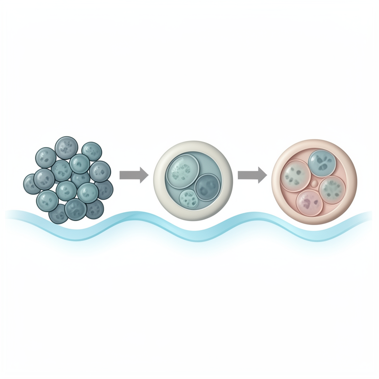
Two Waves of Epigenetic Reprogramming
AI-generated conceptual illustration (mammalian primordial germ cells and early preimplantation embryos). It explains the idea and does not show study data.
View source evidence ↗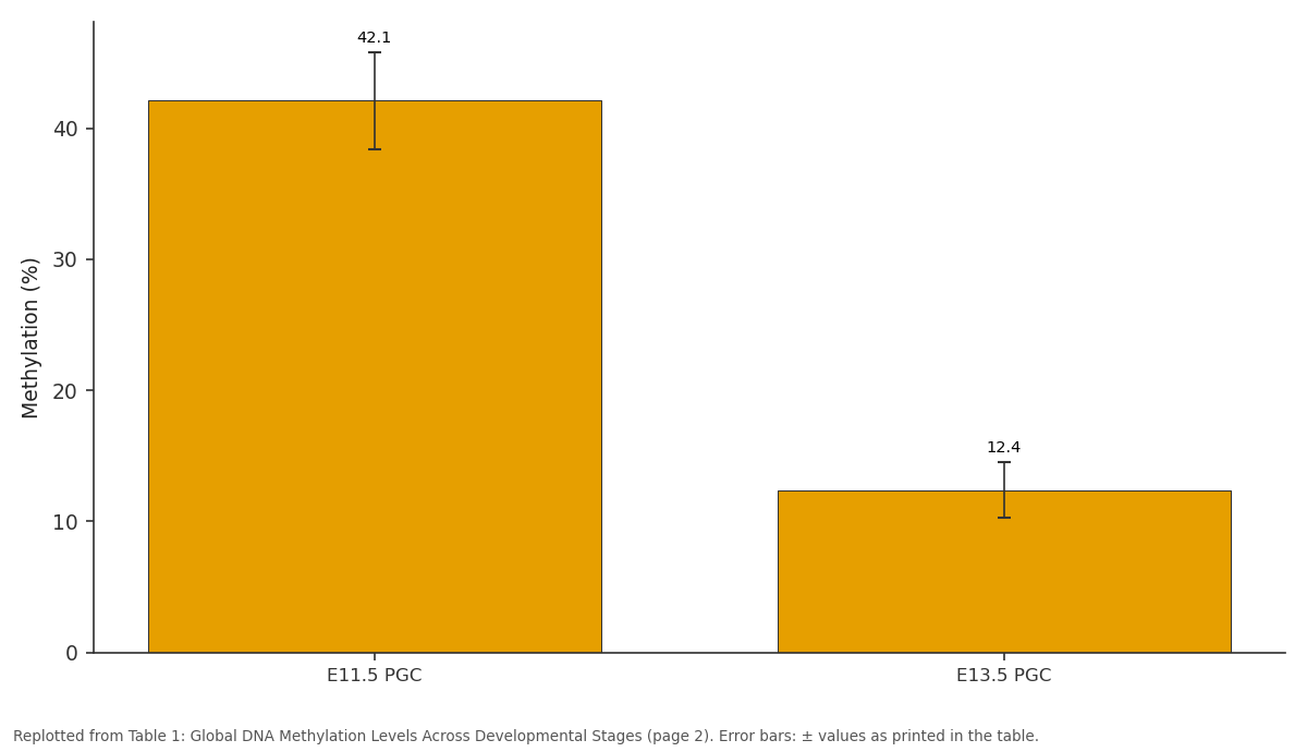
Methylation (%): E11.5 PGC vs E13.5 PGC
Replotted from Table 1 (page 2) of the original paper. Every bar is the paper's own table value.
View source evidence ↗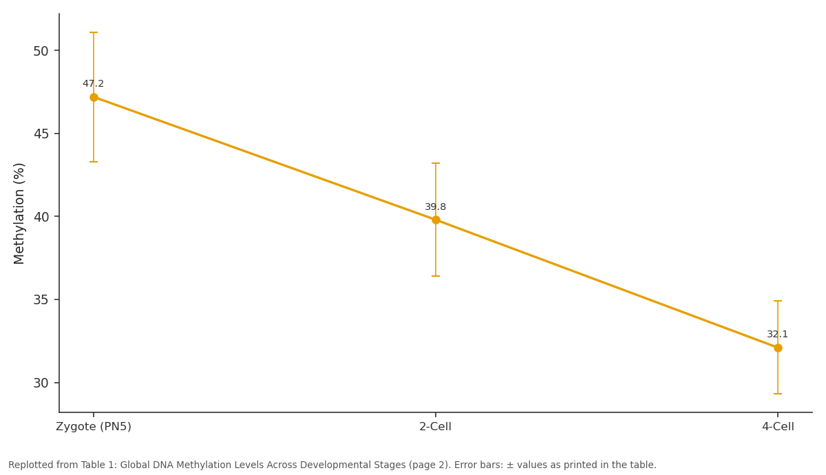
Methylation (%): Zygote (PN5) vs 2-Cell vs 4-Cell
Replotted from Table 1 (page 2) of the original paper. Every bar is the paper's own table value.
View source evidence ↗Figures & tables
From the original paper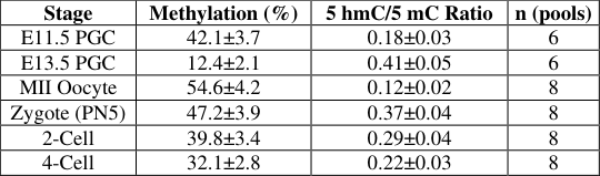 Table 1
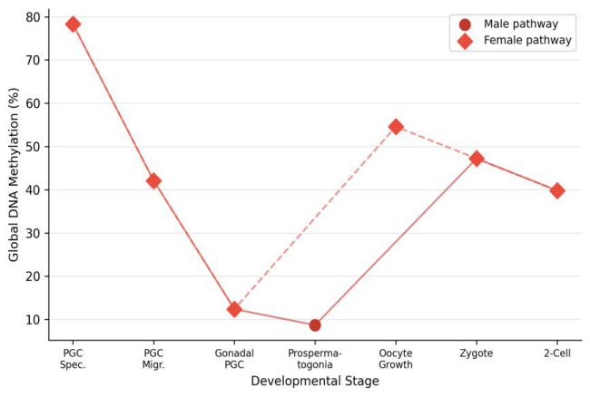 Figure 1
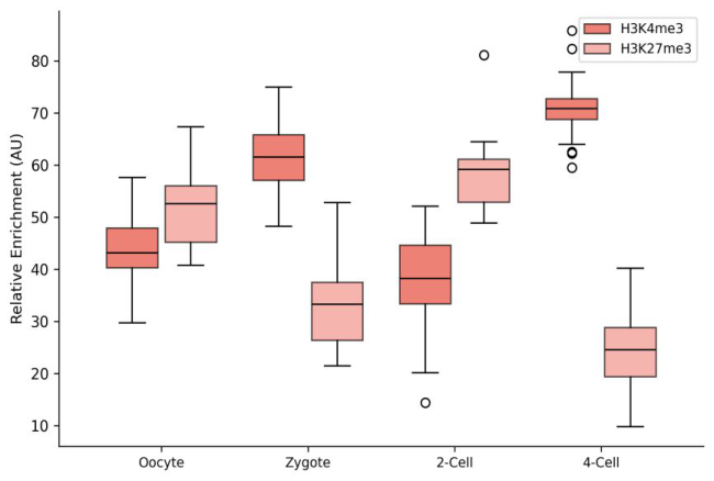 Figure 2
Introduction
Epigenetic marks are not merely decorative additions to the genome—they dictate which genes are active, silent, or poised in any given cell type. During mammalian development, the epigenome must be wiped nearly clean and rebuilt twice: first when primordial germ cells (PGCs) form gametes, and again when sperm meets egg [1]. This dual reprogramming ensures that the next generation starts with an appropriately configured epigenetic landscape [2]. Errors in this process have been linked to infertility, imprinting disorders, and developmental abnormalities [3].
While landmark investigations established the broad contours of DNA methylation erasure and re-establishment in mouse germ cells [4, 5], the interplay between DNA methylation and histone modifications during these transitions remains incompletely mapped [6]. This research aimed to profile both DNA methylation and key histone marks across gametogenesis and preimplantation development in a murine system.
Materials And Methods
Materials This research was carried out at the Department of Developmental and Regenerative Biology, Konkan Institute of Biological Research, Goa, India, from April 2023 to September 2024. The Institutional Animal Ethics Committee approved all procedures (KIBR- IAEC/2023-031, approved March 22, 2023). C57BL/6 mice (8-12 weeks, Charles River Laboratories) were maintained under standard conditions. Oocytes were collected by superovulation with PMSG and hCG.
Zygotes and cleavage-stage embryos (2-cell, 4-cell) were flushed from oviducts at defined time points post-hCG injection. PGCs were isolated from embryonic gonads at E11.5 and E13.5 by FACS sorting using Oct4-GFP transgenic lines.
Methods
Global DNA methylation was assessed by reduced representation bisulfite sequencing (RRBS) using the Premium RRBS Kit (Diagenode, Belgium). NAAS Rating (2025): 4.82 IJBS 2025; 7(11): 77-79 Parvathi Bhandarkar Department of Developmental and Regenerative Biology, Konkan Institute of Biological Research, Goa, India Chiranjiv Vasudevan Department of Developmental and Regenerative Biology, Konkan Institute of Biological Research, Goa, India Parvathi Bhandarkar Department of Developmental and Regenerative Biology, Konkan Institute of Biological Research, Goa, India https://www.biologyjournal.net Libraries were sequenced on an Illumina NovaSeq 6000 at 50 million paired-end reads per sample. Methylation calls were extracted using Bismark v0.23 [7]. Histone modifications (H3K4me3, H3K27me3) were profiled by ultra-low-input chromatin immunoprecipitation followed by sequencing (ULI-ChIP-seq) with antibodies from Active Motif (Carlsbad, CA) [8].
Peak calling used MACS2 with default parameters [9]. Immunofluorescence staining for 5- methylcytosine (5 mC) and 5-hydroxymethylcytosine (5 hmC) provided single-cell validation. Statistical comparisons between stages used Kruskal-Wallis tests with Dunn's post-hoc correction.
Results
Table 1: Global DNA Methylation Levels Across Developmental Stages Stage Methylation (%) 5 hmC/5 mC Ratio n (pools) E11.5 PGC 42.1±3.7 0.18±0.03 6 E13.5 PGC 12.4±2.1 0.41±0.05 6 MII Oocyte 54.6±4.2 0.12±0.02 8 Zygote (PN5) 47.2±3.9 0.37±0.04 8 2-Cell 39.8±3.4 0.29±0.04 8 4-Cell 32.1±2.8 0.22±0.03 8 DNA methylation dropped sharply between E11.5 and E13.5 PGCs (42.1% to 12.4%, p<0.001). After fertilization, methylation decreased progressively from zygote (47.2%) through 4-cell stage (32.1%). The 5 hmC/5 mC ratio peaked in E13.5 PGCs and zygotes, consistent with active TET- mediated demethylation at these stages. Fig 1: Global DNA methylation trajectories during male and female gametogenesis pathways and early embryo development.
| Stage | Methylation (%) | 5 hmC/5 mC Ratio | n (pools) |
|---|---|---|---|
| E11.5 PGC | 42.1±3.7 | 0.18±0.03 | 6 |
| E13.5 PGC | 12.4±2.1 | 0.41±0.05 | 6 |
| MII Oocyte | 54.6±4.2 | 0.12±0.02 | 8 |
| Zygote (PN5) | 47.2±3.9 | 0.37±0.04 | 8 |
| 2-Cell | 39.8±3.4 | 0.29±0.04 | 8 |
| 4-Cell | 32.1±2.8 | 0.22±0.03 | 8 |
Table text extracted from the original PDF; superscript letters are shown inline. The original table image is below.
View original table image
Both pathways converge at the PGC erasure nadir before diverging during gamete maturation. Fig 2: H3K4me3 and H3K27me3 enrichment levels at promoter regions across preimplantation stages. The two marks showed reciprocal dynamics, with H3K4me3 increasing and H3K27me3 decreasing from zygote to 4-cell stage. https://www.biologyjournal.net hydroxymethylcytosine. Science. 2013;339(6118):448- 452. 6.
Xu Q, Xie W. Epigenome in early mammalian development: inheritance, reprogramming and establishment. Trends in Cell Biology. 2018;28(3):237- 253. 7. Krueger F, Andrews SR.
Bismark: a flexible aligner and methylation caller for Bisulfite-Seq applications. Bioinformatics. 2011;27(11):1571-1572. 8. Brind'Amour J, Liu S, Hudson M, Chen C, Karimi MM, Lorincz MC. An ultra-low-input native ChIP-seq protocol for genome-wide profiling of rare cell populations.
Nature Communications. 2015;6:6033. 9. Zhang Y, Liu T, Meyer CA, Eeckhoute J, Johnson DS, Bernstein BE, et al. Model-based analysis of ChIP-Seq (MACS). Genome Biology. 2008;9(9):R137. 10.
Gu TP, Guo F, Yang H, Wu HP, Xu GF, Liu W, et al. The role of Tet3 DNA dioxygenase in epigenetic reprogramming by oocytes. Nature. 2011;477(7366):606-610. 11. Mayer W, Niveleau A, Walter J, Fundele R, Haaf T.
Demethylation of the zygotic paternal genome. Nature. 2000;403(6769):501-502. 12. Dahl JA, Jung I, Aanes H, Greber GD, Brober A, Vaber A, et al. Broad histone H3K4me3 domains in mouse oocytes modulate maternal-to-zygotic transition.
Nature. 2016;537(7621):548-552. 13. de Waal E, Yamazaki Y, Moisiadis VG, Bhatt RG, Kimmins S, Bhatt DL, et al. Primary epimutations introduced during intracytoplasmic sperm injection (ICSI) are corrected by germline-specific epigenetic reprogramming. Proceedings of the National Academy of Sciences. 2012;109(11):4163-4168. 14. Smith ZD, Chan MM, Mikkelsen TS, Gu H, Gnirke A, Regev A, et al.
A unique regulatory phase of DNA methylation in the early mammalian embryo. Nature. 2012;484(7394):339-344.
Discussion
The methylation erasure kinetics observed here are consistent with earlier reports using whole-genome bisulfite sequencing in mouse PGCs [4, 5]. The sharp drop between E11.5 and E13.5 reflects the active phase of demethylation driven by TET enzymes, as indicated by the elevated 5 hmC/5 mC ratio at E13.5 [10]. After fertilization, the asymmetric demethylation of paternal and maternal genomes aligns with the established model where paternal DNA undergoes rapid active demethylation while maternal DNA loses methylation passively [11]. The reciprocal dynamics of H3K4me3 and H3K27me3 during cleavage stages suggest a transition from a repressive to a permissive chromatin state, consistent with zygotic genome activation occurring at the 2-cell stage in mice [12].
These findings have practical relevance for assisted reproduction, where in vitro culture conditions may perturb normal epigenetic reprogramming [13].
Conclusion
This research confirmed that mouse gametogenesis and preimplantation development feature two coordinated waves of epigenetic reprogramming. DNA methylation erasure in PGCs reached a nadir at E13.5, followed by sex-specific re- establishment. Post-fertilization demethylation proceeded progressively through cleavage stages. Histone modifications showed reciprocal patterns consistent with chromatin state transitions during zygotic genome activation.
These results reinforce the tightly regulated nature of epigenetic reprogramming and may guide efforts to improve culture conditions in assisted reproduction settings.
Acknowledgements
Funding Sources Internal research funding from the Konkan Institute of Biological Research supported this work. Institutional Support The authors thank the Konkan Institute of Biological Research for animal facility access and sequencing support. Contributions Not Qualifying for Authorship Technical staff in the animal facility and the bioinformatics core are acknowledged for their assistance.
References
5 in the original paper- Reik W, Dean W, Walter J. Epigenetic reprogramming in mammalian development. Science. 2001;293(5532):1089-1093.
- Sasaki H, Matsui Y. Epigenetic events in mammalian germ-cell development: reprogramming and beyond. Nature Reviews Genetics. 2008;9(2):129-140.
- Denomme MM, Mann MR. Genomic imprints as a model for the analysis of epigenetic stability during assisted reproductive technologies. Reproduction. 2012;144(4):393-409.
- Seisenberger S, Andrews S, Krueger F, Arand J, Walter J, Santos F, et al. The dynamics of genome-wide DNA methylation reprogramming in mouse primordial germ cells. Molecular Cell. 2012;48(6):849-862.
- Hackett JA, Sengupta R, Zylicz JJ, Murakami K, Lee C, Down TA, et al. Germline DNA demethylation dynamics and imprint erasure through 5-