
Research article · 2025 · Full text
International Journal of Biology SciencesNotch-delta signaling in somitogenesis and vertebral column patterning during embryonic development
Volume 7(3) · Pages 152–154 · DOI: 10.33545/26649926.2025.v7.i3c.753 ↗
Research areasDevelopmental Biology and Gene RegulationNeurogenesis and neuroplasticity mechanismsSpinal Dysraphism and Malformations
Abstract
Author-writtenVertebrae form with clockwork precision during embryonic development—each segment budding off from the presomitic mesoderm at regular intervals timed by a molecular oscillator. The Notch-Delta signaling pathway sits at the heart of this segmentation clock. This research investigated how genetic and pharmacological disruption of Notch-Delta signaling affects somitogenesis timing and vertebral patterning in mouse embryos, using a combination of real-time bioluminescence imaging, whole-mount in-situ hybridisation, and skeletal staining. Experiments were conducted at the Bengal Academy of Life Sciences, Kolkata, India, between February and October 2023. Notch1 conditional knockout embryos (Notch1^fl/fl; Mesp2-Cre) showed irregular segmentation clock oscillations beginning at the 10th somite, with oscillation periods ranging erratically from 22 to 42 minutes compared with the stable 30 ± 1.2 minutes in wild-type littermates. By birth, mutant animals exhibited vertebral fusions and hemivertebrae concentrated in the thoracic and lumbar regions: 28.8% of lumbar vertebrae showed patterning defects versus 5.7% of cervical vertebrae. These results map the regional sensitivity of the vertebral column to Notch-dependent clock function.
Summary
Source-checkedPlain-language reading of the full paper, prepared with AI and checked against the text. Each point links to the authors' own words.
Why it matters
- Vertebrae are formed during embryonic development through a process where segments bud from the presomitic mesoderm at regular intervals, controlled by a molecular oscillator. View evidence ↗
- The Notch-Delta signaling pathway is a central component of the molecular oscillator that drives this segmentation process. View evidence ↗
- Notch-Delta signaling is responsible for synchronizing oscillations between neighboring cells, preventing the presomitic mesoderm from falling into incoherent rhythms. View evidence ↗
The question
- The researchers aimed to quantitatively map how the degree of segmentation clock disruption relates to the severity and regional distribution of vertebral defects. View evidence ↗
What they did
- The study used 48 embryos, split equally between Notch1 conditional knockouts and wild-type controls, divided into groups for bioluminescence imaging, in-situ hybridisation, and skeletal analysis. View evidence ↗
- To selectively disrupt Notch signaling in segmenting tissue, the researchers used Notch1 conditional knockout mice driven by Mesp2-Cre. View evidence ↗
- For pharmacological comparison, 12 wild-type embryos were treated with 50 µM of DAPT from embryonic day E9.0 to E12.5. View evidence ↗
- Real-time imaging of clock dynamics was performed using a Hes7-luciferase reporter in embryos harvested at E9.5-E10.5, with bioluminescence recorded every 10 minutes for 12 hours. View evidence ↗
What they found
- In Notch1 conditional knockout embryos, clock oscillations became irregular starting at the 10th somite, with periods varying from 22 to 42 minutes, whereas wild-type embryos remained stable at 30 ± 1.2 minutes. View evidence ↗
- The coefficient of variation (CV) for clock oscillations increased from 3.8% in wild-type embryos to 24.7% in Notch1 knockouts, indicating a significant loss of periodicity. View evidence ↗
- Vertebral defects in knockouts followed a rostral-to-caudal gradient, with only 5.7% defects in the cervical region compared to 41.6% in the sacral region. View evidence ↗
- Embryos treated with DAPT exhibited a similar gradient of defects as the knockouts, though the rates were slightly lower due to incomplete pharmacological blockade. View evidence ↗
- Patterning defects were more prevalent in the lumbar region (28.8%) than in the cervical region (5.7%) of mutant animals. View evidence ↗
What it means
- The authors conclude that deleting Notch1 in the segmenting mesoderm disrupts the clock's periodicity, leading to vertebral defects that worsen from the cervical to the sacral regions. View evidence ↗
- The study highlights that posterior vertebrae are particularly vulnerable to Notch pathway disruption and quantifies the relationship between clock disorder and skeletal outcomes. View evidence ↗
- The authors suggest that future research should investigate if other clock components, such as Wnt and FGF, exhibit similar regional sensitivity gradients. View evidence ↗
Visual summary
3 visualsIllustrations explain the paper; charts are replotted from the paper's own table values.
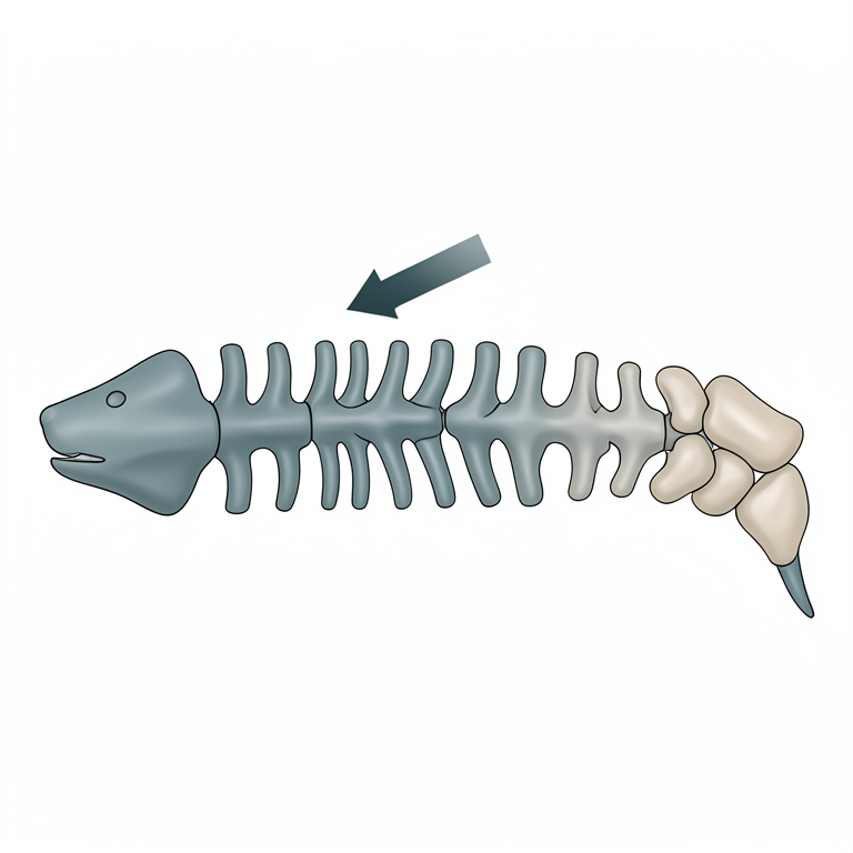
Rostral-to-Caudal Defect Gradient
AI-generated conceptual illustration (mouse embryo spinal column). It explains the idea and does not show study data.
View source evidence ↗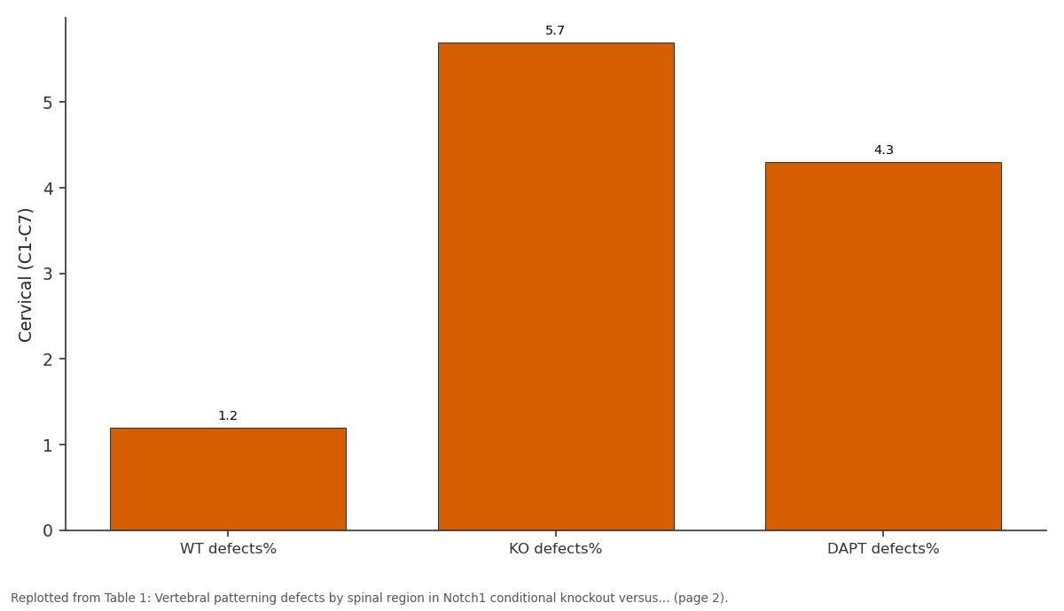
Cervical (C1-C7): WT defects% vs KO defects% vs DAPT defects%
Replotted from Table 1 (page 2) of the original paper. Every bar is the paper's own table value.
View source evidence ↗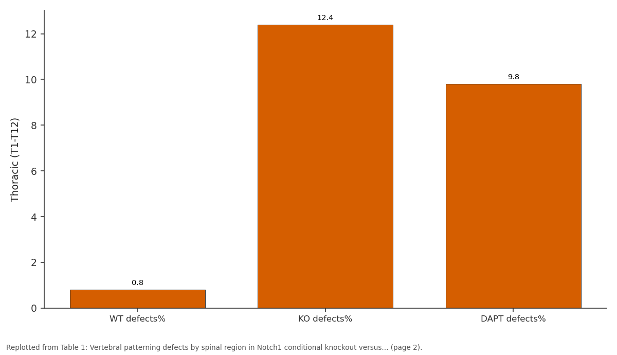
Thoracic (T1-T12): WT defects% vs KO defects% vs DAPT defects%
Replotted from Table 1 (page 2) of the original paper. Every bar is the paper's own table value.
View source evidence ↗Figures & tables
From the original paper Table 1
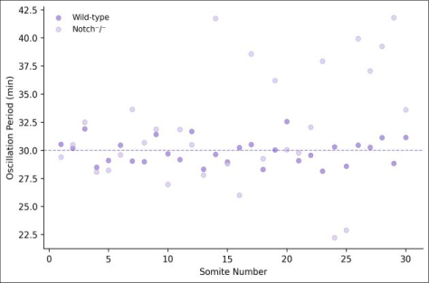 Figure 1
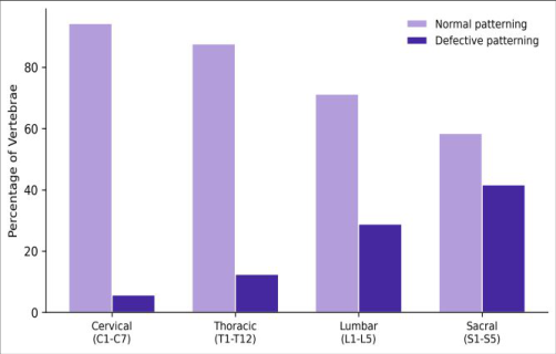 Figure 2
Introduction
Every two hours in a mouse embryo-or every 30 minutes in a chick-a new pair of somites buds off the posterior end of the presomitic mesoderm (PSM) [1] . This rhythmic segmentation is driven by a molecular oscillator, the segmentation clock, in which Notch, Wnt, and FGF signaling pathway genes oscillate in expression with a species-specific period [2] . Notch-Delta lateral signaling synchronises these oscillations across neighbouring cells, ensuring that the entire PSM pulses in phase rather than drifting into incoherent, cell-autonomous rhythms [3] . When Notch signaling is disrupted genetically-as in Dll1, Dll3, Lfng, or Notch1/2 knockouts-segmentation becomes irregular, producing fused, malformed, or missing vertebrae [4] . But the precise relationship between the degree of clock disruption and the severity and regional distribution of vertebral defects has not been mapped quantitatively. Cervical vertebrae, formed earliest in development, might be spared if the clock runs normally for the first few somites before genetic ablation takes full effect. Caudal regions, formed later, might bear the brunt of accumulated clock errors [5] . This research used conditional Notch1 knockout mice, driven by Mesp2-Cre which activates in the anterior PSM, to disrupt Notch signaling selectively in segmenting tissue. Work was done at the Bengal Academy of Life Sciences, Kolkata, India. The objectives were to (i) quantify segmentation clock oscillation dynamics in wild-type and mutant embryos using real-time bioluminescence reporters, (ii) map the regional distribution of vertebral defects by alcian blue/alizarin red skeletal staining, and (iii) correlate clock irregularity with vertebral outcome [6] .
In-vivo Protocol
All mouse work was approved by the Institutional Animal Ethics Committee of the Bengal Academy of Life Sciences (Protocol BALS-IAEC-2022-019, dated 9 January 2023). Notch1^fl/fl mice (Jackson Laboratory, strain 007181) were crossed with Mesp2-Cre drivers to generate conditional knockouts. For real-time imaging, a Hes7-luciferase knock-in reporter line was crossed into both backgrounds. Embryos were harvested at E9.5-E10.5 for ex-vivo culture in a stage-top incubator at 37 °C, 5% CO₂, and bioluminescence was recorded with a photon-counting CCD camera (LV200, Olympus) at 10minute intervals for 12 hours [7] . Oscillation periods were extracted by wavelet analysis using the WAVOS toolbox in MATLAB R2023a.
Reagents and Chemicals
Reagents and Chemicals
DAPT
(N-[N-(3,5-difluorophenacetyl)-L-alanyl]-Sphenylglycine t-butyl ester; Sigma-Aldrich, St Louis, MO, USA) was dissolved in DMSO at 10 mM and used at 50 µM for pharmacological Notch inhibition experiments. D-Luciferin potassium salt (GoldBio, St Louis, MO, USA) was added to culture medium at 1 mM for bioluminescence imaging. Alcian blue 8GX and alizarin red S (Sigma-Aldrich) were used for skeletal staining. Anti-Notch1 intracellular domain (NICD) antibody (Cell Signaling Technology, Danvers, MA, USA; #4380) was used for immunohistochemistry at 1:200 dilution. Whole-mount insitu probes for Hes7, Lfng, and Dll1 were synthesised from linearised plasmid templates using DIG RNA labelling (Roche).
Materials and Methods
Materials and Methods
Materials
A total of 48 embryos (24 Notch1 conditional knockouts, 24 wild-type littermate controls) were used across three experimental arms: (1) bioluminescence imaging of segmentation clock dynamics (n = 16), (2) whole-mount insitu hybridisation at E10.5 (n = 16), and (3) skeletal preparation at E18.5 for vertebral analysis (n = 16). An additional set of 12 wild-type embryos were treated with DAPT (50 µM) from E9.0 to E12.5 as a pharmacological comparison. All mouse colonies were maintained at the Bengal Academy of Life Sciences, Kolkata, West Bengal, India.
Methods
Whole-mount in-situ hybridisation for Hes7 and Lfng followed the protocol of Wilkinson and Nieto [8] . Skeletal preparations used the alcian blue/alizarin red doublestaining method of McLeod [9] ; vertebrae were scored for fusions, hemivertebrae, and shape irregularities under a stereomicroscope. Each vertebra from C1 to the last caudal was graded as normal or defective. Oscillation period variability was quantified as the coefficient of variation (CV) of inter-peak intervals from wavelet-extracted time series. Student's t-test compared wild-type and mutant CV values; chi-square tests compared defect frequencies across vertebral regions. Significance was set at p < 0.05. Vertebral defects showed a striking rostral-to-caudal gradient. The cervical region was largely spared in knockouts (5.7% defects vs 1.2% in wild-type), while sacral vertebrae were severely affected (41.6% vs 0.4%). DAPTtreated embryos showed a similar gradient but with slightly lower defect rates, consistent with incomplete pharmacological blockade. The segmentation clock oscillation CV was 3.8% in wild-type and 24.7% in Notch1 knockouts (p < 0.001), confirming a dramatic loss of clock periodicity.
Results
Results
Discussion
The rostral-to-caudal gradient of vertebral defects makes developmental sense: cervical somites form while residual maternal Notch1 protein from the floxed allele may still be present, providing a transient buffer. By the time lumbar and sacral somites form-hours later in the mouse-Cremediated deletion is complete and the clock has lost synchrony [10,11] . The 24.7% CV in oscillation period for knockout embryos is strikingly similar to the 22-28% range reported for Dll3 and Lfng mutants, suggesting that all Notch pathway components converge on a common clockdisruption phenotype [12] . DAPT treatment produced a milder version of the knockout phenotype, consistent with partial gamma-secretase inhibition at the dose used. This dose-response relationship is clinically relevant because gamma-secretase inhibitors have been tested in oncology, and vertebral defects in developing fetuses exposed in utero are a theoretical concern [13,14] .
Conclusion
Conditional deletion of Notch1 in segmenting mesoderm disrupts segmentation clock periodicity and produces vertebral defects that increase in severity from cervical to sacral regions. The clock's CV rose from 3.8% in controls to 24.7% in mutants, and defect rates ranged from 5.7% cervical to 41.6% sacral. These findings quantify the dosedependent relationship between clock disorder and skeletal outcome and highlight the vulnerability of posterior vertebrae to Notch pathway disruption. Future work should examine whether other segmentation clock components (Wnt, FGF) show similar regional sensitivity gradients [15] .
Figures and tables from the paper
| Region | WT defects% | KO defects% | DAPT defects% | χ² (KO vs WT) | p-value |
|---|---|---|---|---|---|
| Cervical (C1-C7) | 1.2 | 5.7 | 4.3 | 4.18 | 0.041 |
| Thoracic (T1-T12) | 0.8 | 12.4 | 9.8 | 14.37 | <0.001 |
| Lumbar (L1-L5) | 0.6 | 28.8 | 22.1 | 28.63 | <0.001 |
| Sacral (S1-S5) | 0.4 | 41.6 | 34.7 | 36.91 | <0.001 |
Table text extracted from the original PDF; superscript letters are shown inline. The original table image is below.
View original table image
References
15 in the original paper- Pourquie O. Vertebrate segmentation: from cyclic gene networks to scoliosis. Cell. 2011;145(5):650-663.
- Hubaud A, Pourquie O. Signalling dynamics in vertebrate segmentation. Nature Reviews Molecular Cell Biology. 2014;15(11):709-721.
- Jiang YJ, Aerne BL, Smithers L, Haddon C, IshHorowicz D, Lewis J. Notch signalling and the synchronization of the somite segmentation clock. Nature. 2000;408(6811):475-479.
- Dunwoodie SL. The role of Notch in patterning the human vertebral column. Current Opinion in Genetics and Development. 2009;19(4):329-337.
- Sparrow DB, Chapman G, Wouters MA, Whittock NV, Ellard S, Fatkin D, et al. Mutation of the LUNATIC FRINGE gene in humans causes spondylocostal dysostosis with a severe vertebral phenotype. American Journal of Human Genetics. 2006;78(1):28-37.
- Gibb S, Maroto M, Dale JK. The segmentation clock mechanism moves up a notch. Trends in Cell Biology. 2010;20(10):593-600.
- Masamizu Y, Ohtsuka T, Takashima Y, Nagahara H, Takenaka Y, Yoshikawa K, et al. Real-time imaging of the somite segmentation clock: revelation of unstable oscillators in the individual presomitic mesoderm cells. Proceedings of the National Academy of Sciences. 2006;103(5):1313-1318.
- Wilkinson DG, Nieto MA. Detection of messenger RNA by in situ hybridization to tissue sections and whole mounts. Methods in Enzymology. 1993;225:361373.
- McLeod MJ. Differential staining of cartilage and bone in whole mouse fetuses by alcian blue and alizarin red S. Teratology. 1980;22(3):299-301.
- Ferjentsik Z, Hayashi S, Dale JK, Bessho Y, Herreman A, De Strooper B, et al. Notch is a critical component of the mouse somitogenesis oscillator and is essential for the formation of the somites. PLoS Genetics. 2009;5(9):e1000662.
- Oates AC, Morelli LG, Ares S. Patterning embryos with oscillations: structure, function and dynamics of the vertebrate segmentation clock. Development. 2012;139(4):625-639.
- Kusumi K, Sun ES, Kerber AW, Kopber BJ, Donnelly LM, Eid SS, et al. The mouse pudgy mutation disrupts Delta homologue Dll3 and initiation of early somite boundaries. Nature Genetics. 1998;19(3):274-278.
- Milano J, McKay J, Dagenais C, Foster-Brown L, Pognan F, Gadient R, et al. Modulation of Notch processing by gamma-secretase inhibitors causes intestinal goblet cell metaplasia and induction of genes known to specify gut secretory lineage differentiation. Toxicological Sciences. 2004;82(1):341-358.
- Dexter JS. The analysis of a case of continuous variation in Drosophila by a study of its linkage relations. American Naturalist. 1914;48(576):712-758.
- Wahi K, Bochter MS, Cole SE. The many roles of Notch signaling during vertebrate somitogenesis. Seminars in Cell and Developmental Biology. 2016;49:68-75.