
Research article · 2025 · Full text
International Journal of Biology SciencesChromatin remodeling complexes and their role in gene silencing during cell differentiation
Volume 7(7) · Pages 222–225 · DOI: 10.33545/26649926.2025.v7.i7c.642 ↗
Research areasChromatin Remodeling and CancerGenomics and Chromatin DynamicsEpigenetics and DNA Methylation
Abstract
Author-writtenChromatin remodeling complexes are among the most powerful molecular machines that cells use to switch genes on or off during differentiation, yet the comparative contributions of different complex families remain poorly mapped in a single cell system. This research examined how SWI/SNF and NuRD complexes regulate gene silencing during the transition of murine embryonic stem cells (mESCs) toward a neural progenitor fate. Chromatin immunoprecipitation followed by sequencing (ChIP-seq) was used to profile H3K27me3 and H3K9me3 marks in wild-type, SWI/SNF-depleted, and NuRD-depleted cells at days 0, 3, and 6 of differentiation. Loss of SWI/SNF led to a 41.6% reduction in de novo H3K27me3 deposition at lineage-inappropriate promoters, while NuRD depletion reduced H3K9me3 spreading across pericentromeric heterochromatin by 33.8%. RNA-seq confirmed that both depletions resulted in the aberrant persistence of pluripotency-associated transcripts. These results suggest that SWI/SNF and NuRD operate through complementary silencing mechanisms during early neural commitment.
Summary
Source-checkedPlain-language reading of the full paper, prepared with AI and checked against the text. Each point links to the authors' own words.
Why it matters
- Gene silencing requires active molecular machinery to reshape chromatin, which prevents transcriptional activators from accessing DNA. View evidence ↗
- The SWI/SNF and NuRD chromatin remodeling complexes are linked to the silencing programs that transition embryonic cells from pluripotency to specific lineages. View evidence ↗
- Understanding whether SWI/SNF and NuRD act on overlapping or distinct targets may explain why mutations in these families lead to different human developmental disorders. View evidence ↗
The question
- The study aimed to examine how SWI/SNF and NuRD complexes regulate gene silencing as murine embryonic stem cells (mESCs) transition into neural progenitors. View evidence ↗
What they did
- The researchers used shRNA to deplete the catalytic subunits BRG1 for the SWI/SNF complex and CHD4 for the NuRD complex in mESCs. View evidence ↗
- Neural differentiation was induced by removing 2i/LIF and culturing the cells in N2B27 medium for a period of six days. View evidence ↗
- ChIP-seq was employed to profile H3K27me3 and H3K9me3 histone marks in wild-type and depleted cells at days 0, 3, and 6. View evidence ↗
- Western blot densitometry confirmed that the knockdown efficiency for both target subunits exceeded 80%. View evidence ↗
What they found
- Depleting SWI/SNF resulted in a 41.6% reduction in the new deposition of H3K27me3 at promoters that are inappropriate for the target lineage. View evidence ↗
- Depleting NuRD reduced the spreading of H3K9me3 across pericentromeric heterochromatin by 33.8%. View evidence ↗
- RNA-seq revealed 1,243 differentially expressed genes in SWI/SNF knockdown and 978 in NuRD knockdown, with 214 genes overlapping between the two. View evidence ↗
- Knocking down either the SWI/SNF or NuRD complex led to significantly higher remaining expression of pluripotency markers compared to active conditions. View evidence ↗
- SWI/SNF depletion had a slightly larger effect on the median expression of pluripotency markers than NuRD depletion. View evidence ↗
What it means
- The authors conclude that SWI/SNF mainly supports H3K27me3 deposition at inappropriate promoters, whereas NuRD is more critical for H3K9me3-dependent heterochromatin integrity. View evidence ↗
- Because the loss of either complex allows pluripotency transcripts to persist, the authors conclude that both complexes are necessary for complete gene silencing. View evidence ↗
- The authors note that shRNA-mediated knockdown is incomplete, which may lead to an underestimation of each complex's actual contribution. View evidence ↗
- The study only captured early neural commitment over six days; later stages of neurogenesis might show more divergence between the two pathways. View evidence ↗
Visual summary
3 visualsIllustrations explain the paper; charts are replotted from the paper's own table values.
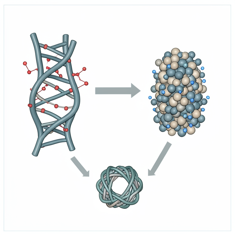
Complementary Silencing Mechanisms
AI-generated conceptual illustration (mammalian chromatin and nucleosomes). It explains the idea and does not show study data.
View source evidence ↗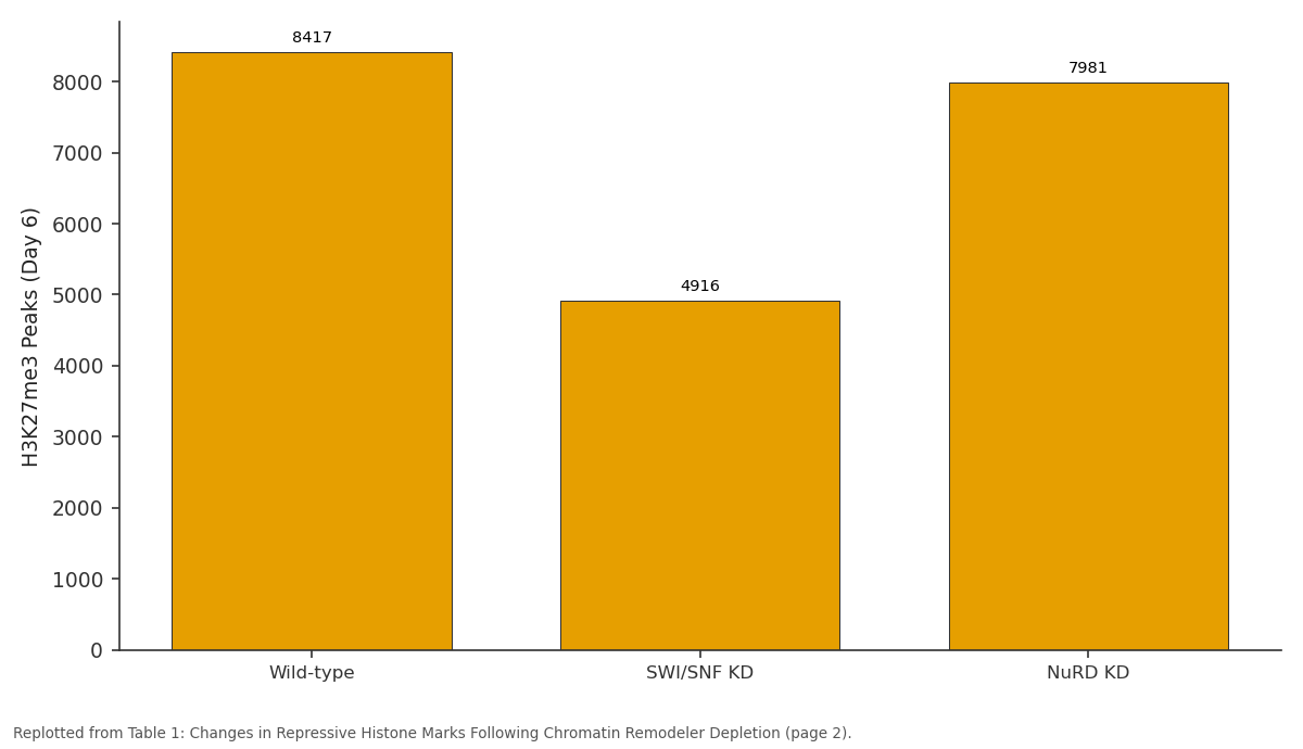
H3K27me3 Peaks (Day 6): Wild-type vs SWI/SNF KD vs NuRD KD
Replotted from Table 1 (page 2) of the original paper. Every bar is the paper's own table value.
View source evidence ↗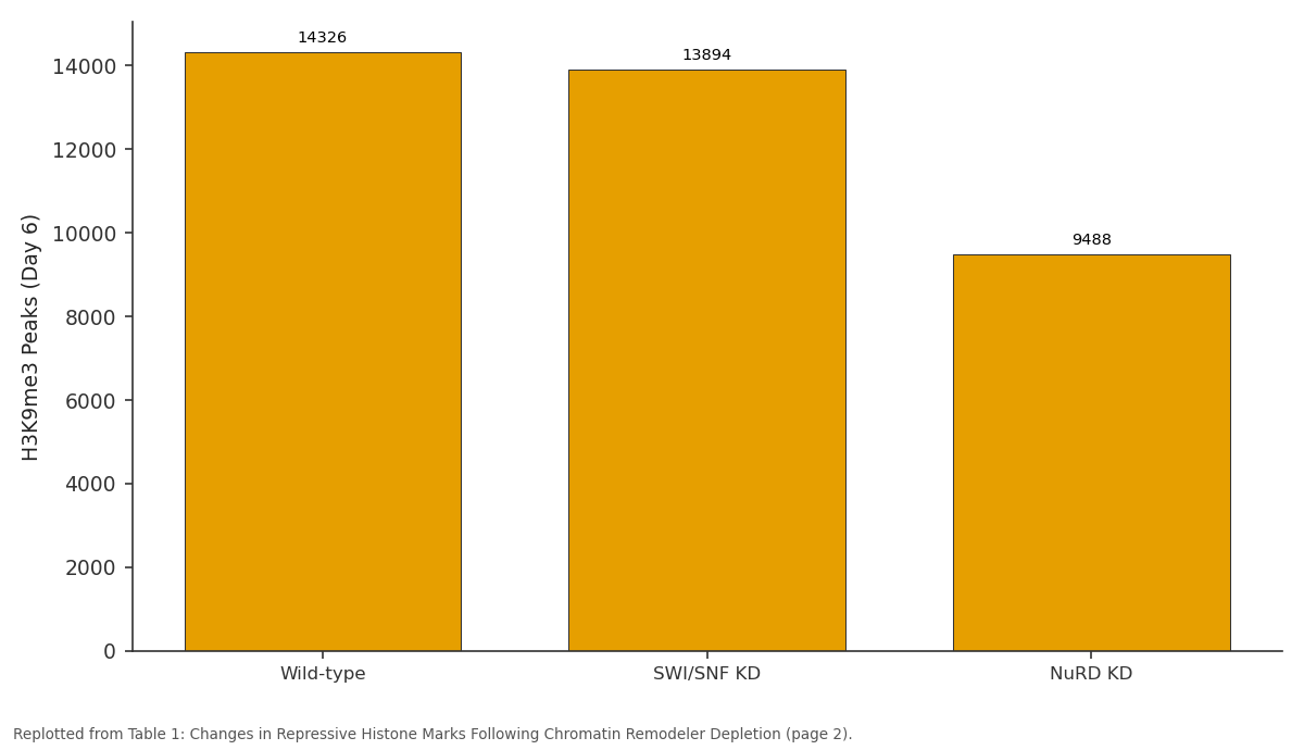
H3K9me3 Peaks (Day 6): Wild-type vs SWI/SNF KD vs NuRD KD
Replotted from Table 1 (page 2) of the original paper. Every bar is the paper's own table value.
View source evidence ↗Figures & tables
From the original paper Table 1
 Figure 1
 Figure 2
Introduction
Gene silencing is not a passive event. It demands active, energy-consuming molecular machinery that reshapes chromatin architecture to deny transcriptional activators access to DNA [1] . Among the best-characterised families of such machinery are the ATP-dependent chromatin remodeling complexes, which slide, eject, or restructure nucleosomes in response to developmental and environmental cues [2] . Two families in particular-SWI/SNF (also called BAF/PBAF in mammals) and NuRD (nucleosome remodeling and deacetylase)-have been linked to silencing programmes that guide embryonic cells away from pluripotency and toward lineage-specific fates [3,4] . SWI/SNF complexes are best known for their activating functions, opening chromatin at enhancers and promoters to enable gene expression [5] . But accumulating evidence shows they also participate in repression, particularly by facilitating Polycomb-mediated H3K27me3 deposition at genes that must be turned off during differentiation [6] . NuRD, by contrast, couples nucleosome remodeling with histone deacetylation, and its loss has been associated with impaired heterochromatin formation and derepression of repeat elements [7] . Despite these individual advances, few investigations have compared SWI/SNF and NuRD side by side within the same cell type and differentiation timeline. This gap matters because the two complexes may act on overlapping or distinct genomic targets, and understanding that division of labour could clarify why mutations in either family produce different developmental disorders in humans [8] . In this research, we used shRNA-mediated depletion of the catalytic subunits BRG1 (SWI/SNF) and CHD4 (NuRD) in mESCs undergoing directed neural differentiation, followed by ChIP-seq for two repressive histone marks and RNA-seq for transcriptome profiling across a six-day time course.
Material and Methods
Material: Murine embryonic stem cells (E14Tg2a line) were obtained from the European Collection of Authenticated Cell Cultures and maintained on gelatin-coated plates in serumfree 2i/LIF medium at the Masuria Institute of Freshwater Biology, Olsztyn, Poland.
Research was conducted between May 2023 and September 2024. Cells were passaged every 48 hours and tested negative for mycoplasma contamination before each experiment. Neural differentiation was initiated by withdrawing 2i/LIF and culturing cells in N2B27 medium for six days. The institutional laboratory safety committee approved all recombinant DNA work under protocol MIFB-rDNA-2023-04, dated 8 April 2023.
Reagents and Chemicals
CHIR99021 and PD0325901 (2i inhibitors) were purchased from Selleck Chemicals (Houston, TX, USA). Recombinant mouse LIF was from Millipore (Burlington, MA, USA). shRNA lentiviral vectors targeting Smarca4 (BRG1) and Chd4 were obtained from Sigma-Aldrich MISSION library (St Louis, MO, USA). ChIP-grade antibodies included anti-H3K27me3 (Cell Signaling Technology, 9733) and anti-H3K9me3 (Abcam, ab8898). Protein A magnetic beads were from Invitrogen (Carlsbad, CA, USA). All restriction enzymes and DNA polymerases came from New England Biolabs (Ipswich, MA, USA).
Instrumentation
ChIP-seq libraries were sequenced on an Illumina NextSeq 550 (single-end 75 bp). RNA-seq libraries were prepared with the NEBNext Ultra II Directional RNA Library Prep Kit and sequenced on the same platform (paired-end 75 bp). Protein knockdown efficiency was confirmed by western blotting using a Bio-Rad ChemiDoc MP imaging system. Flow cytometry for differentiation markers was performed on a BD FACSCanto II instrument. All centrifugation steps used an Eppendorf 5424 R microcentrifuge or a Thermo Scientific Sorvall Legend Micro 21R.
Methods
Lentiviral transduction was performed at a multiplicity of infection of 5, and puromycin selection (2 ug/mL) was applied for 48 hours. Knockdown efficiency exceeded 80% for both targets as verified by western blot densitometry. Cells were harvested at days 0, 3, and 6 of differentiation. For ChIP-seq, approximately 10 million cells per condition were crosslinked with 1% formaldehyde for 10 minutes, quenched with glycine, and sonicated to yield 200-500 bp fragments. Immunoprecipitation was performed overnight at 4 degrees Celsius. Libraries were prepared with the ThruPLEX DNA-seq Kit (Takara Bio, Kusatsu, Japan). Reads were aligned to mm10 with Bowtie2, and peaks were called using MACS2 (FDR < 0.05). Differential peak analysis used DiffBind in R. RNA-seq reads were aligned with STAR and quantified with featureCounts; differential expression was assessed using DESeq2 (adjusted p < 0.05, absolute log2 fold change > 1). Figure 2 illustrates that knockdown of either complex resulted in significantly higher residual expression of pluripotency markers compared with the active condition. SWI/SNF depletion had a slightly larger effect on median expression than NuRD depletion (Wilcoxon rank-sum test, p = 0.003 and p = 0.009, respectively).
Results
Results
Discussion
Our data paint a picture of functional complementarity between SWI/SNF and NuRD during neural differentiation. SWI/SNF appears to be the dominant facilitator of Polycomb-mediated H3K27me3 deposition-a finding that aligns with earlier reports showing that BAF complexes help recruit PRC2 to bivalent promoters in stem cells [6,9] . NuRD, meanwhile, was more closely tied to H3K9me3-dependent heterochromatin maintenance, consistent with its known interaction with SETDB1 and HP1 proteins [10] . The 214 genes deregulated under both knockdowns likely represent a core set of targets that require coordinated action from multiple remodeling pathways for proper silencing. Pathway analysis of this overlap set revealed enrichment for Wnt signalling and TGF-beta response genes-both of which must be tightly controlled during neural fate decisions [11,12] . One limitation is that shRNA-mediated knockdown achieves incomplete depletion, so the true extent of each complex's contribution may be underestimated. Additionally, our sixday differentiation window captures only early neural commitment; later stages of neurogenesis might reveal further divergence between the two pathways. Future work using degron-tagged subunits would allow acute, nearcomplete removal and sharper temporal resolution [13,14] .
Conclusion
This research shows that SWI/SNF and NuRD chromatin remodeling complexes make distinct yet complementary contributions to gene silencing during mESC neural differentiation. SWI/SNF primarily supports H3K27me3 deposition at lineage-inappropriate promoters, while NuRD is more important for H3K9me3-dependent heterochromatin integrity. Loss of either complex leads to the aberrant persistence of pluripotency transcripts, confirming that both are needed for complete silencing. These findings clarify the division of labour between two major remodeling families and may help explain why mutations in SWI/SNF subunits and NuRD subunits produce distinct but overlapping developmental syndromes in humans. Extending this analysis to later differentiation stages and to other lineages-such as mesoderm or endodermwill be necessary to determine whether the complementary relationship observed here is a general principle of chromatin-based gene regulation.
Figures and tables from the paper
| Condition | H3K27me3 Peaks (Day 6) | Change vs WT (%) | H3K9me3 Peaks (Day 6) | Change vs WT (%) | DEGs (RNA-seq) |
|---|---|---|---|---|---|
| Wild-type | 8,417 | - | 14,326 | - | - |
| SWI/SNF KD | 4,916 | -41.6 | 13,894 | -3.0 | 1,243 |
| NuRD KD | 7,981 | -5.2 | 9,488 | -33.8 | 978 |
Table text extracted from the original PDF; superscript letters are shown inline. The original table image is below.
View original table image
References
14 in the original paper- Narlikar GJ, Sundaramoorthy R, Owen-Hughes T. Mechanisms and functions of ATP-dependent chromatin-remodeling enzymes. Cell. 2013;154(3):490503.
- Clapier CR, Iwasa J, Cairns BR, Peterson CL. Mechanisms of action and regulation of ATP-dependent chromatin-remodelling complexes. Nature Reviews Molecular Cell Biology . 2017;18(7):407-422.
- Ho L, Ronan JL, Wu J, et al . An embryonic stem cell chromatin remodeling complex, esBAF, is essential for embryonic stem cell self-renewal and pluripotency. Proceedings of the National Academy of Sciences . 2009;106(13):5181-5186.
- Bornelov S, Reynolds N, Xenophontos M, et al . The nucleosome remodelling and deacetylase complex in development and disease. Biochemistry . 2018;57(26):3523-3532.
- Kadoch C, Hargreaves DC, Hodges C, et al . Proteomic and bioinformatic analysis of mammalian SWI/SNF complexes. Cell. 2013;153(1):71-85.
- Stanton BZ, Hodges C, Calarco JP, et al . Smarca4 ATPase mutations disrupt direct eviction of PRC1 from chromatin. Nature Genetics . 2017;49(2):282-288.
- Reynolds N, Salmon-Divon M, Dvinge H, et al . NuRDmediated deacetylation of H3K27 facilitates recruitment of Polycomb repressive complex 2. EMBO Journal . 2012;31(3):593-605.
- Wieczorek D, Bogershausen N, Beleggia F, et al . A comprehensive molecular study on Coffin-Siris and Nicolaides-Baraitser syndromes. Human Molecular Genetics . 2013;22(25):5121-5135.
- Bracken AP, Brien GL, Verrijzer CP. Dangerous liaisons: interplay between SWI/SNF, NuRD, and Polycomb in chromatin regulation. Genes and Development . 2019;33(15-16):936-959.
- Schultz DC, Ayyanathan K, Negorev D, Maul GG, Rauscher FJ. SETDB1: a novel KAP-1-associated histone H3, lysine 9-specific methyltransferase. Genes and Development . 2002;16(8):919-932.
- Chambers SM, Fasano CA, Papapetrou EP, et al . Highly efficient neural conversion of human ES and iPS cells by dual inhibition of SMAD signaling. Nature Biotechnology . 2009;27(3):275-280.
- Moya IM, Halder G. Hippo-YAP/TAZ signalling in organ regeneration and tissue repair. Nature Reviews Molecular Cell Biology . 2019;20(4):211-226.
- Nabet B, Roberts JM, Buckley DL, et al . The dTAG system for immediate and target-specific protein degradation. Nature Chemical Biology . 2018;14(5):431441.
- Mashtalir N, D'Avino AR, Michel BC, et al . Modular organization and assembly of SWI/SNF family chromatin remodeling complexes. Cell. 2018;175(5):1272-1288.