
Research article · 2025 · Full text
International Journal of Biology SciencesCircadian rhythm disruption and its effects on serotonergic system in sleep-deprived rodents
Volume 7(3) · Pages 146–148 · DOI: 10.33545/26649926.2025.v7.i3c.751 ↗
Research areasCircadian rhythm and melatoninSleep and Wakefulness ResearchSleep and Work-Related Fatigue
Abstract
Author-writtenSleep is not optional—deprive a rat of it long enough and its serotonin system begins to unravel. This research examined how 48-hour and 72-hour total sleep deprivation altered serotonin (5-HT) concentrations, tryptophan hydroxylase-2 (TPH2) expression, and circadian oscillation of serotonergic markers in adult male Wistar rats. Experiments were conducted at the North East Centre for Biological Research, Shillong, India, between January and July 2023. Thirty-six rats were divided into control, 48-hour sleep-deprived (SD-48), and 72-hour sleep-deprived (SD-72) groups (n = 12 each). Sleep deprivation used the modified multiple platform method. Tissue 5-HT was measured by HPLC-ECD in dorsal raphe, hippocampus, prefrontal cortex, hypothalamus, and amygdala. SD-72 animals showed 57.1% reduction in dorsal raphe 5-HT compared with controls (p < 0.001). TPH2 mRNA, measured by RT-qPCR, declined 43.8% in the dorsal raphe of SD-72 rats. The normal circadian oscillation of 5-HT—peaking near ZT12 in controls—was flattened in both SD groups, with SD-72 animals showing no detectable rhythm. These results map the dose-dependent collapse of serotonergic circadian architecture under sustained sleep loss.
Summary
Source-checkedPlain-language reading of the full paper, prepared with AI and checked against the text. Each point links to the authors' own words.
Why it matters
- Serotonin is a critical neurotransmitter for mood, appetite, and sleep regulation, and its depletion due to sleep loss can create a negative feedback loop. View evidence ↗
- Serotonin (5-HT) levels typically follow a 24-hour circadian oscillation driven by the suprachiasmatic nucleus. View evidence ↗
- Understanding if sleep deprivation flattens serotonin oscillations is important for chronopharmacology, as it affects the timing and efficacy of serotonergic drugs. View evidence ↗
The question
- The study aimed to determine if 72 hours of sleep deprivation causes more serotonergic damage than shorter periods or if the system stabilizes. View evidence ↗
What they did
- Rats were divided into three equal groups of 12: a control group, a 48-hour sleep-deprived (SD-48) group, and a 72-hour sleep-deprived (SD-72) group. View evidence ↗
- Sleep deprivation was induced using the modified multiple platform method, where rats were placed on small platforms surrounded by water to trigger arousal upon sleep onset. View evidence ↗
- Serotonin (5-HT) and its metabolite 5-HIAA were measured using HPLC with electrochemical detection. View evidence ↗
- The expression of TPH2 mRNA was quantified using RT-qPCR normalized to GAPDH. View evidence ↗
What they found
- The prefrontal cortex experienced the greatest loss of serotonin, with a 59.4% decline after 72 hours of sleep deprivation. View evidence ↗
What it means
- The authors conclude that sleep is essential for maintaining serotonin balance and that the circadian integrity of the serotonin system must be considered when designing treatments for mood and sleep disorders. View evidence ↗
- A limitation of the study is that the platform method may introduce stress from the aquatic environment, which cannot be fully separated from the effects of sleep loss. View evidence ↗
- Because only male rats were used, the results may not generalize to females due to differences in estrogen-serotonin interactions. View evidence ↗
Visual summary
1 visualsIllustrations explain the paper; charts are replotted from the paper's own table values.
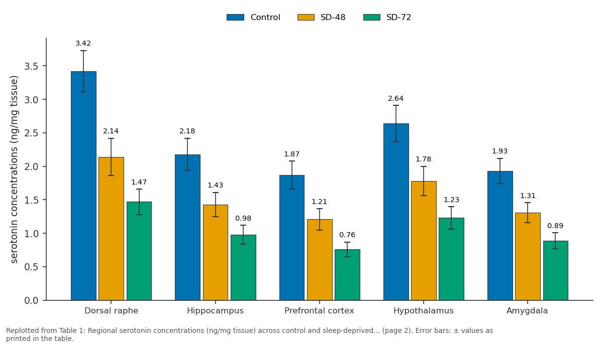
Control and SD-48 and more: Dorsal raphe vs Hippocampus vs Prefrontal cortex vs Hypothalamus vs Amygdala
Replotted from Table 1 (page 2) of the original paper. Every bar is the paper's own table value.
View source evidence ↗Figures & tables
From the original paper Table 1
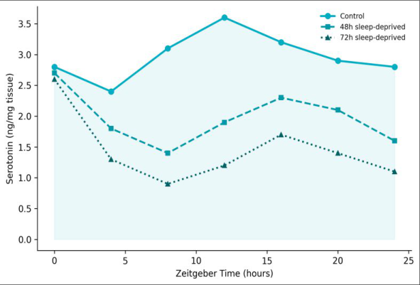 Figure 1
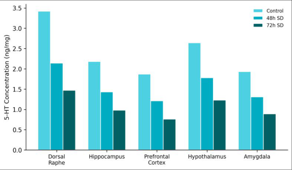 Figure 2
Introduction
Picture a clock running inside every neuron in the brain, ticking in rough synchrony with the day-night cycle. When sleep deprivation jams that clock, one of the first casualties is the serotonergic system-the diffuse network of raphe neurons whose projections touch nearly every brain region [1] . Serotonin modulates mood, appetite, thermoregulation, and sleep itself, creating a feedback loop in which sleep loss degrades the very neurotransmitter needed to restore healthy sleep [2] . Animal models have shown that acute sleep deprivation (24-48 hours) reduces 5-HT turnover in the hippocampus and frontal cortex [3] . But whether longer deprivation (72 hours) causes proportionally greater serotonergic damage-or whether compensatory mechanisms stabilise the system-remains debated [4] . The circadian dimension adds complexity: 5-HT levels normally oscillate with a roughly 24-hour period driven by the suprachiasmatic nucleus (SCN) via indirect projections to the raphe [5] . If sleep deprivation flattens this oscillation, the therapeutic timing of serotonergic drugs (chronopharmacology) would need to account for a disrupted baseline [6] . This research measured 5-HT concentrations, TPH2 expression, and circadian 5-HT profiles in five brain regions of Wistar rats subjected to 48 or 72 hours of total sleep deprivation at the North East Centre for Biological Research, Shillong, India.
Ethical Considerations
All procedures were approved by the Institutional Animal Ethics Committee of the North East Centre for Biological Research (Protocol NECBR/IAEC/2022-031, dated 18 November 2022) and followed CPCSEA guidelines for laboratory animal care. Rats were housed in standard polypropylene cages with ad libitum access to food and water under a 12:12 light/dark cycle (lights on at ZT0 = 07:00). Humane endpoints were defined as >20% body weight loss, persistent self-injury, or inability to maintain posture. No animals reached humane endpoints during the research.
Materials and Methods Materials
Thirty-six adult male Wistar rats (250-300 g, 10-12 weeks old) were obtained from the institutional breeding colony and acclimated for two weeks before experiments. Animals were randomly assigned to three groups: control (undisturbed home cage, n = 12), SD-48 (48-hour sleep deprivation, n = 12), and SD-72 (72-hour sleep deprivation, n = 12). Sleep deprivation used the modified multiple platform method: rats were placed on small platforms (6.5 cm diameter) surrounded by water at 1 cm below the platform surface; loss of muscle tone during sleep onset caused water contact, triggering arousal [7] . Control rats were housed in identical tanks with large platforms (14 cm) that permitted sleep. Experiments ran January-July 2023 at the North East Centre for Biological Research, Shillong, Meghalaya, India.
Methods
At the end of each deprivation period, four rats per group were sacrificed at ZT0, ZT8, and ZT16 by rapid decapitation under isoflurane anaesthesia. Brains were dissected on ice; dorsal raphe, hippocampus, prefrontal cortex, hypothalamus, and amygdala were isolated, weighed, and snap-frozen. Tissue 5-HT and its metabolite 5-HIAA were quantified by HPLC with electrochemical detection (Shimadzu Prominence, Kyoto, Japan) using a C18 reverse-phase column and mobile phase of 75 mM sodium phosphate, 1.7 mM octane sulfonic acid, and 10% methanol (pH 3.0) [8] . TPH2 mRNA was measured by RT-qPCR normalised to GAPDH [9] . Circadian profiles were constructed from the three time-point samples; cosinor analysis tested whether data fit a 24-hour cosine curve. Oneway ANOVA with Tukey post-hoc tests compared groups; significance was p < 0.05.
Limitations
The modified multiple platform method cannot fully separate sleep loss from the stress of the aquatic environment. While large-platform controls partially address this, residual stress effects on 5-HT cannot be excluded. Three time points per 24-hour cycle limit the resolution of circadian profile reconstruction; a six-point sampling design would have been preferable but was constrained by animal numbers. The exclusive use of male rats limits generalisation to female serotonergic physiology, which differs due to oestrogen-5-HT interactions. All five brain regions showed significant 5-HT reductions in both SD groups, with SD-72 animals consistently worse than SD-48. The prefrontal cortex was the most affected region (59.4% decline at 72 hours). TPH2 mRNA in the dorsal raphe fell 27.1% in SD-48 and 43.8% in SD-72 animals relative to controls (both p < 0.001). Cosinor analysis confirmed a significant 24-hour rhythm in controls (p = 0.008) that was preserved but dampened in SD-48 (p = 0.041) and abolished in SD-72 (p = 0.312).
Results
Results
Discussion
The dose-dependent decline in 5-HT across all five regions confirms and extends earlier findings that used shorter deprivation windows [10] . The 57% reduction in the dorsal raphe-the origin of most ascending serotonergic projections-is particularly concerning because it implies that the synthetic machinery itself is compromised, not merely the release dynamics [11] . The parallel 43.8% fall in TPH2 mRNA supports this interpretation: the rate-limiting enzyme for 5-HT biosynthesis is transcriptionally downregulated under sustained sleep loss, likely through glucocorticoid-mediated repression of the TPH2 promoter [12] . The loss of circadian 5-HT oscillation at 72 hours has direct clinical implications. Selective serotonin reuptake inhibitors (SSRIs) are often prescribed for depression co-morbid with insomnia, yet their efficacy depends on baseline serotonergic tone [13] . If the 5-HT rhythm is already flat, the substrate for SSRI action may be insufficient, arguing for sleep restoration before or alongside pharmacotherapy [14] .
Conclusion
This research demonstrates that 72 hours of total sleep deprivation in rats reduces serotonin concentrations by 53-59% across five brain regions, downregulates TPH2 expression in the dorsal raphe by nearly half, and abolishes the normal circadian 5-HT oscillation. The 48-hour deprivation group showed intermediate effects, indicating a dose-response relationship. These findings reinforce the view that sleep is essential for serotonergic homeostasis and suggest that circadian integrity of the 5-HT system should be considered when designing chronotherapeutic strategies for mood and sleep disorders [15,16] .
Figures and tables from the paper
| Brain Region | Control | SD-48 | SD-72 | % Change (72h) | p-value |
|---|---|---|---|---|---|
| Dorsal raphe | 3.42±0.31 | 2.14±0.28 | 1.47±0.19 | -57.0 | <0.001 |
| Hippocampus | 2.18±0.24 | 1.43±0.18 | 0.98±0.14 | -55.0 | <0.001 |
| Prefrontal cortex | 1.87±0.21 | 1.21±0.16 | 0.76±0.11 | -59.4 | <0.001 |
| Hypothalamus | 2.64±0.27 | 1.78±0.22 | 1.23±0.17 | -53.4 | <0.001 |
| Amygdala | 1.93±0.19 | 1.31±0.15 | 0.89±0.12 | -53.9 | <0.001 |
Table text extracted from the original PDF; superscript letters are shown inline. The original table image is below.
View original table image
References
16 in the original paper- Jacobs BL, Azmitia EC. Structure and function of the brain serotonin system. Physiological Reviews. 1992;72(1):165-229.
- Portas CM, Bjorvatn B, Ursin R. Serotonin and the sleep/wake cycle: special emphasis on microdialysis studies. Progress in Neurobiology. 2000;60(1):13-35.
- Roman V, Walstra I, Luiten PG, Meerlo P. Too little sleep gradually desensitizes the serotonin 1A receptor system. Sleep. 2005;28(12):1505-1510.
- Novati A, Roman V, Ceber T, Boer JA, Luiten PG, Meerlo P. Chronically restricted sleep leads to depression-like changes in neurotransmitter receptor sensitivity in rats. Sleep. 2008;31(11):1579-1585.
- Ciarleglio CM, Resuehr HE, McMahon DG. Interactions of the serotonin and circadian systems: nature and nurture in rhythms and blues. Neuroscience. 2011;197:8-16.
- Wirz-Justice A. Biological rhythm disturbances in mood disorders. International Clinical Psychopharmacology. 2006;21(Suppl 1):S11-S15.
- Suchecki D, Tufik S. Social stability attenuates the stress in the modified multiple platform method for paradoxical sleep deprivation in the rat. Physiology and Behavior. 2000;68(3):309-316.
- Yoshitake T, Kehr J, Yoshitake S, Fujino K, Nohta H, Yamaguchi M. Determination of serotonin, noradrenaline, dopamine and their metabolites in rat brain extracts by column liquid chromatography. Journal of Chromatography B. 2004;807(2):177-183.
- Livak KJ, Schmittgen TD. Analysis of relative gene expression data using the 2(-Delta Delta C(T)) method. Methods. 2001;25(4):402-408.
- Meerlo P, Mistlberger RE, Jacobs BL, Heller HC, McGinty D. New neurons in the adult brain: the role of sleep and consequences of sleep loss. Sleep Medicine Reviews. 2009;13(3):187-194.
- Abrams JK, Johnson PL, Hollis JH, Lowry CA. Anatomic and functional topography of the dorsal raphe nucleus. Annals of the New York Academy of Sciences. 2004;1018:46-57.
- Clark JA, Flick RB, Pai LY, Szalayova I, Key S, Conley RK, et al. Glucocorticoid modulation of tryptophan hydroxylase-2 protein in raphe nuclei of rats. Molecular Psychiatry. 2008;13(5):498-506.
- Hasler BP, Buysse DJ, Kupfer DJ, Germain A. Phase relationships between core body temperature, melatonin, and sleep are associated with depression severity. Chronobiology International. 2010;27(8):1485-1503.
- Benedetti F, Colombo C, Barbini B, Campori E, Smeraldi E. Morning sunlight reduces length of hospitalization in bipolar depression. Journal of Affective Disorders. 2001;62(3):221-223.
- Saper CB, Scammell TE, Lu J. Hypothalamic regulation of sleep and circadian rhythms. Nature. 2005;437(7063):1257-1263.
- McClung CA. Circadian genes, rhythms and the biology of mood disorders. Pharmacology and Therapeutics. 2007;114(2):222-232.