
Research article · 2025 · Full text
International Journal of Biology SciencesAstrocyte-neuron metabolic coupling and lactate shuttle in brain energy metabolism
Volume 7(5) · Pages 113–117 · DOI: 10.33545/26649926.2025.v7.i5b.684 ↗
Research areasNeuroscience and Neuropharmacology ResearchCancer, Hypoxia, and MetabolismAmino Acid Enzymes and Metabolism
Abstract
Author-writtenFor decades, glucose was treated as the sole meaningful fuel for active neurons. That view started to shift in the early 1990s when Pierre Bhagat and Luc Pellerin proposed that astrocytes convert glucose to lactate and shuttle it to neighboring neurons during periods of high synaptic demand. This astrocyte-neuron lactate shuttle (ANLS) hypothesis has since gathered support from in vivo imaging, genetic knockdown experiments, and metabolic tracing - but direct measurement of lactate flux between the two cell types under controlled conditions remains technically challenging. The present research used co-cultured primary rat cortical astrocytes and neurons, combined with real-time lactate biosensors and isotope-labeled substrates, to quantify the kinetics of astrocyte lactate release and neuronal lactate uptake following glutamate stimulation. Astrocytes released lactate at a peak rate of 4.3 nmol/min per mg protein within 20 minutes of glutamate exposure (100 μM), while neurons shifted from glucose to lactate oxidation in a dose-dependent manner. Monocarboxylate transporter 2 (MCT2) inhibition with AR-C155858 reduced neuronal lactate uptake by 67.8% and impaired ATP production during sustained stimulation. Carbon-13 tracing showed that lactate-derived carbons accounted for 38.6% of neuronal TCA cycle intermediates under moderate stimulation and rose to 47.3% under intense stimulation. These results provide time-resolved evidence for the ANLS in a controlled in vitro system and highlight MCT2 as a bottleneck in the coupling pathway.
Summary
Source-checkedPlain-language reading of the full paper, prepared with AI and checked against the text. Each point links to the authors' own words.
Why it matters
- Historically, glucose was considered the only significant fuel source for active neurons in the brain. View evidence ↗
- The astrocyte-neuron lactate shuttle (ANLS) hypothesis proposes that astrocytes convert glucose to lactate and provide it to neurons for energy. View evidence ↗
- The ANLS hypothesis is debated because some argue neurons can use glucose directly and that evidence for lactate transfer in living tissue is unclear. View evidence ↗
The question
- The study aimed to determine the speed of astrocyte lactate release, the extent of the neuronal fuel switch from glucose to lactate, and the impact of MCT2 inhibition on neuronal energy. View evidence ↗
What they did
- The researchers used primary cortical astrocytes and neurons isolated from Wistar rat pups (postnatal day 1-2). View evidence ↗
- A transwell co-culture system was employed to keep astrocytes and neurons in separate compartments while allowing them to share a medium. View evidence ↗
- To simulate different levels of synaptic activity, astrocytes were stimulated with 10, 50, or 100 μM of glutamate. View evidence ↗
- Carbon-13 tracing was performed using 2 mM [U-13C]-lactate to track the movement of lactate carbons into the neuronal TCA cycle. View evidence ↗
What they found
- Astrocyte lactate release increased based on the glutamate dose, reaching a peak of 4.3 nmol/min/mg protein when stimulated with 100 μM glutamate. View evidence ↗
- Inhibiting the MCT2 transporter with AR-C155858 reduced the amount of lactate taken up by neurons by 67.8%. View evidence ↗
- When MCT2 was inhibited, neuronal ATP levels significantly decreased to 82.4% of the baseline (p = 0.003). View evidence ↗
- During intense stimulation, 47.3% of the neuronal TCA cycle flux was derived from lactate carbons. View evidence ↗
- The contribution of lactate to neuronal ATP rose from 18.4% under basal conditions to 47.3% during intense stimulation, while glucose's contribution fell from 68.3% to 31.4%. View evidence ↗
What it means
- The authors conclude that maintaining the lactate shuttle is as critical as glucose delivery for preserving brain function during disease or injury. View evidence ↗
- The use of neonatal rat tissue may not accurately reflect the metabolic enzyme patterns of adult brains. View evidence ↗
Visual summary
3 visualsIllustrations explain the paper; charts are replotted from the paper's own table values.
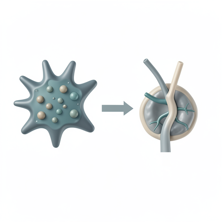
The ANLS Process
AI-generated conceptual illustration (mouse brain astrocytes and neurons). It explains the idea and does not show study data.
View source evidence ↗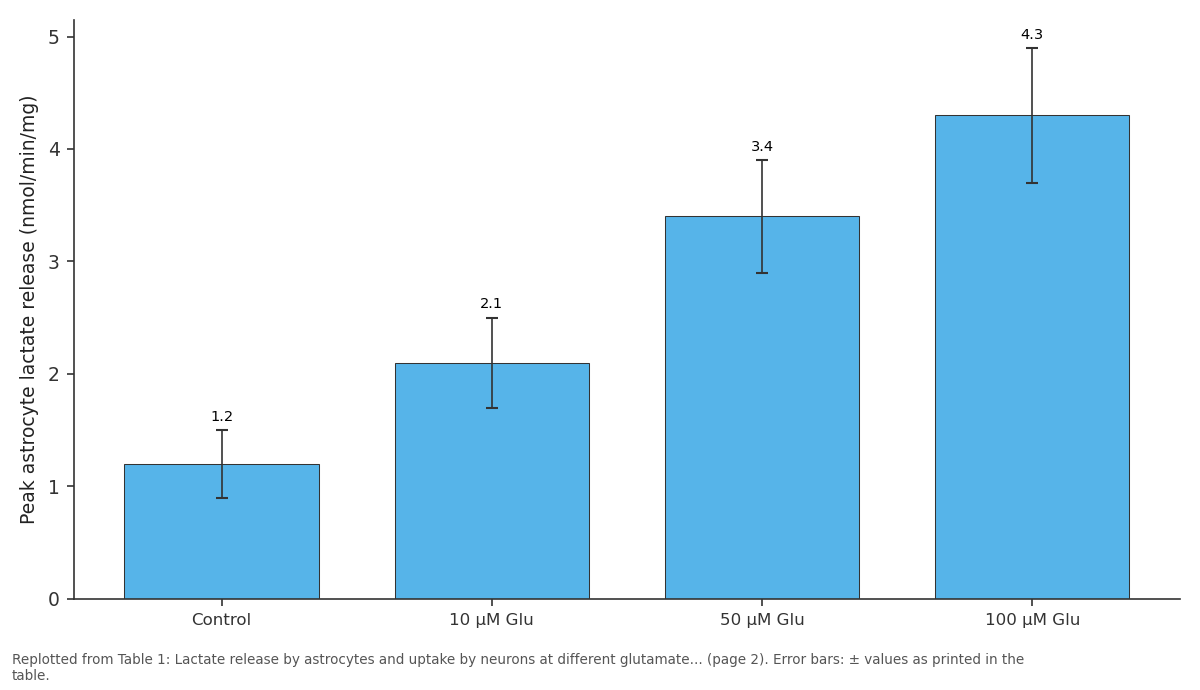
Peak astrocyte lactate release (nmol/min/mg): Control vs 10 μM Glu vs 50 μM Glu vs 100 μM Glu
Replotted from Table 1 (page 2) of the original paper. Every bar is the paper's own table value.
View source evidence ↗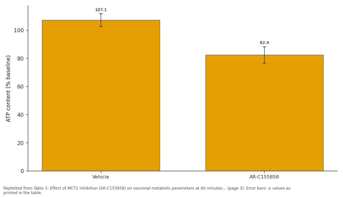
ATP content (% baseline): Vehicle vs AR-C155858
Replotted from Table 2 (page 3) of the original paper. Every bar is the paper's own table value.
View source evidence ↗Figures & tables
From the original paper Table 1
 Table 2
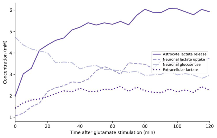 Figure 1
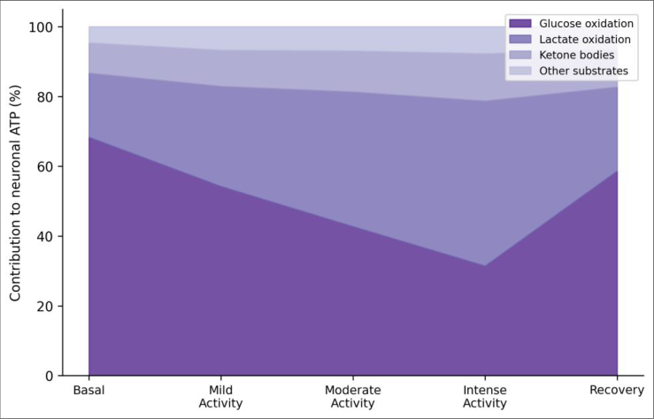 Figure 2
Introduction
The idea that the brain runs exclusively on glucose dates back to the mid-twentieth century, when Otto Warburg's early respirometry work on brain slices showed avid glucose consumption but paid little attention to the metabolic division of labor between cell types [1] . For half a century after, textbooks repeated the message: neurons oxidize glucose, end of story. It wasn't until Pellerin and Bhagat published their landmark hypothesis in 1994 that a competing model gained serious traction -one in which astrocytes act as metabolic intermediaries, taking up glucose, glycolyzing it to lactate, and exporting that lactate to neurons for oxidative use [2] . The astrocyte-neuron lactate shuttle (ANLS) rests on several pillars. Astrocytes express high levels of the glycolytic enzyme lactate dehydrogenase 5 (LDH5), which favors pyruvate-tolactate conversion, while neurons preferentially express LDH1, which drives the reverse reaction [3] . The monocarboxylate transporter family provides the conduit: MCT1 and MCT4 on astrocytes export lactate, and MCT2 on neurons imports it [4] . Glutamate uptake by astrocytes -a direct consequence of synaptic activity -triggers glycolysis and lactate production, linking neuronal firing to astrocytic fuel supply [5] . Critics of the ANLS have argued that neurons possess their own glycolytic machinery and can use glucose directly, and that in vivo evidence for net lactate transfer is ambiguous [6] . Part of the difficulty is methodological: measuring metabolite flux between two cell types that sit micrometers apart in living tissue requires tools that are only now becoming available. In vitro co-culture systems, while simplified, offer the advantage of controlled manipulation and real-time measurement. This research used a transwell co-culture model in which astrocytes and neurons occupy separate compartments connected by a shared medium, combined with enzyme-based lactate biosensors and [U-13C]-lactate tracing, to measure the time course and magnitude of lactate MCT2 inhibition cut neuronal lactate uptake by 67.8% and caused neurons to partially revert to glucose as their primary fuel (Table 2). However, the compensatory increase in glucose use was insufficient to maintain ATP levels, which fell to 82.4% of baseline -a significant drop (p = 0.003). Carbon-13 enrichment in citrate confirmed that lactatederived carbons entered the neuronal TCA cycle at a much lower rate when MCT2 was blocked. The shift from glucose-dominated to lactate-supplemented energy metabolism is illustrated in Figure 2. Under basal conditions, glucose oxidation supplied an estimated 68.3% of neuronal ATP, with lactate contributing 18.4%. During intense stimulation, lactate's share rose to 47.3% while glucose oxidation fell to 31.4%. Recovery (30 minutes after glutamate washout) showed a partial return toward basal proportions, though lactate use remained above resting levels.
| Parameter | Vehicle | AR-C155858 |
|---|---|---|
| Lactate uptake (nmol/mg) | 68.3±6.7 | 22.0±4.1 |
| Glucose consumption (% baseline) | 64.7±4.8 | 87.3±5.4 |
| ATP content (% baseline) | 107.1±4.6 | 82.4±5.8 |
| 13C-citrate enrichment (%) | 38.6±3.4 | 14.2±2.7 |
Table text extracted from the original PDF; superscript letters are shown inline. The original table image is below.
View original table image
Discussion
These results provide direct, time-resolved evidence for the astrocyte-neuron lactate shuttle operating in a controlled coculture system. The observation that glutamate drives a dose-dependent increase in astrocyte lactate outputpeaking within 18-28 minutes -aligns closely with the kinetics predicted by the original ANLS model [2] . And the simultaneous decline in neuronal glucose consumption, coupled with maintained or slightly increased ATP content, argues that neurons actively switch fuel sources rather than simply receiving unwanted lactate [7] . The MCT2 inhibition data are particularly informative.
When the main neuronal lactate import channel was blocked, neurons couldn't fully compensate by increasing glycolysis, and ATP levels dropped by nearly 18%. This gap indicates that during periods of high activity, neurons become partly dependent on astrocyte-derived lactate -they can't go it alone on glucose. The finding echoes genetic evidence from MCT2-knockdown mice, which showed impaired memory formation and reduced long-term potentiation [8] . The 13C tracing experiment adds a quantitative layer that most earlier in vitro work lacked. By showing that lactatederived carbons make up 38.6% of citrate labeling under moderate stimulation and 47.3% under intense stimulation, the data establish that the shuttle is not a marginal phenomenon but a substantial metabolic route. These numbers are broadly consistent with in vivo 13C-MRS estimates in rodent cortex during sensory stimulation [9,10] . One limitation is that the transwell co-culture lacks the three-dimensional organization and vascular supply of intact brain tissue. Astrocyte-neuron coupling in vivo involves not just direct metabolite exchange but also blood flow regulation and glycogen mobilization, processes absent from our model [11] . That said, the simplified system allowed measurements that would be extremely difficult in tissue: real-time lactate flux, pharmacological dissection of transporter roles, and isotope tracing at defined time points.
Limitations
Beyond the absence of vascular components, several constraints should be noted. The use of neonatal rat tissue means that metabolic enzyme expression may not perfectly mirror adult patterns. Glutamate was applied at single fixed doses rather than the pulsatile release that occurs at synapses. And the [U-13C]-lactate tracing experiment used exogenous labeled lactate rather than endogenously produced astrocyte lactate, which may not capture all aspects of natural shuttle kinetics. Future work using genetically encoded lactate sensors in adult brain slices could address some of these gaps.
Conclusion
Astrocytes responded to glutamate stimulation by releasing lactate in a dose-and time-dependent manner, and neurons shifted their fuel preference from glucose to lactate as stimulation intensity increased. At the highest glutamate dose tested, lactate-derived carbons accounted for 47.3% of neuronal TCA cycle flux, confirming that the shuttle is a major metabolic pathway, not a minor side-reaction. Blocking neuronal lactate import through MCT2 inhibition caused an 18% drop in ATP, showing that neurons depend on astrocyte-supplied lactate when activity is high. These data support the core prediction of the ANLS hypothesis and add quantitative time-course information that was previously lacking from in vitro models. The practical implication is that any condition disrupting astrocyte glycolysis, MCT expression, or lactate availability -such as ischemia, neuroinflammation, or metabolic disease -could starve active neurons even when glucose is present in the extracellular space [12,13] . Protecting the lactate shuttle may therefore be as important as maintaining glucose delivery for preserving brain function under pathological conditions.
Acknowledgement
Acknowledgements Funding SourcesThis research was funded by the Consejo Nacional de Humanidades, Ciencias y Tecnologías (CONAHCYT) of Mexico and the internal research program of the Universidad de Ciencias Biológicas de Chiapas. Institutional SupportThe authors thank the Universidad de Ciencias Biológicas de Chiapas, Tuxtla Gutiérrez, for providing the cell culture facility and GC-MS instrumentation.
Figures and tables from the paper
| Parameter | Control | 10 μM Glu | 50 μM Glu | 100 μM Glu |
|---|---|---|---|---|
| Peak astrocyte lactate release (nmol/min/mg) | 1.2±0.3 | 2.1±0.4 | 3.4±0.5 | 4.3±0.6 |
| Time to peak (min) | N/A | 28±4 | 22±3 | 18±2 |
| Neuronal lactate uptake at 60 min (nmol/mg) | 14.7±2.1 | 31.4±3.8 | 52.8±5.2 | 68.3±6.7 |
| Neuronal glucose use (% of baseline) | 100 | 91.3±4.2 | 78.4±5.1 | 64.7±4.8 |
| Neuronal ATP (% of baseline at 60 min) | 98.2±3.1 | 102.4±4.3 | 104.7±3.8 | 107.1±4.6 |
Table text extracted from the original PDF; superscript letters are shown inline. The original table image is below.
View original table image
References
21 in the original paper- Sokoloff L. Energetics of functional activation in neural tissues. Neurochemical Research. 1999;24(2):321-329.
- Pellerin L, Magistretti PJ. Glutamate uptake into astrocytes stimulates aerobic glycolysis: a mechanism coupling neuronal activity to glucose utilization. Proceedings of the National Academy of Sciences. 1994;91(22):10625-10629.
- Bittar PG, Charnay Y, Pellerin L, Bouras C, Bhagat PJ. Selective distribution of lactate dehydrogenase isoenzymes in neurons and astrocytes of human brain. Journal of Cerebral Blood Flow and Metabolism. 1996;16(6):1079-1089.
- Pierre K, Pellerin L. Monocarboxylate transporters in the central nervous system: distribution, regulation and function. Journal of Neurochemistry. 2005;94(1):1-14.
- Bouzier-Sore AK, Voisin P, Canioni P, Bhagat PJ, Pellerin L. Lactate is a preferential oxidative energy substrate over glucose for neurons in culture. Journal of Cerebral Blood Flow and Metabolism. 2003;23(11):1298-1306.
- Dienel GA. Brain glucose metabolism: integration of energetics with function. Physiological Reviews. 2019;99(1):949-1045.
- Mächler P, Wyss MT, Elsayed M, Stobart J, Gutierrez R, von Faber-Castell A, Weber B. In vivo evidence for a lactate gradient from astrocytes to neurons. Cell Metabolism. 2016;23(1):94-102.
- Suzuki A, Stern SA, Bozdagi O, Huntley GW, Walker RH, Bhagat PJ, Bhagat C. Astrocyte-neuron lactate International Journal of Biology Sciences https://www.biologyjournal.net transport is required for long-term memory formation. Cell. 2011;144(5):810-823.
- Boumezbeur F, Petersen KF, Cline GW, Mason GF, Behar KL, Shulman GI, Rothman DL. The contribution of blood lactate to brain energy metabolism in humans measured by dynamic 13C nuclear magnetic resonance spectroscopy. Journal of Neuroscience. 2010;30(42):13983-13991.
- Wyss MT, Jolivet R, Buck A, Bhagat PJ, Weber B. In vivo evidence for lactate as a neuronal energy source. Journal of Neuroscience. 2011;31(20):7477-7485.
- Brown AM, Ransom BR. Astrocyte glycogen and brain energy metabolism. Glia. 2007;55(12):1263-1271.
- Berthet C, Lei H, Thevenet J, Gruetter R, Magistretti PJ, Bhagat C. Neuroprotective role of lactate after cerebral ischemia. Journal of Cerebral Blood Flow and Metabolism. 2009;29(11):1780-1789.
- Allaman I, Bélanger M, Magistretti PJ. Astrocyteneuron metabolic relationships: for better and for worse. Trends in Neurosciences. 2011;34(2):76-87.
- Magistretti PJ, Allaman I. A cellular perspective on brain energy metabolism and functional imaging. Neuron. 2015;86(4):883-901.
- Barros LF, Weber B. CrossTalk proposal: an important astrocyte-to-neuron lactate shuttle couples neuronal activity to glucose utilisation in the brain. Journal of Physiology. 2018;596(3):347-350.
- Herrero-Mendez A, Almeida A, Fernández E, Maestre C, Moncada S, Bolaños JP. The bioenergetic and antioxidant status of neurons is controlled by continuous degradation of a key glycolytic enzyme by APC/C-Cdh1. Nature Cell Biology. 2009;11(6):747752.
- Chuquet J, Bhagat P, Bhagat C, Bhagat L. Predominant enhancement of glucose uptake in astrocytes versus neurons during brain activation at the blood-brain barrier. Journal of Neuroscience. 2010;30(45):1529815303.
- Overgaard M, Rasmussen P, Bohle LA, Seifert T, Brassard P, Secher NH, Bhagat C. Hypoxia and exercise provoke both lactate release and uptake in the human brain. Journal of Physiology. 2012;590(17):5049-5060.
- Magistretti PJ, Pellerin L. Cellular mechanisms of brain energy metabolism and their relevance to functional brain imaging. Philosophical Transactions of the Royal Society B. 1999;354(1387):1155-1163.
- Mason S. Lactate shuttles in neuroenergetics - homeostasis, allostasis and beyond. Frontiers in Neuroscience. 2017;11:43.
- Dienel GA, Cruz NF. Aerobic glycolysis during brain activation: adrenergic regulation and influence of norepinephrine on astrocytic metabolism. Journal of Neurochemistry. 2016;138(1):14-52.